Epiregulin is a member of the EGF family of growth factors which includes, among others, epidermal growth factor (EGF), transforming growth factor (TGF)-alpha, amphiregulin (ARG), HB (heparin-binding)-EGF, betacellulin, and the various heregulins. All EGF family members are synthesized as transmembrane precursors and are converted to soluble forms by proteolytic cleavage. Epiregulin was originally purified from the mouse fibroblast-derived tumor cell line NIH3T3/T7. The human epiregulin cDNA encodes a 169 amino acid (aa) residues transmembrane precursor with a 29 aa signal peptide, a 21 aa transmembrane domain and a 21 aa cytoplasmic domain. The putative soluble mature Epiregulin comprising the EGF-like domain (aa residues 64-104) is formed by proteolytic removal of the propeptide regions. There is 85% aa sequence homology between human and mouse epiregulins. Epiregulin is expressed primarily in the placenta and macrophages. High level expression has also been detected in various carcinomas. Epiregulin specifically binds EGF R (ErbB1) and ErbB4 but not ErbB2 and ErbB3. It activates the homodimers of both ErbB1 and ErbB4. In addition, epiregulin can also activate all possible heteromeric combinations of the four ErbB family members. Epiregulin stimulates the proliferation of fibroblasts, smooth muscle cells and hepatocytes. It has been shown to be an autocrine growth factor for epidermal keratinocytes as well as mesangial cells. Epiregulin has also been shown to inhibit growth of several epithelial tumor cells. In addition, Epiregulin has been implicated in the implantation process during pregnancy.

Key Product Details
Species Reactivity
Validated:
Cited:
Applications
Validated:
Cited:
Label
Antibody Source
Product Specifications
Immunogen
Val63-Leu108
Accession # O14944
Specificity
Clonality
Host
Isotype
Endotoxin Level
Scientific Data Images for Human Epiregulin Antibody
Cell Proliferation Induced by Epiregulin and Neutralization by Human Epiregulin Antibody.
Recombinant Human Epiregulin (Catalog # 1195-EP) stimulates proliferation in the Balb/3T3 mouse embryonic fibroblast cell line in a dose-dependent manner (orange line). Proliferation elicited by Recombinant Human Epiregulin is neutralized (green line) by increasing concentrations of Goat Anti-Human Epiregulin Antigen Affinity-purified Polyclonal Antibody (Catalog # AF1195). The ND50 is typically ≤1.5 µg/mL.
Human Epiregulin ELISA Standard Curve.
Recombinant Human Epiregulin protein was serially diluted 2-fold and captured by Rat Anti-Human Epiregulin Monoclonal Antibody (Catalog # MAB14252) coated on a Clear Polystyrene Microplate (Catalog # DY990). Goat Anti-Human Epiregulin Antigen Affinity-purified Polyclonal Antibody (Catalog # AF1195) was biotinylated and incubated with the protein captured on the plate. Detection of the standard curve was achieved by incubating Streptavidin-HRP (Catalog # DY998) followed by Substrate Solution (Catalog # DY999) and stopping the enzymatic reaction with Stop Solution (Catalog # DY994).
Detection of Human Epiregulin by Immunohistochemistry
EREG expression correlates with MMP-1 levels in human DCIS lesions. a qRT-PCR analysis of MMP-1 gene expression in MCF10A and MCF10DCIS cells. Expression levels of MMP-1 are normalized to CYBP. b Immunoblot analysis of MMP-1 in conditioned media obtained from MCF10A and MCF10DCIS cells. Loading was assessed by Coomassie staining (Additional file 1: Figure S1C). c qRT-PCR analysis of MMP-1 gene expression in MCF10DCIS cells expressing either NT or EREG shRNA constructs. Expression levels of MMP-1 are normalized to CYBP. d Immunohistochemistry of normal breast tissue and DCIS stained with an anti-EREG antibody. Scale bars represent 50 μm. e Normal and DCIS human samples were stained with an anti-MMP-1 antibody. Representative images of normal and varying levels of MMP-1 staining in DCIS lesions are shown. f Quantification of the percent of samples staining positive for EREG. g Quantification of the percent of samples staining positive for MMP-1. Scale bars represent 50 μm. *p < 0.05. ***p < 0.0001 Image collected and cropped by CiteAb from the following open publication (https://pubmed.ncbi.nlm.nih.gov/26215578), licensed under a CC-BY license. Not internally tested by R&D Systems.
Detection of Epiregulin by Immunocytochemistry/ Immunofluorescence
EREG increased IL-8 production from cancer cells through EGFR signaling to induce tumor neovascularization. (A) Representative images of the change in angiogenic factors in both Huh7 and HepG2 cells with/without stimulation with EREG. IL-8 levels in Huh7 cells only increased during stimulation with rEREG. Red box showed the expression of IL-8. The graph for the intensity of IL-8 in the angiogenesis array kit in the untreated and EREG-treated groups (B) in Huh7 and (C) in HepG2. The graph for the expression level of IL-8 by RT-PCR in the untreated and EREG-treated groups (D) in Huh7 and (E) in HepG2. (F) The expression level of IL-8 in Huh7 cells in the monoculture group (left graph) and the group co-cultured with LX-2 (right graph) under the LPS stimulation. (G) Change in IL-8 expression levels when treated with/without anti-EREG antibodies under LPS present. (H) Representative images of the change in angiogenic factors in LX-2 with/without stimulation with LPS. (I) The graph for the intensity of IL-8 measured by the angiogenesis array kit in the untreated and LPS-treated groups in LX-2. Significance was determined using a one-way analysis of variance for group comparisons. * p < 0.05, ** p < 0.01, **** p < 0.0001, ns; not significant. Image collected and cropped by CiteAb from the following open publication (https://www.mdpi.com/1422-0067/25/8/4405), licensed under a CC-BY license. Not internally tested by R&D Systems.
Detection of Epiregulin by Western Blot
EREG promoted cell proliferation and migration/invasion activity of liver cancer cells expressing EGFR in vitro. (A) Expression levels of EGFR in each HCC line (Huh7, JHH, and HepG2). EGFR was expressed in Huh7 and JHH cells but not in HepG2 cells. Cell proliferation of (B) Huh7 cells and (C) HepG2 cells was stimulated with different concentrations of EREG (0, 1, 10, 50, and 100 ng/mL). (D) Cell proliferation of Huh7 co-cultured with LX-2 was measured by the WST-1 assay when stimulated with 100 and 1000 ng/mL of LPS. (E) Change in the phosphorylation of ERK1/2 in Huh7 stimulated with EREG and in combination with EREG and EGFR inhibitors (AG1478; 100 ng/mL). (F) Cell proliferation of Huh7 co-cultured with LX-2 with LPS stimulation and a combination of LPS and anti-EREG antibodies. Significance was determined using a one-way analysis of variance for group comparisons. *; p < 0.05, **; p < 0.01. Image collected and cropped by CiteAb from the following open publication (https://www.mdpi.com/1422-0067/25/8/4405), licensed under a CC-BY license. Not internally tested by R&D Systems.
Detection of Epiregulin by Immunocytochemistry/ Immunofluorescence
Change in EREG expression and production in LX-2 cells by LPS stimulation. (A) Expression level of EREG in LX-2 stimulated with different concentrations of LPS (0, 1, 10, and 100 ng/mL) measured by RT-PCR. (B) Expression of EREG in LX-2 cells measured by RT-PCR and (C) production of EREG protein level by enzyme-linked immuno-sorbent assay (ELISA) in the supernatant with LX-2 cells cultured with/without 100 ng/mL of LPS for 24 h. (D) EREG expression in LX-2 cells stimulated with/without recombinant human EREG protein (100 ng/mL) by RT-PCR. (E) Representative images of immunostaining for EREG in LX-2 cells compared between the conditions stimulated with/without LPS stimulation. Magnification, 60×. (F) Gene expression levels of other members in the EGF family in LX-2 cells with/without LPS stimulation. Graphs showed the mean ± SEM. *; p < 0.05, ***; p < 0.001, ns; not significant. Image collected and cropped by CiteAb from the following open publication (https://www.mdpi.com/1422-0067/25/8/4405), licensed under a CC-BY license. Not internally tested by R&D Systems.Applications for Human Epiregulin Antibody
ELISA
This antibody functions as an ELISA detection antibody when paired with Rat Anti-Human Epiregulin Monoclonal Antibody (Catalog # MAB14252).
This product is intended for assay development on various assay platforms requiring antibody pairs. We recommend the Human Epiregulin DuoSet ELISA Kit (Catalog # DY1195-05) for convenient development of a sandwich ELISA.
Western Blot
Sample: Recombinant Human Epiregulin (Catalog # 1195-EP)
Neutralization
Reviewed Applications
Read 1 review rated 4 using AF1195 in the following applications:
Formulation, Preparation, and Storage
Purification
Reconstitution
Reconstitute at 0.2 mg/mL in sterile PBS. For liquid material, refer to CoA for concentration.
Formulation
*Small pack size (-SP) is supplied either lyophilized or as a 0.2 µm filtered solution in PBS.
Shipping
Stability & Storage
- 12 months from date of receipt, -20 to -70 °C as supplied.
- 1 month, 2 to 8 °C under sterile conditions after reconstitution.
- 6 months, -20 to -70 °C under sterile conditions after reconstitution.
Calculators
Background: Epiregulin
Additional Epiregulin Products
Product Documents for Human Epiregulin Antibody
Certificate of Analysis
To download a Certificate of Analysis, please enter a lot or batch number in the search box below.
Note: Certificate of Analysis not available for kit components.
Product Specific Notices for Human Epiregulin Antibody
For research use only
Related Research Areas
Citations for Human Epiregulin Antibody
 Powered by Bioz
Customer Reviews for Human Epiregulin Antibody (1)
Have you used Human Epiregulin Antibody?
Submit a review and receive an Amazon gift card!
$25/€18/£15/$25CAN/¥2500 Yen for a review with an image
$10/€7/£6/$10CAN/¥1110 Yen for a review without an image
Submit a review
Customer Images
-
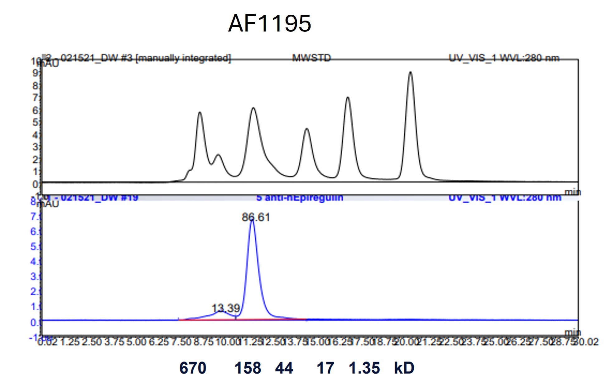
Application: characterize. 86% monomer.Sample Tested: Red blood cellsSpecies: GoatVerified Customer | Posted 08/15/2025
 Bio-Techne ResponseThis review reflects a new species or application tested on a primary antibody.
There are no reviews that match your criteria.
Protocols
Find general support by application which include: protocols, troubleshooting, illustrated assays, videos and webinars.
- Cellular Response to Hypoxia Protocols
- ELISA Sample Preparation & Collection Guide
- ELISA Troubleshooting Guide
- How to Run an R&D Systems DuoSet ELISA
- How to Run an R&D Systems Quantikine ELISA
- How to Run an R&D Systems Quantikine™ QuicKit™ ELISA
- Quantikine HS ELISA Kit Assay Principle, Alkaline Phosphatase
- Quantikine HS ELISA Kit Principle, Streptavidin-HRP Polymer
- R&D Systems Quality Control Western Blot Protocol
- Sandwich ELISA (Colorimetric) – Biotin/Streptavidin Detection Protocol
- Sandwich ELISA (Colorimetric) – Direct Detection Protocol
- Troubleshooting Guide: ELISA
- Troubleshooting Guide: Western Blot Figures
- Western Blot Conditions
- Western Blot Protocol
- Western Blot Protocol for Cell Lysates
- Western Blot Troubleshooting
- Western Blot Troubleshooting Guide
- View all Protocols, Troubleshooting, Illustrated assays and Webinars