FoxF1 belongs to a large family of proteins that share a common forkhead/winged helix DNA binding domain. FoxF1 is implicated in pulmonary morphogenesis, specifically in development of the lung mesenchyme. Experiments in mice indicate that haploinsufficiency of FoxF1 can lead to perinatal lethality due to pulmonary abnormalities. In addition expression of FoxF1 can be induced by hedgehog ligands and appears to regulate expression of BMP-4.

Key Product Details
Validated by
Biological Validation
Species Reactivity
Validated:
Human
Cited:
Human, Mouse, Frog - Xenopus (African Clawed Frog), Transgenic Mouse
Applications
Validated:
Western Blot
Cited:
Immunohistochemistry, Immunohistochemistry-Paraffin, Western Blot, Immunocytochemistry, Immunoprecipitation, Chromatin Immunoprecipitation (ChIP), IHC-
Label
Unconjugated
Antibody Source
Polyclonal Goat IgG
Loading...
Product Specifications
Immunogen
E. coli-derived recombinant human FoxF1
Met154-Met379
Accession # Q12946
Met154-Met379
Accession # Q12946
Specificity
Detects human FoxF1 in Western blots.
Clonality
Polyclonal
Host
Goat
Isotype
IgG
Scientific Data Images for Human FoxF1 Antibody
Detection of Human FoxF1 by Western Blot.
Western blot shows lysates of HeLa human cervical epithelial carcinoma cell line, Jurkat human acute T cell leukemia cell line, and PC-3 human prostate cancer cell line. PVDF membrane was probed with 1 µg/mL of Goat Anti-Human FoxF1 Antigen Affinity-purified Polyclonal Antibody (Catalog # AF4798) followed by HRP-conjugated Anti-Goat IgG Secondary Antibody (Catalog # HAF017). A specific band was detected for FoxF1 at approximately 50 kDa (as indicated). This experiment was conducted under reducing conditions and using Immunoblot Buffer Group 1.
Detection of Human FoxF1 by Western Blot.
Western blot shows lysates of WI-38 human lung fibroblast cell line. PVDF membrane was probed with 1 µg/mL of Goat Anti-Human FoxF1 Antigen Affinity-purified Polyclonal Antibody (Catalog # AF4798) followed by HRP-conjugated Anti-Goat IgG Secondary Antibody (Catalog # HAF017). A specific band was detected for FoxF1 at approximately 44 kDa (as indicated). This experiment was conducted under reducing conditions and using Immunoblot Buffer Group 1.
Detection of Human FoxF1 by Western Blot
Loss of FOXF1 promotes fundamental cellular processes in LR-MSCs. (A) mRNA was isolated from fibrotic and non-fibrotic LR-MSCs derived from bronchoalveolar lavage fluid of transplant patients, and FOXF1 expression was analyzed by real-time PCR. Values: Means ± SEM; n = 9 (non-Fib-MSCs); n = 8 (Fib-MSCs); **p < 0.0034. (B) Protein lysates from fibrotic and non-fibrotic LR-MSCs were analyzed for FOXF1 and GAPDH by western blotting. Graph shows densitometry analyses of these immunoblots. Values: Means ± SEM; n = 16; **p < 0.0086. (C) LR-MSCs were transfected with scrambled or FOXF1-specific siRNA and confirmed by real-time PCR. Values: Means ± SEM; n = 7; ***p < 0.0005. (D) Protein lysates from (A) were subjected to immunoblotting with antibodies against FOXF1 and GAPDH. (E–I) Gene regulation due to FOXF1 silencing was analyzed by Affymetrix gene array in 3 lines of LR-MSCs. Data reflects fold changes ≥ 1.5, and an adjusted p < 0.01. (E) Diagram showing the number of upregulated and downregulated genes. (F) Gene–gene interaction network (using STRING database) showing associations due to FOXF1-silencing. (G–I) Heatmaps showing two-fold Log changes are presented for positive regulation of cell cycle ((G) GO:0045787), inflammatory response ((H) GO:0006954), and regulation of cell migration ((I) GO:0030334). Note: Full length blots for Fig. 1B and Fig. 1D are provided in Supplementary Fig. S1 and S2. Image collected and cropped by CiteAb from the following publication (https://pubmed.ncbi.nlm.nih.gov/33277571), licensed under a CC-BY license. Not internally tested by R&D Systems.
Detection of Human FoxF1 by Western Blot
ATX-dependent cell migration in FOXF1-silenced LR-MSCs. (A) LR-MSCs were transfected with scrambled or FOXF1-specific siRNA. 24 h later, these LR-MSCs were transfected with scrambled or ATX-specific siRNA. Immunoblotting was performed to confirm RNAi-mediated FOXF1 and ATX silencing efficacy. n = 5 per group. (B) Migration assay was conducted in LR-MSCs transfected with scrambled or siRNA specific to FOXF1, ATX, or both FOXF1- and ATX-specific siRNA. Representative images of cell migration are shown. (C) Quantification of (B), n = 5, ***p < 0.0003. (D) LR-MSCs transfected with scrambled or FOXF1-specific siRNA were treated with the ATX inhibitor, PF-8380 (1 μM) and migration assay was performed. Values: Means ± SEM. n = 5, **p < 0.0142, *p < 0.0325. (E,F) Migration assays are shown with LR-MSCs transfected with scrambled or FOXF1-specific siRNA, and then treated with the LPA1 inihibitor, VPC12249 (1 μM) (E), or subjected to lentivirus-mediated shRNA interference against LPAR1 (F). Values: Means ± SEM. n = 5. ***p < 0.0002, ****p < 0.0001. Note: Full length blots for Fig. 3A are provided in Supplementary Fig. S5. Image collected and cropped by CiteAb from the following publication (https://pubmed.ncbi.nlm.nih.gov/33277571), licensed under a CC-BY license. Not internally tested by R&D Systems.Applications for Human FoxF1 Antibody
Application
Recommended Usage
Western Blot
1 µg/mL
Sample: HeLa human cervical epithelial carcinoma cell line, Jurkat human acute T cell leukemia cell line, PC‑3 human prostate cancer cell line, WI‑38 human lung fibroblast cell line.
Sample: HeLa human cervical epithelial carcinoma cell line, Jurkat human acute T cell leukemia cell line, PC‑3 human prostate cancer cell line, WI‑38 human lung fibroblast cell line.
Reviewed Applications
Read 1 review rated 4 using AF4798 in the following applications:
Formulation, Preparation, and Storage
Purification
Antigen Affinity-purified
Reconstitution
Reconstitute at 0.2 mg/mL in sterile PBS. For liquid material, refer to CoA for concentration.
Loading...
Formulation
Lyophilized from a 0.2 μm filtered solution in PBS with Trehalose. *Small pack size (SP) is supplied either lyophilized or as a 0.2 µm filtered solution in PBS.
Shipping
Lyophilized product is shipped at ambient temperature. Liquid small pack size (-SP) is shipped with polar packs. Upon receipt, store immediately at the temperature recommended below.
Stability & Storage
Use a manual defrost freezer and avoid repeated freeze-thaw cycles.
- 12 months from date of receipt, -20 to -70 °C as supplied.
- 1 month, 2 to 8 °C under sterile conditions after reconstitution.
- 6 months, -20 to -70 °C under sterile conditions after reconstitution.
Calculators
Background: FoxF1
Long Name
Forkhead Box F1
Alternate Names
FKHL5, FREAC-1
Gene Symbol
FOXF1
UniProt
Additional FoxF1 Products
Product Documents for Human FoxF1 Antibody
Certificate of Analysis
To download a Certificate of Analysis, please enter a lot or batch number in the search box below.
Note: Certificate of Analysis not available for kit components.
Product Specific Notices for Human FoxF1 Antibody
For research use only
Related Research Areas
Citations for Human FoxF1 Antibody
 Powered by Bioz
Customer Reviews for Human FoxF1 Antibody (1)
4 out of 5
1 Customer Rating
Have you used Human FoxF1 Antibody?
Submit a review and receive an Amazon gift card!
$25/€18/£15/$25CAN/¥2500 Yen for a review with an image
$10/€7/£6/$10CAN/¥1110 Yen for a review without an image
Submit a review
Customer Images
Showing
1
-
1 of
1 review
Showing All
Filter By:
-
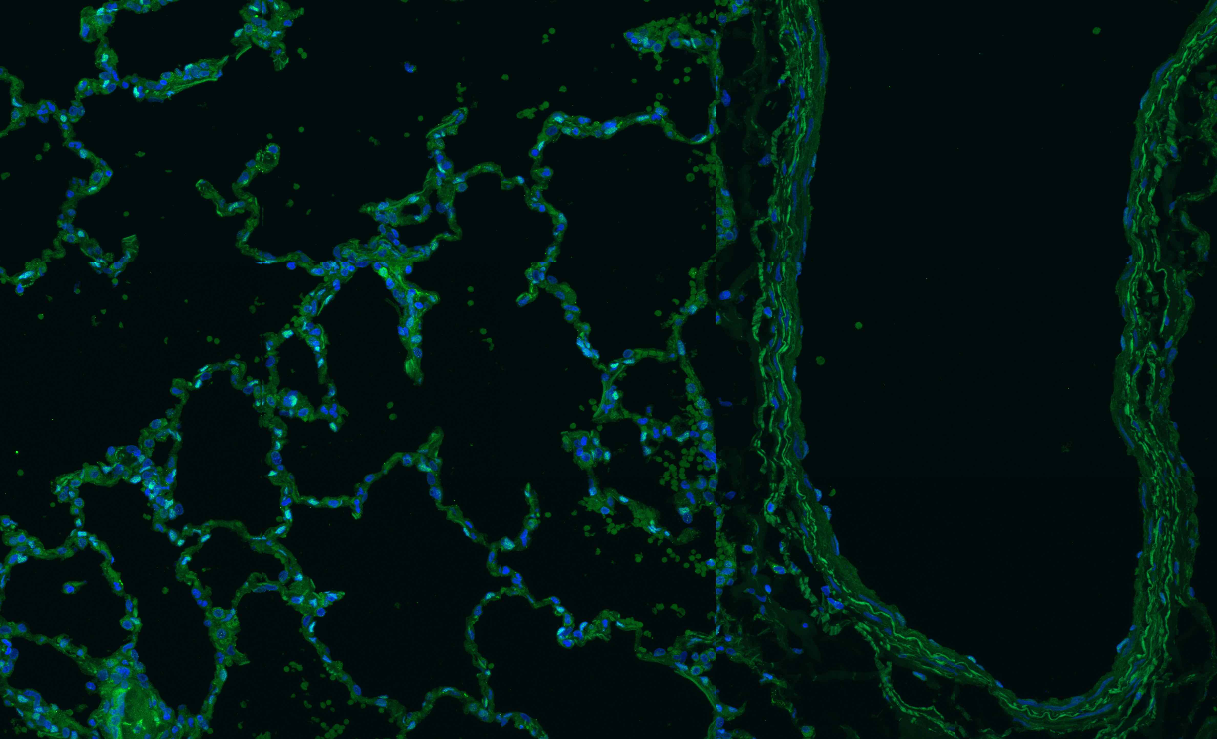
Application: Immunocytochemistry/ImmunofluorescenceSample Tested: human lungSpecies: HumanVerified Customer | Posted 07/01/2018

There are no reviews that match your criteria.
Protocols
Find general support by application which include: protocols, troubleshooting, illustrated assays, videos and webinars.
- Cellular Response to Hypoxia Protocols
- R&D Systems Quality Control Western Blot Protocol
- Troubleshooting Guide: Western Blot Figures
- Western Blot Conditions
- Western Blot Protocol
- Western Blot Protocol for Cell Lysates
- Western Blot Troubleshooting
- Western Blot Troubleshooting Guide
- View all Protocols, Troubleshooting, Illustrated assays and Webinars
Loading...
Associated Pathways
