The galectins constitute a large family of carbohydrate-binding proteins with specificity for N-acetyl-lactosamine-containing glycoproteins. At least 14 mammalian galectins, which share structural similarities in their carbohydrate recognition domains (CRD), have been identified. The galectins have been classified into the prototype galectins (-1, -2, -5, -7, -10, -11, -13, -14), which contain one CRD and exist either as a monomer or a noncovalent homodimer; the chimera galectins (Galectin-3) containing one CRD linked to a nonlectin domain; and the tandem-repeat galectins (-4, -6, -8, -9, -12) consisting of two CRDs joined by a linker peptide. Galectins lack a classical signal peptide and can be localized to the cytosolic compartments where they have intracellular functions. However, via one or more as yet unidentified non-classical secretory pathways, galectins can also be secreted to function extracellularly. Individual members of the galectin family have different tissue distribution profiles and exhibit subtle differences in their carbohydrate-binding specificities. Each family member may preferentially bind to a unique subset of cell-surface glycoproteins (1-4). Human Galectin-7 is a prototype monomeric galectin. It is specifically expressed in stratified epithelia, notably in epidermis, but is barely detectable in epidermal tumors and significantly down regulated or absent from squamous carconima cell lines. The Galectin-7 gene is induced by tumor suppressor protein p53 transcriptional activity following genotoxic events. A pro-apoptotic protein, Galectin-7 functions intracellularly upstream of JNK activation and cytochrome-c release. This protein has been shown to increase the susceptibility of keratinocytes to UVB induced apoptosis, an essential processss in the maintenance of epidermal homeostasis. Cell lines transfected with the Galectin-7 gene localized the protein in the nucleus and intracellularly. Human and mouse Galectin-7 share 79% amino acid homology (4-6).

Key Product Details
Species Reactivity
Validated:
Human
Cited:
Human
Applications
Validated:
Immunohistochemistry, Western Blot, ELISA Capture (Matched Antibody Pair), Simple Western
Cited:
Immunohistochemistry, Immunohistochemistry-Paraffin, Immunocytochemistry
Label
Unconjugated
Antibody Source
Polyclonal Goat IgG
Loading...
Product Specifications
Immunogen
E. coli-derived recombinant human Galectin‑7
Ser2-Phe136
Accession # NP_002298
Ser2-Phe136
Accession # NP_002298
Specificity
Detects human Galectin-7 in ELISAs and Western blots. In sandwich immunoassays, approximately 50% cross-reactivity with recombinant mouse (rm) Galectin-7 is observed and less than 0.5% cross-reactivity with recombinant human (rh) Galectin-1, rmGalectin-3, rhGalectin-4, and rhGalectin-8 is observed.
Clonality
Polyclonal
Host
Goat
Isotype
IgG
Scientific Data Images for Human Galectin-7 Antibody
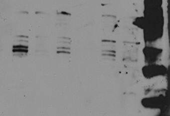
Detection of Human Galectin‑7 by Western Blot.
Western blot shows lysates of human skin tissue. PVDF membrane was probed with 1 µg/mL of Goat Anti-Human Galectin-7 Antigen Affinity-purified Polyclonal Antibody (Catalog # AF1339) followed by HRP-conjugated Anti-Goat IgG Secondary Antibody (Catalog # HAF017). A specific band was detected for Galectin-7 at approximately 15 kDa (as indicated). This experiment was conducted under reducing conditions and using Immunoblot Buffer Group 1.
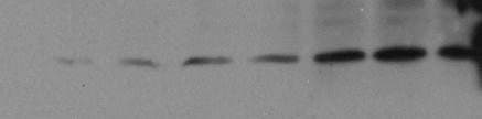
Detection of Human Galectin‑7 by Simple WesternTM.
Simple Western lane view shows lysates of HEK001 human epidermal keratinocyte cell line, loaded at 0.2 mg/mL. A specific band was detected for Galectin-7 at approximately 21 kDa (as indicated) using 10 µg/mL of Goat Anti-Human Galectin-7 Antigen Affinity-purified Polyclonal Antibody (Catalog # AF1339) followed by 1:50 dilution of HRP-conjugated Anti-Goat IgG Secondary Antibody (Catalog # HAF109). This experiment was conducted under reducing conditions and using the 12-230 kDa separation system.
Human Galectin-7 ELISA Standard Curve
Recombinant Human Galectin‑7 (Catalog # 1339-GA) was serially diluted and captured by Goat Anti-Human Galectin‑7 Antigen Affinity-purified Polyclonal Antibody (Catalog # AF1339) coated on a Clear Polystyrene Microplate (Catalog # DY990). Goat Anti-Human Galectin‑7 Antigen Affinity-purified Polyclonal Antibody (Catalog # AF1339) was biotinylated and incubated with the protein captured on the plate. Detection of the standard curve was achieved by incubating Streptavidin-HRP (Catalog # DY998)
Human Galectin-7 ELISA Standard Curve
Recombinant Human Galectin‑7 (Catalog # 1339-GA) was serially diluted and captured by Goat Anti-Human Galectin‑7 Antigen Affinity-purified Polyclonal Antibody (Catalog # AF1339) coated on a Clear Polystyrene Microplate (Catalog # DY990). Goat Anti-Human Galectin‑7 Antigen Affinity-purified Polyclonal Antibody (Catalog # AF1339) was biotinylated and incubated with the protein captured on the plate. Detection of the standard curve was achieved by incubating Streptavidin-HRP (Catalog # DY998)Applications for Human Galectin-7 Antibody
Application
Recommended Usage
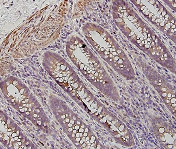
Immunohistochemistry
5-15 µg/mL
Sample: Immersion fixed paraffin-embedded sections of human skin
Sample: Immersion fixed paraffin-embedded sections of human skin
Simple Western
10 µg/mL
Sample: HEK001 human epidermal keratinocyte cell line
Sample: HEK001 human epidermal keratinocyte cell line
Western Blot
1 µg/mL
Sample: Human skin tissue
Sample: Human skin tissue
Human Galectin-7 Sandwich Immunoassay
Please Note: Optimal dilutions of this antibody should be experimentally determined.
Reviewed Applications
Read 4 reviews rated 4.3 using AF1339 in the following applications:
Formulation, Preparation, and Storage
Purification
Antigen Affinity-purified
Reconstitution
Reconstitute at 0.2 mg/mL in sterile PBS. For liquid material, refer to CoA for concentration.
Loading...
Formulation
Lyophilized from a 0.2 μm filtered solution in PBS with Trehalose. *Small pack size (SP) is supplied either lyophilized or as a 0.2 µm filtered solution in PBS.
Shipping
Lyophilized product is shipped at ambient temperature. Liquid small pack size (-SP) is shipped with polar packs. Upon receipt, store immediately at the temperature recommended below.
Stability & Storage
Use a manual defrost freezer and avoid repeated freeze-thaw cycles.
- 12 months from date of receipt, -20 to -70 °C as supplied.
- 1 month, 2 to 8 °C under sterile conditions after reconstitution.
- 6 months, -20 to -70 °C under sterile conditions after reconstitution.
Calculators
Background: Galectin-7
References
- Rabinovich, A. et al. (2002) TRENDS in Immunol. 23:313.
- Rabinovich, A. et al. (2002) J. Leukocyte Biology 71:741.
- Hughes, R.C. (2002) Biochimie 83:667.
- R&D Systems Cytokine Bulletin; Summer 2002.
- Bernerd, F. et al. (1999) Proc. Natl. Acad. Sci. USA 96:11329.
- Kuwabara, I. et al. (2002) J. Biol. Chem. 277:3487.
Alternate Names
GAL7, Galectin7, LGALS7
Gene Symbol
LGALS7
UniProt
Additional Galectin-7 Products
Product Documents for Human Galectin-7 Antibody
Certificate of Analysis
To download a Certificate of Analysis, please enter a lot or batch number in the search box below.
Note: Certificate of Analysis not available for kit components.
Product Specific Notices for Human Galectin-7 Antibody
For research use only
Related Research Areas
Citations for Human Galectin-7 Antibody
 Powered by Bioz
Customer Reviews for Human Galectin-7 Antibody (4)
4.3 out of 5
4 Customer Ratings
Have you used Human Galectin-7 Antibody?
Submit a review and receive an Amazon gift card!
$25/€18/£15/$25CAN/¥2500 Yen for a review with an image
$10/€7/£6/$10CAN/¥1110 Yen for a review without an image
Submit a review
Customer Images
Showing
1
-
4 of
4 reviews
Showing All
Filter By:
-
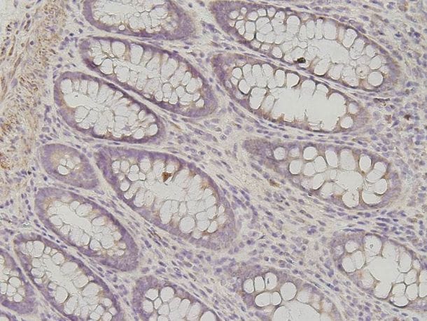
Application: ImmunohistochemistrySample Tested: Colon tissueSpecies: HumanVerified Customer | Posted 10/26/2015At 1:500 the antibody is very efficient in detecting galectin-7. <br />Specificity: Specific<br />Sensitivity: Sensitive<br />Buffer: PBS<br />Dilution: 1:500

-
Application: Western BlotSample Tested: Cancer cell lysatesVerified Customer | Posted 10/26/2015First run with 1:1000 for difference cancer cell lysates gave many bands, which could be isoforms detected. <br />Specificity: Reasonably specific<br />Sensitivity: Reasonably sensitive<br />Buffer: BSA<br />Dilution: 1:1000
-
Application: ImmunohistochemistrySample Tested: Colon tissueSpecies: HumanVerified Customer | Posted 10/26/2015At 1:1000 there was low detection levels of Gal-7. <br />Specificity: Not Specific<br />Sensitivity: Reasonably sensitive<br />Buffer: PBS<br />Dilution: 1:1000
-
Application: Western BlotSample Tested: Cancer cell lysatesSpecies: HumanVerified Customer | Posted 10/26/2015At higher dilutions, only the main form was visible. <br />Specificity: Specific<br />Sensitivity: Sensitive<br />Buffer: Loading buffer<br />Dilution: 1:2000
There are no reviews that match your criteria.
Protocols
Find general support by application which include: protocols, troubleshooting, illustrated assays, videos and webinars.
- Antigen Retrieval Protocol (PIER)
- Antigen Retrieval for Frozen Sections Protocol
- Appropriate Fixation of IHC/ICC Samples
- Cellular Response to Hypoxia Protocols
- Chromogenic IHC Staining of Formalin-Fixed Paraffin-Embedded (FFPE) Tissue Protocol
- Chromogenic Immunohistochemistry Staining of Frozen Tissue
- ClariTSA™ Fluorophore Kits
- Detection & Visualization of Antibody Binding
- Fluorescent IHC Staining of Frozen Tissue Protocol
- Graphic Protocol for Heat-induced Epitope Retrieval
- Graphic Protocol for the Preparation and Fluorescent IHC Staining of Frozen Tissue Sections
- Graphic Protocol for the Preparation and Fluorescent IHC Staining of Paraffin-embedded Tissue Sections
- Graphic Protocol for the Preparation of Gelatin-coated Slides for Histological Tissue Sections
- IHC Sample Preparation (Frozen sections vs Paraffin)
- Immunofluorescent IHC Staining of Formalin-Fixed Paraffin-Embedded (FFPE) Tissue Protocol
- Immunohistochemistry (IHC) and Immunocytochemistry (ICC) Protocols
- Immunohistochemistry Frozen Troubleshooting
- Immunohistochemistry Paraffin Troubleshooting
- Preparing Samples for IHC/ICC Experiments
- Preventing Non-Specific Staining (Non-Specific Binding)
- Primary Antibody Selection & Optimization
- Protocol for Heat-Induced Epitope Retrieval (HIER)
- Protocol for Making a 4% Formaldehyde Solution in PBS
- Protocol for VisUCyte™ HRP Polymer Detection Reagent
- Protocol for the Preparation & Fixation of Cells on Coverslips
- Protocol for the Preparation and Chromogenic IHC Staining of Frozen Tissue Sections
- Protocol for the Preparation and Chromogenic IHC Staining of Frozen Tissue Sections - Graphic
- Protocol for the Preparation and Chromogenic IHC Staining of Paraffin-embedded Tissue Sections
- Protocol for the Preparation and Chromogenic IHC Staining of Paraffin-embedded Tissue Sections - Graphic
- Protocol for the Preparation and Fluorescent IHC Staining of Frozen Tissue Sections
- Protocol for the Preparation and Fluorescent IHC Staining of Paraffin-embedded Tissue Sections
- Protocol for the Preparation of Gelatin-coated Slides for Histological Tissue Sections
- R&D Systems Quality Control Western Blot Protocol
- TUNEL and Active Caspase-3 Detection by IHC/ICC Protocol
- The Importance of IHC/ICC Controls
- Troubleshooting Guide: Immunohistochemistry
- Troubleshooting Guide: Western Blot Figures
- Western Blot Conditions
- Western Blot Protocol
- Western Blot Protocol for Cell Lysates
- Western Blot Troubleshooting
- Western Blot Troubleshooting Guide
- View all Protocols, Troubleshooting, Illustrated assays and Webinars
Loading...