
Gas6 (Growth Arrest Specific 6) is a multimodular protein that is upregulated by a wide variety of cell types in response to growth arrest (1). Gas6 and the structurally related Protein S are vitamin K-dependent and have an extensively gamma -carboxylated N-terminal Gla domain, four EGF‑like repeats, and a C-terminal region with homology to steroid hormone binding globulin (SHBG) (2). Human Gas6 is a 75 kDa protein that shares 77-79% aa sequence identity with mouse and rat Gas6, and 43% aa identity with human protein S (over the region expressed). Alternate splicing generates isoforms that lack the Gla domain and/or the spacer between the EGF‑like and SHBG regions. Gas6 binds and induces signaling through the receptor tyrosine kinases Axl, Dtk, and Mer (3-5). Human Gas6 interacts with both mouse and rat orthologs of these receptors (1). The full length isoform may be cleaved, resulting in release of the free SHBG region which can independently activate Axl (6). Shed soluble forms of Axl and Mer bind Gas6 and function as decoy receptors (7, 8). Gas6 induces a variety of responses, including prevention of apoptosis (9), cell proliferation (10), platelet-mediated thrombosis (11), retinal epithelial cell phagocytosis of outer rod segments (12), inhibition of VEGF-induced endothelial cell chemotaxis (13), and the differentiation and expansion of NK cell precursors (14). The affinity of Gas6 for phosphatidylserine likely contributes to its role in promoting the phagocytosis of apoptotic cells (15). Several of these effects have been shown to require gamma -carboxylation of the Gla domain (12, 16).

Scientific images may be resized, cropped, or adjusted for brightness or contrast for presentation purposes. These updates are limited to presentation and do not affect the underlying scientific interpretation of the data. Where available, additional source data may be provided upon request.
Key Product Details
Species Reactivity
Validated:
Cited:
Applications
Validated:
Cited:
Label
Antibody Source
Product Specifications
Immunogen
Asp118-Ala678
Accession # NP_000811
Specificity
Clonality
Host
Isotype
Endotoxin Level
Scientific Data Images for Human Gas6 Antibody
Detection of Gas6 by Western Blot
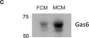
TAMs and CAFs are the main sources of Gas6 in pancreatic tumors. (A) KPCluc/zsGreen (zsGreen) -derived tumor cells (FC1242luc/zsGreen) were orthotopically implanted into the pancreas of syngeneic recipient (C57/BL6) mice. Tumors were harvested and digested at day 23 after implantation and tumor cells, non-immune stromal cells, M1-like and M2-like macrophages were sorted by flow cytometry. Gas6 mRNA levels were quantified in CD45–/zsGreen+ tumor cells, CD45–/zsGreen– non-immune stromal cells, CD45+/F4/80+/CD206– M1-like macrophages and CD45+/F4/80+/CD206+ M2-like macrophages sorted by flow cytometry from murine pancreatic tumors. Values shown are the mean and SD (n = 3). (B) Quantification of Gas6 mRNA expression levels in ex vivo mouse primary isolated macrophages and pancreatic fibroblasts from naïve mice. Values shown are the mean and SD (n = 3). (C) Immunoblotting analysis of Gas6 secreted protein present in mouse macrophage conditioned media (MCM) and pancreatic fibroblast conditioned media (FCM). (D) Images show phospho-Axl, alpha SMA (fibroblast marker) and CD68 (pan-macrophage marker) staining in naïve mouse pancreas and in serial sections of mouse PDA tissues. Scale bar = 50 μm. Image collected and cropped by CiteAb from the following open publication (https://pubmed.ncbi.nlm.nih.gov/32174917), licensed under a CC-BY license. Not internally tested by R&D Systems.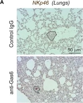
Detection of Gas6 by Immunohistochemistry
Gas6 blockade increases NK cell numbers in metastatic lungs and in tumor draining lymph nodes. (A) Immunohistochemical staining of NK cells in metastatic lungs from pancreatic tumor bearing mice treated with control IgG or anti-Gas6 antibody. Lesions indicated by dashed line and NK cells by red asterisk. Scale bar, 50 μm. (B) Quantification of NK cells in metastatic lung tissues from control IgG and anti-Gas6 treated mice. Values shown are the mean and SEM (n = 6 mice in IgG treatment group, n = 7 mice in anti-Gas6 treatment group). **p ≤ 0.01, using unpaired student T-test. (C) Immunofluorescent staining of NK cells in mesenteric lymph nodes from pancreatic tumor bearing mice treated with control IgG or anti-Gas6 antibody. NK marker NKp46 is shown in red, Ki67 is shown in green and nuclei were stained with DAPI (in blue). Scale bar, 50 μm. (D) Quantification of NK cells in tumor draining lymph nodes from control IgG and anti-Gas6 treated mice. Values shown are the mean and SEM (n = 6 mice IgG treatment group and n = 7 mice anti-Gas6 treatment group, 3–6 fields/ mouse tissue were quantified). *p ≤ 0.05, using unpaired student T-test. Image collected and cropped by CiteAb from the following open publication (https://pubmed.ncbi.nlm.nih.gov/32174917), licensed under a CC-BY license. Not internally tested by R&D Systems.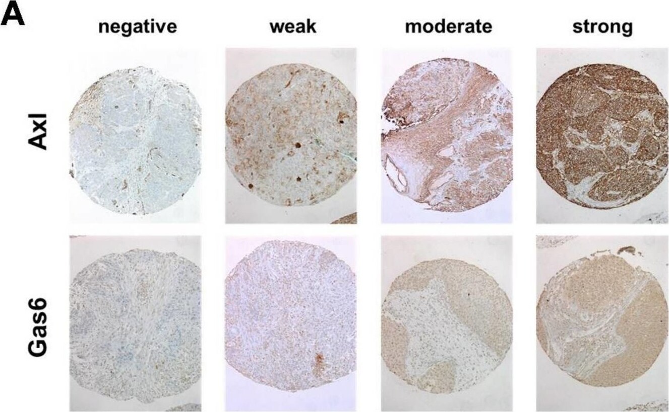
Detection of Gas6 by Immunohistochemistry
Immunohistochemical staining. (A) Representative examples of expression score categories. (B) Distribution of expression scores in the whole HNSCC population and specific subtypes. neg.—negative, we.—weak, mod.—moderate, str.—strong, IHC—immunohistochemistry. Image collected and cropped by CiteAb from the following open publication (https://pubmed.ncbi.nlm.nih.gov/35406601), licensed under a CC-BY license. Not internally tested by R&D Systems.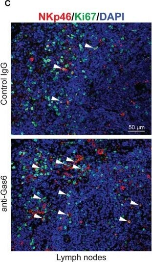
Detection of Gas6 by Immunohistochemistry
Gas6 blockade increases NK cell numbers in metastatic lungs and in tumor draining lymph nodes. (A) Immunohistochemical staining of NK cells in metastatic lungs from pancreatic tumor bearing mice treated with control IgG or anti-Gas6 antibody. Lesions indicated by dashed line and NK cells by red asterisk. Scale bar, 50 μm. (B) Quantification of NK cells in metastatic lung tissues from control IgG and anti-Gas6 treated mice. Values shown are the mean and SEM (n = 6 mice in IgG treatment group, n = 7 mice in anti-Gas6 treatment group). **p ≤ 0.01, using unpaired student T-test. (C) Immunofluorescent staining of NK cells in mesenteric lymph nodes from pancreatic tumor bearing mice treated with control IgG or anti-Gas6 antibody. NK marker NKp46 is shown in red, Ki67 is shown in green and nuclei were stained with DAPI (in blue). Scale bar, 50 μm. (D) Quantification of NK cells in tumor draining lymph nodes from control IgG and anti-Gas6 treated mice. Values shown are the mean and SEM (n = 6 mice IgG treatment group and n = 7 mice anti-Gas6 treatment group, 3–6 fields/ mouse tissue were quantified). *p ≤ 0.05, using unpaired student T-test. Image collected and cropped by CiteAb from the following open publication (https://pubmed.ncbi.nlm.nih.gov/32174917), licensed under a CC-BY license. Not internally tested by R&D Systems.Applications for Human Gas6 Antibody
Blockade of Receptor-ligand Interaction
Western Blot
Sample: Recombinant Human Gas6 (Catalog # 885-GS)
Reviewed Applications
Read 2 reviews rated 5 using AB885 in the following applications:
Formulation, Preparation, and Storage
Purification
Reconstitution
Reconstitute at 1 mg/mL in sterile PBS.
Formulation
Shipping
Stability & Storage
- 12 months from date of receipt, -20 to -70 °C as supplied.
- 1 month, 2 to 8 °C under sterile conditions after reconstitution.
- 6 months, -20 to -70 °C under sterile conditions after reconstitution.
Calculators
Background: Gas6
References
- Hafizi, S. and B. Dahlback (2006) FEBS J. 273:5231.
- Manfioletti, G. et al. (1993) Mol. Cell Biol. 13:4976.
- Stitt, T.N. et al. (1995) Cell 80:661.
- Ohashi, K. et al. (1995) J. Biol. Chem. 270:22681.
- Nagata, K. et al. (1996) J. Biol. Chem. 271:30022.
- Goruppi, S. et al. (1997) FEBS Lett. 415:59.
- Sather, S. et al. (2007) Blood 109:1026.
- Budagian, V. et al. (2005) Mol. Cell. Biol. 25:9324.
- Shankar, S.L. et al. (2006) J. Neurosci. 26:5638.
- Yanagita, M. et al. (2002) J. Clin. Invest. 110:239.
- Gould, W.R. et al. (2005) J. Thromb. Haemost. 3:733.
- Hall, M.O. et al. (2002) Exp. Eye Res. 75:391.
- Gallicchio, M. et al. (2005) Blood 105:1970.
- Caraux, A. et al. (2006) Nat. Immunol. 7:747.
- Wu, Y. et al. (2005) J. Cell Sci. 118:539.
- Hasanbasic, I. et al. (2005) J. Thromb. Haemost. 3:2790.
Long Name
Alternate Names
Gene Symbol
UniProt
Additional Gas6 Products
Product Documents for Human Gas6 Antibody
Certificate of Analysis
To download a Certificate of Analysis, please enter a lot or batch number in the search box below.
Note: Certificate of Analysis not available for kit components.
Product Specific Notices for Human Gas6 Antibody
For research use only
Related Research Areas
Citations for Human Gas6 Antibody
 Powered by Bioz
Customer Reviews for Human Gas6 Antibody (2)
Have you used Human Gas6 Antibody?
Submit a review and receive an Amazon gift card!
$25/€18/£15/$25CAN/¥2500 for a review with an image
$10/€7/£6/$10CAN/¥1110 for a review without an image
Submit a review
Customer Images

-
Application: Western BlotSample Tested: Cell Culture MediaSpecies: HumanVerified Customer | Posted 06/17/2017
-
Application: Flow CytometrySample Tested: Mesenchymal stem cellsSpecies: HumanVerified Customer | Posted 01/10/2017

There are no reviews that match your criteria.
Protocols
Find general support by application which include: protocols, troubleshooting, illustrated assays, videos and webinars.
- Cellular Response to Hypoxia Protocols
- R&D Systems Quality Control Western Blot Protocol
- Troubleshooting Guide: Western Blot Figures
- Western Blot Conditions
- Western Blot Protocol
- Western Blot Protocol for Cell Lysates
- Western Blot Troubleshooting
- Western Blot Troubleshooting Guide
- View all Protocols, Troubleshooting, Illustrated assays and Webinars