Hepsin, also known as TMPRSS1, is a Type II membrane protein with an extracellular serine protease domain (1). It is most highly expressed in liver but is also present in many other tissues; most notably lung, kidney, and skeletal muscle (2). A soluble form of Hepsin lacking the transmembrane domain has been identified (3). Hepsin is capable of activating Factor VII and may initiate blood coagulation at the cell surface (4). Hepsin is over-expressed in various human tumors including prostate (5) and is considered to be a biomarker for prostate cancer (6). Recombinant human (rh) Hepsin was expressed as a secreted, soluble protein lacking its cytosolic and transmembrane domains. The D161E and R162K mutations were introduced into the prosequence to improve expression of the rhHepsin.

Key Product Details
Species Reactivity
Human
Applications
Immunohistochemistry
Label
Unconjugated
Antibody Source
Monoclonal Mouse IgG1 Clone # 506230
Loading...
Product Specifications
Immunogen
Mouse myeloma cell line NS0-derived recombinant human Hepsin
Ser46-Leu417
Accession # P05981
Ser46-Leu417
Accession # P05981
Specificity
Detects human Hepsin in direct ELISAs.
Clonality
Monoclonal
Host
Mouse
Isotype
IgG1
Scientific Data Images for Human Hepsin Antibody (506230)
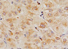
Hepsin in Human Liver.
Hepsin was detected in immersion fixed paraffin-embedded sections of human liver using Mouse Anti-Human Hepsin Monoclonal Antibody (Catalog # MAB47761) at 25 µg/mL overnight at 4 °C. Tissue was stained using the Anti-Mouse HRP-DAB Cell & Tissue Staining Kit (brown; Catalog # CTS002) and counterstained with hematoxylin (blue). Specific labeling was localized to the cytoplasm of hepatocytes. View our protocol for Chromogenic IHC Staining of Paraffin-embedded Tissue Sections.Applications for Human Hepsin Antibody (506230)
Application
Recommended Usage
Immunohistochemistry
8-25 µg/mL
Sample: Immersion fixed paraffin-embedded sections of human liver
Sample: Immersion fixed paraffin-embedded sections of human liver
Reviewed Applications
Read 1 review rated 5 using MAB47761 in the following applications:
Formulation, Preparation, and Storage
Purification
Protein A or G purified from hybridoma culture supernatant
Reconstitution
Reconstitute at 0.5 mg/mL in sterile PBS. For liquid material, refer to CoA for concentration.
Loading...
Formulation
Lyophilized from a 0.2 μm filtered solution in PBS with Trehalose. *Small pack size (SP) is supplied either lyophilized or as a 0.2 µm filtered solution in PBS.
Shipping
Lyophilized product is shipped at ambient temperature. Liquid small pack size (-SP) is shipped with polar packs. Upon receipt, store immediately at the temperature recommended below.
Stability & Storage
Use a manual defrost freezer and avoid repeated freeze-thaw cycles.
- 12 months from date of receipt, -20 to -70 °C as supplied.
- 1 month, 2 to 8 °C under sterile conditions after reconstitution.
- 6 months, -20 to -70 °C under sterile conditions after reconstitution.
Calculators
Background: Hepsin
References
- Leytus, S.P. et al. (1988) Biochemistry 27:1067.
- Tsuji, A. et al. (1991) J. Biol. Chem. 266:16948.
- Li, Y. et al. (2005) Biomed. Biochim. Acta 1681:157.
- Kazama, Y. et al. (1995) J. Biol. Chem. 270:66.
- Dhanasekaran, S.M. et al. (2001) Nature 412:822.
- Wu, Q. and G. Parry (2007) Front. Biosci. 12:5052.
Alternate Names
HPN, TMPRSS1
Gene Symbol
HPN
UniProt
Additional Hepsin Products
Product Documents for Human Hepsin Antibody (506230)
Certificate of Analysis
To download a Certificate of Analysis, please enter a lot or batch number in the search box below.
Note: Certificate of Analysis not available for kit components.
Product Specific Notices for Human Hepsin Antibody (506230)
For research use only
Related Research Areas
Citations for Human Hepsin Antibody (506230)
 Powered by Bioz
Customer Reviews for Human Hepsin Antibody (506230) (1)
5 out of 5
1 Customer Rating
Have you used Human Hepsin Antibody (506230)?
Submit a review and receive an Amazon gift card!
$25/€18/£15/$25CAN/¥2500 Yen for a review with an image
$10/€7/£6/$10CAN/¥1110 Yen for a review without an image
Submit a review
Customer Images
Showing
1
-
1 of
1 review
Showing All
Filter By:
-
Application: ImmunohistochemistrySample Tested: Liver tissueSpecies: HumanVerified Customer | Posted 12/26/2021

There are no reviews that match your criteria.
Protocols
Find general support by application which include: protocols, troubleshooting, illustrated assays, videos and webinars.
- Antigen Retrieval Protocol (PIER)
- Antigen Retrieval for Frozen Sections Protocol
- Appropriate Fixation of IHC/ICC Samples
- Cellular Response to Hypoxia Protocols
- Chromogenic IHC Staining of Formalin-Fixed Paraffin-Embedded (FFPE) Tissue Protocol
- Chromogenic Immunohistochemistry Staining of Frozen Tissue
- ClariTSA™ Fluorophore Kits
- Detection & Visualization of Antibody Binding
- Fluorescent IHC Staining of Frozen Tissue Protocol
- Graphic Protocol for Heat-induced Epitope Retrieval
- Graphic Protocol for the Preparation and Fluorescent IHC Staining of Frozen Tissue Sections
- Graphic Protocol for the Preparation and Fluorescent IHC Staining of Paraffin-embedded Tissue Sections
- Graphic Protocol for the Preparation of Gelatin-coated Slides for Histological Tissue Sections
- IHC Sample Preparation (Frozen sections vs Paraffin)
- Immunofluorescent IHC Staining of Formalin-Fixed Paraffin-Embedded (FFPE) Tissue Protocol
- Immunohistochemistry (IHC) and Immunocytochemistry (ICC) Protocols
- Immunohistochemistry Frozen Troubleshooting
- Immunohistochemistry Paraffin Troubleshooting
- Preparing Samples for IHC/ICC Experiments
- Preventing Non-Specific Staining (Non-Specific Binding)
- Primary Antibody Selection & Optimization
- Protocol for Heat-Induced Epitope Retrieval (HIER)
- Protocol for Making a 4% Formaldehyde Solution in PBS
- Protocol for VisUCyte™ HRP Polymer Detection Reagent
- Protocol for the Preparation & Fixation of Cells on Coverslips
- Protocol for the Preparation and Chromogenic IHC Staining of Frozen Tissue Sections
- Protocol for the Preparation and Chromogenic IHC Staining of Frozen Tissue Sections - Graphic
- Protocol for the Preparation and Chromogenic IHC Staining of Paraffin-embedded Tissue Sections
- Protocol for the Preparation and Chromogenic IHC Staining of Paraffin-embedded Tissue Sections - Graphic
- Protocol for the Preparation and Fluorescent IHC Staining of Frozen Tissue Sections
- Protocol for the Preparation and Fluorescent IHC Staining of Paraffin-embedded Tissue Sections
- Protocol for the Preparation of Gelatin-coated Slides for Histological Tissue Sections
- TUNEL and Active Caspase-3 Detection by IHC/ICC Protocol
- The Importance of IHC/ICC Controls
- Troubleshooting Guide: Immunohistochemistry
- View all Protocols, Troubleshooting, Illustrated assays and Webinars
Loading...