Interleukin 10, also known as cytokine synthesis inhibitory factor (CSIF), is the charter member of the IL-10 family of alpha -helical cytokines that also includes IL-19,
IL‑20, IL-22, IL-24, and IL-26/AK155 (1, 2). IL-10 is secreted by many activated hematopoietic cell types as well as hepatic stellate cells, keratinocytes, and placental cytotrophoblasts (2‑5). Mature human IL-10 shares 72%‑86% amino acid sequence identity with bovine, canine, equine, feline, mouse, ovine, porcine, and rat IL-10. Whereas human IL-10 is active on mouse cells, mouse IL-10 does not act on human cells (6, 7). IL-10 is a 178 amino acid molecule that contains two intrachain disulfide bridges and is expressed as a 36 kDa noncovalently associated homodimer (6, 8, 9). The IL-10 dimer binds to two IL-10 R alpha /IL-10 R1 chains, resulting in recruitment of two IL-10 R beta /IL-10 R2 chains and activation of a signaling cascade involving JAK1, TYK2, and STAT3 (10). IL-10 R beta does not bind IL-10 by itself but is required for signal transduction (1). IL-10 R beta also associates with IL‑20 R alpha, IL-22 R alpha, or IL-28 R alpha to form the receptor complexes for IL-22, IL-26, IL-28, and IL‑29
(11‑13). IL-10 is a critical molecule in the control of viral infections and allergic and autoimmune inflammation (14‑16). It promotes phagocytic uptake and Th2 responses but suppresses antigen presentation and Th1 proinflammatory responses (2).

Key Product Details
Species Reactivity
Human
Applications
Immunohistochemistry
Label
Unconjugated
Antibody Source
Monoclonal Rat IgG1 Clone # 997935
Loading...
Product Specifications
Immunogen
Human embryonic kidney cell, HEK293-derived human IL-10
Met1-Asn178
Accession # P22301
Met1-Asn178
Accession # P22301
Specificity
Detects human IL-10 in direct ELISAs.
Clonality
Monoclonal
Host
Rat
Isotype
IgG1
Scientific Data Images for Human IL‑10 Antibody
IL‑10 in Human Tonsil.
IL-10 was detected in immersion fixed paraffin-embedded sections of human tonil using Rat Anti-Human IL-10 Monoclonal Antibody (Catalog # MAB92101) at 0.5 µg/mL for 1 hour at room temperature followed by incubation with the Anti-Rat IgG VisUCyte™ HRP Polymer Antibody (Catalog # VC005). Before incubation with the primary antibody, tissue was subjected to heat-induced epitope retrieval using Antigen Retrieval Reagent-Basic (Catalog # CTS013). Tissue was stained using DAB (brown) and counterstained with hematoxylin (blue). Specific staining was localized to cytoplasm in lymphocytes. View our protocol for IHC Staining with VisUCyte HRP Polymer Detection Reagents.Applications for Human IL‑10 Antibody
Application
Recommended Usage
Immunohistochemistry
0.5-25 µg/mL
Sample: Immersion fixed paraffin-embedded sections of human tonsil
Sample: Immersion fixed paraffin-embedded sections of human tonsil
Reviewed Applications
Read 1 review rated 5 using MAB92101 in the following applications:
Formulation, Preparation, and Storage
Purification
Protein A or G purified from cell culture supernatant
Reconstitution
Reconstitute at 0.5 mg/mL in sterile PBS. For liquid material, refer to CoA for concentration.
Loading...
Formulation
Lyophilized from a 0.2 μm filtered solution in PBS with Trehalose. *Small pack size (SP) is supplied either lyophilized or as a 0.2 µm filtered solution in PBS.
Shipping
Lyophilized product is shipped at ambient temperature. Liquid small pack size (-SP) is shipped with polar packs. Upon receipt, store immediately at the temperature recommended below.
Stability & Storage
Use a manual defrost freezer and avoid repeated freeze-thaw cycles.
- 12 months from date of receipt, -20 to -70 °C as supplied.
- 1 month, 2 to 8 °C under sterile conditions after reconstitution.
- 6 months, -20 to -70 °C under sterile conditions after reconstitution.
Calculators
Background: IL-10
References
- Pestka, S. et al. (2004) Annu. Rev. Immunol. 22:929.
- O’Garra, A. and P. Vieira (2007) Nat. Rev. Immunol. 7:425.
- Mathurin, P. et al. (2002) Am. J. Physiol. Gastrointest. Liver Physiol. 282:G981.
- Grewe, M. et al. (1995) J. Invest. Dermatol. 104:3.
- Szony, B.J. et al. (1999) Mol. Hum. Reprod. 5:1059.
- Vieira, P. et al. (1991) Proc. Natl. Acad. Sci. 88:1172.
- Hsu, D.-H. et al. (1990) Science 250:830.
- Windsor, W.T. et al. (1993) Biochemistry 32:8807.
- Syto, R. et al. (1998) Biochemistry 37:16943.
- Kotenko, S.V. et al. (1997) EMBO J. 16:5894.
- Kotenko, S.V. et al. (2000) J. Biol. Chem. 276:2725.
- Hor, S. et al. (2004) J. Biol. Chem. 279:33343.
- Sheppard, P. et al. (2003) Nat. Immunol. 4:63.
- Fitzgerald, D.C. et al. (2007) Nat. Immunol. 8:1372.
- Wu, K. et al. (2007) Cell. Mol. Immunol. 4:269.
- Blackburn, S.D. and E.J. Wherry (2007) Trends Microbiol. 15:143.
Long Name
Interleukin 10
Alternate Names
CSIF, GVHDS, IL10, IL10A, TGIF
Entrez Gene IDs
Gene Symbol
IL10
UniProt
Additional IL-10 Products
Product Documents for Human IL‑10 Antibody
Certificate of Analysis
To download a Certificate of Analysis, please enter a lot or batch number in the search box below.
Note: Certificate of Analysis not available for kit components.
Product Specific Notices for Human IL‑10 Antibody
For research use only
Related Research Areas
Citations for Human IL‑10 Antibody
 Powered by Bioz
Customer Reviews for Human IL‑10 Antibody (1)
5 out of 5
1 Customer Rating
Have you used Human IL‑10 Antibody?
Submit a review and receive an Amazon gift card!
$25/€18/£15/$25CAN/¥2500 Yen for a review with an image
$10/€7/£6/$10CAN/¥1110 Yen for a review without an image
Submit a review
Customer Images
Showing
1
-
1 of
1 review
Showing All
Filter By:
-
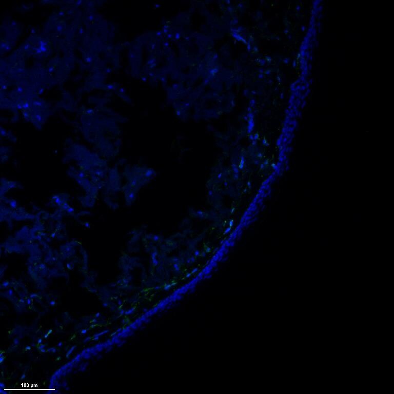
Application: Immunohistochemistry-FrozenSample Tested: Frozen human skin sectionsSpecies: HumanVerified Customer | Posted 12/14/2018Rt anti-IL10 (1:200, green) stained normal human skin, counterstained with DAPI (blue)Sections fixed, permeablized, blocked with 5% BSA, and stained with antibody diluted 1:200 in blocking solution

There are no reviews that match your criteria.
Protocols
Find general support by application which include: protocols, troubleshooting, illustrated assays, videos and webinars.
- Antigen Retrieval Protocol (PIER)
- Antigen Retrieval for Frozen Sections Protocol
- Appropriate Fixation of IHC/ICC Samples
- Cellular Response to Hypoxia Protocols
- Chromogenic IHC Staining of Formalin-Fixed Paraffin-Embedded (FFPE) Tissue Protocol
- Chromogenic Immunohistochemistry Staining of Frozen Tissue
- ClariTSA™ Fluorophore Kits
- Detection & Visualization of Antibody Binding
- Fluorescent IHC Staining of Frozen Tissue Protocol
- Graphic Protocol for Heat-induced Epitope Retrieval
- Graphic Protocol for the Preparation and Fluorescent IHC Staining of Frozen Tissue Sections
- Graphic Protocol for the Preparation and Fluorescent IHC Staining of Paraffin-embedded Tissue Sections
- Graphic Protocol for the Preparation of Gelatin-coated Slides for Histological Tissue Sections
- IHC Sample Preparation (Frozen sections vs Paraffin)
- Immunofluorescent IHC Staining of Formalin-Fixed Paraffin-Embedded (FFPE) Tissue Protocol
- Immunohistochemistry (IHC) and Immunocytochemistry (ICC) Protocols
- Immunohistochemistry Frozen Troubleshooting
- Immunohistochemistry Paraffin Troubleshooting
- Preparing Samples for IHC/ICC Experiments
- Preventing Non-Specific Staining (Non-Specific Binding)
- Primary Antibody Selection & Optimization
- Protocol for Heat-Induced Epitope Retrieval (HIER)
- Protocol for Making a 4% Formaldehyde Solution in PBS
- Protocol for VisUCyte™ HRP Polymer Detection Reagent
- Protocol for the Preparation & Fixation of Cells on Coverslips
- Protocol for the Preparation and Chromogenic IHC Staining of Frozen Tissue Sections
- Protocol for the Preparation and Chromogenic IHC Staining of Frozen Tissue Sections - Graphic
- Protocol for the Preparation and Chromogenic IHC Staining of Paraffin-embedded Tissue Sections
- Protocol for the Preparation and Chromogenic IHC Staining of Paraffin-embedded Tissue Sections - Graphic
- Protocol for the Preparation and Fluorescent IHC Staining of Frozen Tissue Sections
- Protocol for the Preparation and Fluorescent IHC Staining of Paraffin-embedded Tissue Sections
- Protocol for the Preparation of Gelatin-coated Slides for Histological Tissue Sections
- TUNEL and Active Caspase-3 Detection by IHC/ICC Protocol
- The Importance of IHC/ICC Controls
- Troubleshooting Guide: Immunohistochemistry
- View all Protocols, Troubleshooting, Illustrated assays and Webinars
Loading...
Associated Pathways


