Interleukin12 (IL-12) is a key mediator of cellular-immunity and induces the differentiation of Th1 cells from precursor T helper cells. The biological activities of IL-12 are mediated through the high-affinity receptor complex composed of two subunits designated IL-12 R beta 1 and IL-12 R beta 2. Individually, IL-12 R beta 1 and IL-12 R beta 2 bind IL‑12 with low affinity. Co-expression of both subunits confers high-affinity binding and is required for IL-12 activity. Both IL-12 receptor subunits are type I transmembrane proteins that share similarities with the gp130/G-CSF R subgroup in the cytokine receptor superfamily. IL-12 R beta 1 cDNA encodes a 662 amino acid (aa) protein with a putative 23 aa signal peptide that is cleaved to generate the mature protein with a 522 aa extracellular domain, a 25 aa transmembrane domain and a 92 aa cytoplasmic region. Expression of IL-12 R beta 1 is detected in activated T cells, NK cells and B cells. The expression of IL‑12 R beta 2 is more restricted and appears to be limited to Th2 cells.
Human IL-12 R beta 1 Antibody (69310)
R&D Systems | Catalog # MAB839

Key Product Details
Species Reactivity
Validated:
Human
Cited:
Human
Applications
Validated:
Flow Cytometry, CyTOF-ready
Cited:
Flow Cytometry
Label
Unconjugated
Antibody Source
Monoclonal Mouse IgG1 Clone # 69310
Loading...
Product Specifications
Immunogen
Mouse myeloma cell line NS0-derived recombinant human IL-12 R beta 1
Cys24-Glu540
Accession # P42701
Cys24-Glu540
Accession # P42701
Specificity
Detects human IL-12 R beta 1 in direct ELISAs. In direct ELISAs, no cross-reactivity with recombinant mouse IL-12 R beta 1 is observed.
Clonality
Monoclonal
Host
Mouse
Isotype
IgG1
Scientific Data Images for Human IL-12 R beta 1 Antibody (69310)
Detection of IL‑12 R beta 1 in Daudi cells by Flow Cytometry
Daudi cells were stained with Mouse Anti-Human IL‑12 R beta 1 Monoclonal Antibody (Catalog # MAB839, filled histogram) or isotype control antibody (Catalog # MAB002, open histogram) followed by Phycoerythrin-conjugated Anti-Mouse IgG Secondary Antibody (Catalog # F0102B). View our protocol for Staining Membrane-associated Proteins.Applications for Human IL-12 R beta 1 Antibody (69310)
Application
Recommended Usage
CyTOF-ready
Ready to be labeled using established conjugation methods. No BSA or other carrier proteins that could interfere with conjugation.
Flow Cytometry
0.25 µg/106 cells
Sample: Human peripheral blood mononuclear cells treated with PHA; Daudi human Burkitt's lymphoma cell line
Sample: Human peripheral blood mononuclear cells treated with PHA; Daudi human Burkitt's lymphoma cell line
Reviewed Applications
Read 1 review rated 4 using MAB839 in the following applications:
Flow Cytometry Panel Builder
Bio-Techne Knows Flow Cytometry
Save time and reduce costly mistakes by quickly finding compatible reagents using the Panel Builder Tool.
Advanced Features
- Spectra Viewer - Custom analysis of spectra from multiple fluorochromes
- Spillover Popups - Visualize the spectra of individual fluorochromes
- Antigen Density Selector - Match fluorochrome brightness with antigen density

Formulation, Preparation, and Storage
Purification
Protein A or G purified from ascites
Reconstitution
Reconstitute at 0.5 mg/mL in sterile PBS. For liquid material, refer to CoA for concentration.
Loading...
Formulation
Lyophilized from a 0.2 μm filtered solution in PBS with Trehalose. *Small pack size (SP) is supplied either lyophilized or as a 0.2 µm filtered solution in PBS.
Shipping
Lyophilized product is shipped at ambient temperature. Liquid small pack size (-SP) is shipped with polar packs. Upon receipt, store immediately at the temperature recommended below.
Stability & Storage
Use a manual defrost freezer and avoid repeated freeze-thaw cycles.
- 12 months from date of receipt, -20 to -70 °C as supplied.
- 1 month, 2 to 8 °C under sterile conditions after reconstitution.
- 6 months, -20 to -70 °C under sterile conditions after reconstitution.
Calculators
Background: IL-12 R beta 1
References
- Gately, M.K. et al. (1998) Annu. Rev. Immunol. 16:495.
Long Name
Interleukin 12 Receptor beta 1
Alternate Names
CD212, IL-12Rb1, IL12R beta 1, IL12RB1
Gene Symbol
IL12RB1
UniProt
Additional IL-12 R beta 1 Products
Product Documents for Human IL-12 R beta 1 Antibody (69310)
Certificate of Analysis
To download a Certificate of Analysis, please enter a lot or batch number in the search box below.
Note: Certificate of Analysis not available for kit components.
Product Specific Notices for Human IL-12 R beta 1 Antibody (69310)
For research use only
Citations for Human IL-12 R beta 1 Antibody (69310)
 Powered by Bioz
Customer Reviews for Human IL-12 R beta 1 Antibody (69310) (1)
4 out of 5
1 Customer Rating
Have you used Human IL-12 R beta 1 Antibody (69310)?
Submit a review and receive an Amazon gift card!
$25/€18/£15/$25CAN/¥2500 Yen for a review with an image
$10/€7/£6/$10CAN/¥1110 Yen for a review without an image
Submit a review
Customer Images
Showing
1
-
1 of
1 review
Showing All
Filter By:
-
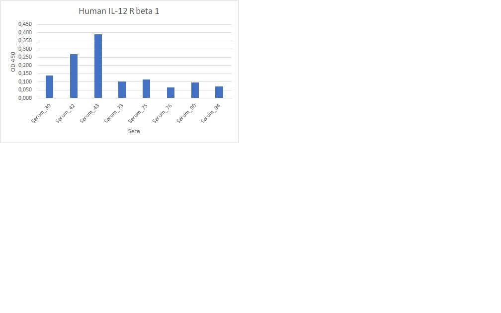
Application: ELISASample Tested: Serum and PlasmaSpecies: HumanVerified Customer | Posted 02/13/2023workign good with 1:5 dilution for the detection of human IL12 R beta in the serum or plasma samples.

There are no reviews that match your criteria.
Protocols
Find general support by application which include: protocols, troubleshooting, illustrated assays, videos and webinars.
- 7-Amino Actinomycin D (7-AAD) Cell Viability Flow Cytometry Protocol
- Extracellular Membrane Flow Cytometry Protocol
- Flow Cytometry Protocol for Cell Surface Markers
- Flow Cytometry Protocol for Staining Membrane Associated Proteins
- Flow Cytometry Staining Protocols
- Flow Cytometry Troubleshooting Guide
- Intracellular Flow Cytometry Protocol Using Alcohol (Methanol)
- Intracellular Flow Cytometry Protocol Using Detergents
- Intracellular Nuclear Staining Flow Cytometry Protocol Using Detergents
- Intracellular Staining Flow Cytometry Protocol Using Alcohol Permeabilization
- Intracellular Staining Flow Cytometry Protocol Using Detergents to Permeabilize Cells
- Propidium Iodide Cell Viability Flow Cytometry Protocol
- Protocol for Liperfluo
- Protocol for the Characterization of Human Th22 Cells
- Protocol for the Characterization of Human Th9 Cells
- Protocol: Annexin V and PI Staining by Flow Cytometry
- Protocol: Annexin V and PI Staining for Apoptosis by Flow Cytometry
- Troubleshooting Guide: Fluorokine Flow Cytometry Kits
- View all Protocols, Troubleshooting, Illustrated assays and Webinars
Loading...
Associated Pathways


