MIF (or macrophage migration inhibitory factor) was the first lymphokine/cytokine to be recognized in the pregenomics era (1, 2). Regardless, it is one of the least understood of all inflammatory mediators (1, 3). Human MIF is a 12.5 kDa, 115 amino acid (aa) nonglycosylated polypeptide that is synthesized without a signal sequence (4-7). Secretion occurs nonclassically via an ABCA1 transporter (8). The initiating Met is removed, leaving Pro as the first amino acid. The molecule consists of two alpha -helices and six beta -strands, four of which form a beta -sheet. The two remaining beta -strands interact with other MIF molecules, creating a trimer (2, 9, 10). Structure-function studies suggest MIF is bifunctional with segregated topology. The N- and C-termini mediate enzyme activity (in theory). Phenylpyruvate tautomerase activity (enol-to-keto) has been demonstrated and is dependent upon Pro at position #1 (11). Amino acids 50-65 have also been suggested to contain thiol-protein oxidoreductase activity (12). MIF has proinflammatory cytokine activity centered around aa’s 49-65. On fibroblasts, MIF induces, IL-1, IL-8, and MMP expression; on macrophages, MIF stimulates NO production and TNF-alpha release following IFN-gamma activation (13, 14). MIF apparently acts through CD74 and CD44, likely in some form of trimeric interaction (15, 16). Human MIF is active on mouse cells (14). Human MIF is 90%, 94%, 95%, and 90% aa identical to mouse, bovine, porcine, and rat MIF, respectively.

Key Product Details
Species Reactivity
Validated:
Human
Cited:
Human, Mouse, Bovine, Transgenic Mouse
Applications
Validated:
Western Blot, Simple Western
Cited:
Immunohistochemistry, Immunohistochemistry-Paraffin, Western Blot, Neutralization, Immunocytochemistry, Immunoprecipitation, ELISA Capture, ELISA Development, ELISA Development (Capture), Luminex Development
Label
Unconjugated
Antibody Source
Monoclonal Mouse IgG1 Clone # 12302
Loading...
Product Specifications
Immunogen
E. coli-derived recombinant human MIF
Pro2-Ala115
Accession # P14174
Pro2-Ala115
Accession # P14174
Specificity
Detects human MIF in Western blots. In Western blots, this antibody shows approximately 25% cross-reactivity with recombinant mouse MIF.
Clonality
Monoclonal
Host
Mouse
Isotype
IgG1
Scientific Data Images for Human MIF Antibody (12302)
Detection of Human MIF by Western Blot.
Western blot shows lysate of K562 human chronic myelogenous leukemia cell line. PVDF membrane was probed with 1 µg/mL of Mouse Anti-Human MIF Monoclonal Antibody (Catalog # MAB289) followed by HRP-conjugated Anti-Mouse IgG Secondary Antibody (Catalog # HAF018). A specific band was detected for MIF at approximately 12 kDa (as indicated). This experiment was conducted under reducing conditions and using Immunoblot Buffer Group 1.
Detection of Human MIF by Simple WesternTM.
Simple Western lane view shows lysates of Exosome Standards (HEK293) (NBP3-11684), Exosome Standards (LNCaP) (NBP3-11687) and K562 human chronic myelogenous leukemia cell line, loaded at 0.5 mg/ml. A specific band was detected for MIF at approximately 11 kDa (as indicated) using 10 µg/ml of Mouse Anti-Human MIF Monoclonal Antibody (Catalog # MAB289) followed by HRP-conjugated Goat Anti-Mouse Secondary Antibody (Catalog # 042-205). This experiment was conducted under reducing conditions and using the 2-40kDa separation system.
Detection of Humean MIF by Simple WesternTM.
Simple Western lane view shows lysates of THP‑1 human acute monocytic leukemia cell line and U937 human histiocytic lymphoma cell line, loaded at 0.2 mg/mL. A specific band was detected for MIF at approximately 12 kDa (as indicated) using 10 µg/mL of Mouse Anti-Human MIF Monoclonal Antibody (Catalog # MAB289). This experiment was conducted under reducing conditions and using the 12-230 kDa separation system.
Detection of MIF by Western Blot
Effect of treatments on MIF secretion, intracellular content and gene expression. A. MIF secretion – Conditioned medium following 24 h growth was assayed for MIF content by ELISA. Data are expressed as culture medium MIF concentration in pg per 105 cells. All treatments decreased MIF in the culture media. B. Western blot of anti-MIF treated conditioned medium following 24 h treatment- 12 kDa MIF band is present in all samples. Lane 1, DMEM 1% BSA; Lane 2, non-specific mouse IgG1; Lane 3, anti-MIF antibody treatment. C. Intracellular MIF content – Changes in intracellular MIF 24 h post treatment. Data are expressed as cleared cell lysate MIF concentration in pg per 105 cells. Note that HA treatment increased, while MIF antisense treatment decrease the concentration of MIF in the cell lysates. Anti-MIF treatment did not produce a significant effect. D. MIF gene expression -MIF mRNA content was quantified in cells following 24 h and the indicated treatment. Data are expressed as a relative intensity ratio. PCR band intensity was determined from the formula, total intensity = area × average intensity. The relative intensity ratio is determined from the total intensity of gene specific PCR product band divided by the 18 S rRNA band intensity (internal standard). All treatments resulted in a decrease in MIF mRNA with the greatest effects seen following MIF anti-sense treatment. (* – p < 0.05, ** – p < 0.01, *** – p < 0.001). Image collected and cropped by CiteAb from the following open publication (https://pubmed.ncbi.nlm.nih.gov/15248897), licensed under a CC-BY license. Not internally tested by R&D Systems.Applications for Human MIF Antibody (12302)
Application
Recommended Usage
Simple Western
10 µg/mL
Sample: THP‑1 human acute monocytic leukemia cell line and U937 human histiocytic lymphoma cell line
Sample: THP‑1 human acute monocytic leukemia cell line and U937 human histiocytic lymphoma cell line
Western Blot
1 µg/mL
Sample: K562 human chronic myelogenous leukemia cell line
Sample: K562 human chronic myelogenous leukemia cell line
Reviewed Applications
Read 8 reviews rated 4.4 using MAB289 in the following applications:
Formulation, Preparation, and Storage
Purification
Protein A or G purified from hybridoma culture supernatant
Reconstitution
Reconstitute at 0.5 mg/mL in sterile PBS. For liquid material, refer to CoA for concentration.
Loading...
Formulation
Lyophilized from a 0.2 μm filtered solution in PBS with Trehalose. See Certificate of Analysis for details.
*Small pack size (-SP) is supplied either lyophilized or as a 0.2 µm filtered solution in PBS.
*Small pack size (-SP) is supplied either lyophilized or as a 0.2 µm filtered solution in PBS.
Shipping
Lyophilized product is shipped at ambient temperature. Liquid small pack size (-SP) is shipped with polar packs. Upon receipt, store immediately at the temperature recommended below.
Stability & Storage
Use a manual defrost freezer and avoid repeated freeze-thaw cycles.
- 12 months from date of receipt, -20 to -70 °C as supplied.
- 1 month, 2 to 8 °C under sterile conditions after reconstitution.
- 6 months, -20 to -70 °C under sterile conditions after reconstitution.
Calculators
Background: MIF
References
- Norand, E.F. and M. Leech (2005) Front. Biosci. 10:12.
- Donn, R.P. and D.W. Ray (2004) J. Endocrinol. 182:1.
- Calandra, T. and T. Roger (2003) Nat. Rev. Immunol. 3:791.
- Kozak, C.A. et al. (1995) Genomics 27:405.
- Weiser, W.Y. et al. (1989) Proc. Natl. Acad. Sci. USA 86:7522.
- Paralkar, V. and G. Wistow (1994) Genomics 19:48.
- Wistow, G.J. et al. (1993) Proc. Natl. Acad. Sci. USA 90:1272.
- Flieger, O. et al. (2003) FEBS Lett. 551:78.
- Philo, J.S. et al. (2004) Biophys. Chem. 108:77.
- Sun, H-W. et al. (1996) Protein Eng. 9:631.
- Stamps, S.L. et. al. (2000) Biochemistry 39:9671.
- Nguyen, M.T. et al. (2003) J. Biol. Chem. 278:33654.
- Sato, A. et al. (2003) Dev. Comp. Immunol. 27:401.
- Bernhagen, J. et al. (1994) Biochemistry 33:14144.
- Leng, L. et al. (2003) J. Exp. Med. 197:1467.
- Meyer-Siegler, K.L. and P.L. Vera (2005) J. Urol. 173:615.
Long Name
Macrophage Migration Inhibitory Factor
Alternate Names
EC 5.3.2.1, EC 5.3.3.12, GIFmacrophage migration inhibitory factor, GLIF, Glycosylation-inhibiting factor, L-dopachrome isomerase, L-dopachrome tautomerase, macrophage migration inhibitory factor (glycosylation-inhibiting factor), MMIF, Phenylpyruvate tautomerase
Gene Symbol
MIF
UniProt
Additional MIF Products
Product Documents for Human MIF Antibody (12302)
Certificate of Analysis
To download a Certificate of Analysis, please enter a lot or batch number in the search box below.
Note: Certificate of Analysis not available for kit components.
Product Specific Notices for Human MIF Antibody (12302)
For research use only
Related Research Areas
Citations for Human MIF Antibody (12302)
 Powered by Bioz
Customer Reviews for Human MIF Antibody (12302) (8)
4.4 out of 5
8 Customer Ratings
Have you used Human MIF Antibody (12302)?
Submit a review and receive an Amazon gift card!
$25/€18/£15/$25CAN/¥2500 Yen for a review with an image
$10/€7/£6/$10CAN/¥1110 Yen for a review without an image
Submit a review
Customer Images

Showing
1
-
5 of
8 reviews
Showing All
Filter By:
-
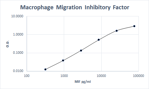
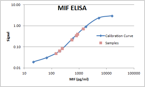
Application: ELISASample Tested: SerumSpecies: PrimateVerified Customer | Posted 11/14/2022Used as a non-human primate capture molecule

-
Application: Western BlotSample Tested: THP-1 human acute monocytic leukemia cell lineSpecies: HumanVerified Customer | Posted 10/07/2021Western blot. THP-1 human acute monocytic leukemia cell line.
-
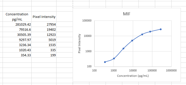
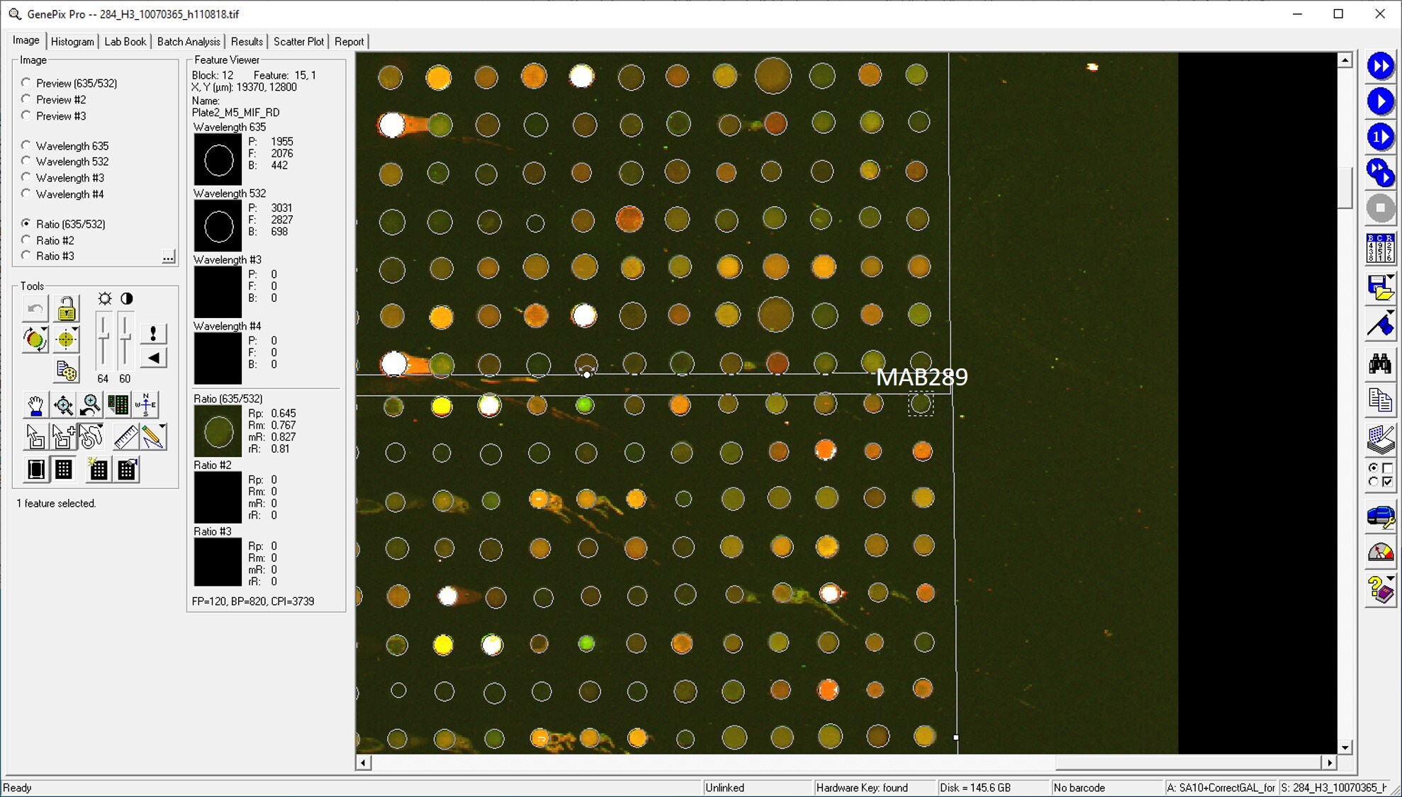
Application: MicroarraysSample Tested: EDTA PlasmaSpecies: HumanVerified Customer | Posted 01/14/2021
-
Application: ELISASample Tested: Serum and PlasmaSpecies: HumanVerified Customer | Posted 07/19/2019
-
Application: MicroarraySample Tested: EDTA PlasmaSpecies: HumanVerified Customer | Posted 11/20/2018
-
Application: MicroarraysSample Tested: EDTA PlasmaSpecies: HumanVerified Customer | Posted 11/14/2018
-
Application: ELISASample Tested: Serum and PlasmaSpecies: HumanVerified Customer | Posted 11/08/2018
-
Application: ELISASample Tested: Serum and PlasmaSpecies: HumanVerified Customer | Posted 11/07/2017This antibody was used as a matched pair with AF289 to build a sandwich ELISA to measure MIF in human serum and plasma. The antibody worked well.
There are no reviews that match your criteria.
Protocols
Find general support by application which include: protocols, troubleshooting, illustrated assays, videos and webinars.
- Cellular Response to Hypoxia Protocols
- R&D Systems Quality Control Western Blot Protocol
- Troubleshooting Guide: Western Blot Figures
- Western Blot Conditions
- Western Blot Protocol
- Western Blot Protocol for Cell Lysates
- Western Blot Troubleshooting
- Western Blot Troubleshooting Guide
- View all Protocols, Troubleshooting, Illustrated assays and Webinars
Loading...