Contactin-2 (CNTN2), also called TAG1 (transient axonal glycoprotein), TAX1 (transiently-expressed axonal glycoprotein), or axonin-1, is a 135 kDa glycosyl-phosphatidylinositol (GPI)- anchored cell adhesion molecule that belongs to the contactin subfamily within the immunoglobulin (Ig) protein superfamily (1-3). Mouse Contactin-2 cDNA encodes a 30 amino acid (aa) signal peptide, a 984 aa mature secreted protein with 6 Ig-like domains followed by 4 fibronectin type III-like repeats, and a 26 aa C-terminal GPI anchor pro-sequence. GPI-specific phospholipase activity can release soluble, active Contactin-2 from the membrane (2). Mature mouse Contactin-2 shares approximately 93%, 97%, and 77% aa sequence identity with human, rat, and chicken Contactin-2, respectively. During development, Contactin-2 is expressed by a subset of neuronal populations in the central nervous system (CNS) and peripheral nervous system (PNS), particularly during initial phases of axon outgrowth (3-5). Both the 135 kDa form and a 90 kDa form are also upregulated in response to CNS injury in the adult (6). Data support a role for Contactin-2 in axon pathfinding, neurite outgrowth and adhesion, especially in the CNS (3-6). In mature myelinated fibers, Contactin-2 is expressed by oligodendrocytes and Schwann cells, which are myelinating glial cells of the CNS and PNS, respectively (7, 8). It is enriched in the juxtaparanodal regions, where it recruits contactin-associated protein 2 (caspr2), a transmembrane neurexin involved in cell adhesion and intercellular communication (7-10). The axonal Contactin-2 interacts in cis with caspr2 and in trans with another Contactin-2 on the glial membrane (8). This ternary complex is required for the accumulation and organization of K+ channels in the juxtaparanodes (9).

Key Product Details
Species Reactivity
Validated:
Human, Mouse, Rat
Cited:
Human, Mouse, Rat, Hamster - Mesocricetus auratus (Golden Hamster), Primate, Transgenic Mouse
Applications
Validated:
Immunohistochemistry, Western Blot, Simple Western
Cited:
Immunohistochemistry, Immunohistochemistry-Frozen, Western Blot, Immunocytochemistry, Mass Spectrometry
Label
Unconjugated
Antibody Source
Polyclonal Goat IgG
Loading...
Product Specifications
Immunogen
Mouse myeloma cell line NS0-derived recombinant mouse Contactin-2/TAG1
Gln31-Ser1014
Accession # Q61330
Gln31-Ser1014
Accession # Q61330
Specificity
Detects mouse and rat Contactin-2 in direct ELISAs. Detects human, mouse, and rat Contactin-2 in Western blots.
Clonality
Polyclonal
Host
Goat
Isotype
IgG
Scientific Data Images for Contactin-2/TAG1 Antibody
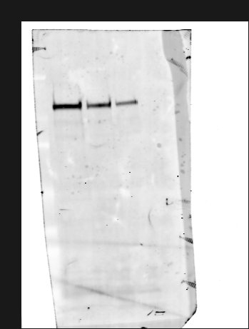
Detection of Human, Mouse, and Rat Contactin‑2/TAG1 by Western Blot.
Western blot shows lysates of human cerebellum tissue, mouse brain tissue, and rat brain tissue. PVDF membrane was probed with 1 µg/mL of Goat Anti-Human/Mouse/Rat Contactin-2/TAG1 Antigen Affinity-purified Polyclonal Antibody (Catalog # AF4439) followed by HRP-conjugated Anti-Goat IgG Secondary Antibody (Catalog # HAF017). A specific band was detected for Contactin-2/TAG1 at approximately 135 kDa (as indicated). This experiment was conducted under reducing conditions and using Immunoblot Buffer Group 1.
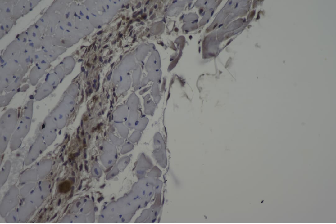
Contactin‑2/TAG1 in Mouse Embryo.
Contactin-2/TAG1 was detected in immersion fixed frozen sections of mouse embryo (E13) using Goat Anti-Human/Mouse/Rat Contactin-2/TAG1 Antigen Affinity-purified Polyclonal Antibody (Catalog # AF4439) at 15 µg/mL overnight at 4 °C. Tissue was stained using the Anti-Goat HRP-DAB Cell & Tissue Staining Kit (brown; Catalog # CTS008) and counterstained with hematoxylin (blue). Specific staining was localized to muscle cells in proximity to ribs. View our protocol for Chromogenic IHC Staining of Frozen Tissue Sections.
Detection of Mouse Contactin‑2/TAG1 by Simple WesternTM.
Simple Western lane view shows lysates of mouse spinal cord tissue, loaded at 0.2 mg/mL. A specific band was detected for Contactin-2/TAG1 at approximately 162 kDa (as indicated) using 10 µg/mL of Goat Anti-Human/Mouse/Rat Contactin-2/TAG1 Antigen Affinity-purified Polyclonal Antibody (Catalog # AF4439) followed by 1:50 dilution of HRP-conjugated Anti-Goat IgG Secondary Antibody (Catalog # HAF109). This experiment was conducted under reducing conditions and using the 12-230 kDa separation system.
Detection of Mouse Human/Mouse/Rat Contactin-2/TAG1 Antibody by Western Blot
Quantitative proteomics in Rab35 cKO P0 hippocampus.a Volcano plot of the TMT-based quantitative proteomes identifying the dysregulated proteins in Rab35 cKO hippocampus in comparison with the control hippocampus (n = 5 mice per genotype). b Number of proteins identified as significantly dysregulated and as either membrane traffic-related or neuronal migration-related. c, d Western blot analysis of control and Rab35 cKO P0 hippocampi using anti-contactin-2, anti-CHL1, and anti-actin antibodies. e, f Quantification of contactin-2 (c) and CHL1 (d) protein levels in control and Rab35 cKO P0 hippocampi. Band intensities of the indicated proteins were normalized to those of actin (n = 9 mice per genotype). Unpaired Student’s t-test; e, p = 0.0153; f, p = 0.0095. g, h Levels of contactin-2 (g) and CHL1 (h) were quantified by targeted MS using the PRM method (n = 5 mice per genotype). Unpaired Student’s t-test; gp = 0.0053; hp = 0.0229. i Western blot analysis of control and Rab35 cKO P0 hippocampus using anti-N-cadherin and anti-actin antibodies. j Quantification of N-cadherin protein levels in the control and Rab35 cKO P0 hippocampus (n = 9 mice per genotypes). Unpaired Student’s t-test, p = 0.9020. k Representative images of DIV 2 hippocampal primary neurons stained for contactin-2 (green), rhodamine-phalloidin (magenta) and DAPI (blue). Scale bar, 20 μm. l Quantification of contactin-2 intensity at the somatic plasma membrane in control (n = 4) and Rab35-deficient (n = 4) cells. Thirty neurons from four different cultures per genotype were analyzed. Mann–Whitney U-test, p = 0.0286. Data represent the mean ± SEM; n.s. not significant (p > 0.05); *p < 0.05; **p < 0.01. Image collected and cropped by CiteAb from the following publication (https://pubmed.ncbi.nlm.nih.gov/37085665), licensed under a CC-BY license. Not internally tested by R&D Systems.Applications for Contactin-2/TAG1 Antibody
Application
Recommended Usage
Immunohistochemistry
5-15 µg/mL
Sample:
Sample:
Immersion fixed frozen sections of mouse embryo (E13)
Simple Western
10 µg/mL
Sample: Mouse spinal cord tissue
Sample: Mouse spinal cord tissue
Western Blot
1 µg/mL
Sample: Human cerebellum tissue, Mouse brain tissue, and Rat brain tissue
Sample: Human cerebellum tissue, Mouse brain tissue, and Rat brain tissue
Reviewed Applications
Read 2 reviews rated 4.5 using AF4439 in the following applications:
Formulation, Preparation, and Storage
Purification
Antigen Affinity-purified
Reconstitution
Reconstitute at 0.2 mg/mL in sterile PBS. For liquid material, refer to CoA for concentration.
Loading...
Formulation
Lyophilized from a 0.2 μm filtered solution in PBS with Trehalose. *Small pack size (SP) is supplied either lyophilized or as a 0.2 µm filtered solution in PBS.
Shipping
Lyophilized product is shipped at ambient temperature. Liquid small pack size (-SP) is shipped with polar packs. Upon receipt, store immediately at the temperature recommended below.
Stability & Storage
Use a manual defrost freezer and avoid repeated freeze-thaw cycles.
- 12 months from date of receipt, -20 to -70 °C as supplied.
- 1 month, 2 to 8 °C under sterile conditions after reconstitution.
- 6 months, -20 to -70 °C under sterile conditions after reconstitution.
Calculators
Background: Contactin-2/TAG1
References
- Wolfer, D. and R.J. Giger (1994) Swissprot Accession # Q61330.
- Hasler, T.H. et al. (1993) Eur. J. Biochem. 211:329.
- Karagogeos, D. (2003) Front. Biosci. 8:s1304.
- Liu, Y. and M.C. Halloran (2005) J. Neurosci. 25:10556.
- Denaxa, M. et al. (2005) Dev. Biol. 288:87.
- Soares, S. et al. (2005) Eur. J. Neurosci. 21:1169.
-
Traka, M. et al. (2002) J. Neurosci. 22:3016.
-
Poliak, S. and E. Peles (2003) Nat. Rev. Neurosci. 4:968.
-
Traka, M. et al. (2003) J. Cell Biol. 162:1161.
-
Poliak, S. et al. (2003) J. Cell Biol. 162:1149.
Alternate Names
Axonin-1, AXT, CNTN2, Contactin2, TAG1, TAX1
Gene Symbol
CNTN2
UniProt
Additional Contactin-2/TAG1 Products
Product Documents for Contactin-2/TAG1 Antibody
Certificate of Analysis
To download a Certificate of Analysis, please enter a lot or batch number in the search box below.
Note: Certificate of Analysis not available for kit components.
Product Specific Notices for Contactin-2/TAG1 Antibody
For research use only
Related Research Areas
Citations for Contactin-2/TAG1 Antibody
 Powered by Bioz
Customer Reviews for Contactin-2/TAG1 Antibody (2)
4.5 out of 5
2 Customer Ratings
Have you used Contactin-2/TAG1 Antibody?
Submit a review and receive an Amazon gift card!
$25/€18/£15/$25CAN/¥2500 Yen for a review with an image
$10/€7/£6/$10CAN/¥1110 Yen for a review without an image
Submit a review
Customer Images
Showing
1
-
2 of
2 reviews
Showing All
Filter By:
-
Application: ImmunohistochemistrySample Tested: mouse heartSpecies: MouseVerified Customer | Posted 08/05/2020

-
Application: Western BlotSample Tested: Brain tissueSpecies: MouseVerified Customer | Posted 04/26/201680, 40 and 20ug of total mouse brain homogenate was separated by SDS-PAGE and transfered to PVDF membrane Block: 1% BSA, 1% FSG PBS-T 1h RT Primary: 1:1000, 1% BSA, 1% FSG PBS-T O/N 4oC Secondary: 1:5000,1% BSA, 1% FSG PBS-T 2h RT (Secondary - DaG 800, Licor)
There are no reviews that match your criteria.
Protocols
Find general support by application which include: protocols, troubleshooting, illustrated assays, videos and webinars.
- Antigen Retrieval Protocol (PIER)
- Antigen Retrieval for Frozen Sections Protocol
- Appropriate Fixation of IHC/ICC Samples
- Cellular Response to Hypoxia Protocols
- Chromogenic IHC Staining of Formalin-Fixed Paraffin-Embedded (FFPE) Tissue Protocol
- Chromogenic Immunohistochemistry Staining of Frozen Tissue
- ClariTSA™ Fluorophore Kits
- Detection & Visualization of Antibody Binding
- Fluorescent IHC Staining of Frozen Tissue Protocol
- Graphic Protocol for Heat-induced Epitope Retrieval
- Graphic Protocol for the Preparation and Fluorescent IHC Staining of Frozen Tissue Sections
- Graphic Protocol for the Preparation and Fluorescent IHC Staining of Paraffin-embedded Tissue Sections
- Graphic Protocol for the Preparation of Gelatin-coated Slides for Histological Tissue Sections
- IHC Sample Preparation (Frozen sections vs Paraffin)
- Immunofluorescent IHC Staining of Formalin-Fixed Paraffin-Embedded (FFPE) Tissue Protocol
- Immunohistochemistry (IHC) and Immunocytochemistry (ICC) Protocols
- Immunohistochemistry Frozen Troubleshooting
- Immunohistochemistry Paraffin Troubleshooting
- Preparing Samples for IHC/ICC Experiments
- Preventing Non-Specific Staining (Non-Specific Binding)
- Primary Antibody Selection & Optimization
- Protocol for Heat-Induced Epitope Retrieval (HIER)
- Protocol for Making a 4% Formaldehyde Solution in PBS
- Protocol for VisUCyte™ HRP Polymer Detection Reagent
- Protocol for the Preparation & Fixation of Cells on Coverslips
- Protocol for the Preparation and Chromogenic IHC Staining of Frozen Tissue Sections
- Protocol for the Preparation and Chromogenic IHC Staining of Frozen Tissue Sections - Graphic
- Protocol for the Preparation and Chromogenic IHC Staining of Paraffin-embedded Tissue Sections
- Protocol for the Preparation and Chromogenic IHC Staining of Paraffin-embedded Tissue Sections - Graphic
- Protocol for the Preparation and Fluorescent IHC Staining of Frozen Tissue Sections
- Protocol for the Preparation and Fluorescent IHC Staining of Paraffin-embedded Tissue Sections
- Protocol for the Preparation of Gelatin-coated Slides for Histological Tissue Sections
- R&D Systems Quality Control Western Blot Protocol
- TUNEL and Active Caspase-3 Detection by IHC/ICC Protocol
- The Importance of IHC/ICC Controls
- Troubleshooting Guide: Immunohistochemistry
- Troubleshooting Guide: Western Blot Figures
- Western Blot Conditions
- Western Blot Protocol
- Western Blot Protocol for Cell Lysates
- Western Blot Troubleshooting
- Western Blot Troubleshooting Guide
- View all Protocols, Troubleshooting, Illustrated assays and Webinars
Loading...