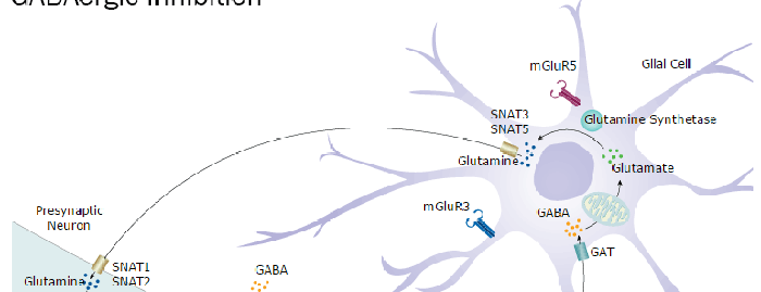
GABAB R1 (GABA-B receptor subunit 1; also GABA-BR1, GABBR1 and GB1) is a multispan member of the GABA-B receptor subfamily, GPCR-3 family of proteins. It forms an obligatory heterodimer with GABA-BR2, creating a G-protein metabotropic GABA receptor that inhibits adenylyl cyclase activity and activates K+ channels. Presynaptically, this blocks neurotransmitter release; postsynaptically, it lowers neuron excitability. Rat GABAB R1 is 991 amino acids (aa) in length. It is a 7‑transmembrane glycoprotein that contains a 16 aa signal sequence, an extended N‑terminal extracellular region (aa 17-590) that contains two SUSHI domains (aa 29‑158), and a long C-terminal cytoplasmic domain (aa 885-991). There are several splice variants with predicted molecular weights ranging from 90 to 111 kDa and multiple glycosylation sites. The 991 aa isoform described above is called GABAB R1e (R1e). There is also a 960 aa, 130 kDa isoform that shows a deletion of aa 771‑801. This variant (R1a) is associated with postsynaptic membranes. A third isoform (R1b) is 844 aa in length and 100 kDa in size, and possesses both a deletion of aa 771-801, and a 47 aa substitution for aa 1-163. This variant is presynaptic in location. Two other isoforms are variants of GABAB R1b. Each show the same N‑terminal substitution, with a fourth isoform (R1c) retaining aa 771-801, and a fifth isoform (R1d) deleting aa 771-801, coupled to a 25 aa substitution for aa 935‑991. Over aa 17-586, rat GABAB R1e/a shares 99% aa identity with both mouse and human GABAB R1.

Key Product Details
Species Reactivity
Human, Mouse, Rat
Applications
Immunohistochemistry, Western Blot
Label
Unconjugated
Antibody Source
Polyclonal Sheep IgG
Loading...
Product Specifications
Immunogen
Chinese hamster ovary cell line CHO-derived recombinant rat GABAB R1
Gly17-Leu586
Accession # Q9Z0U4
Gly17-Leu586
Accession # Q9Z0U4
Specificity
Detects recombinant mouse and rat GABAB R1 in direct ELISAs and Western blots.Detects human and rat GABAB R1 in Western blots. In direct ELISAs, less than 1% cross-reactivity with recombinant rat GABAB R2 is observed.
Clonality
Polyclonal
Host
Sheep
Isotype
IgG
Scientific Data Images for GABA-B R1 Antibody
Detection of Human and Rat GABABR1 by Western Blot.
Western blot shows lysates of IMR-32 human neuroblastoma cell line and rat embryonic hippocampal neurons. PVDF membrane was probed with 1 µg/mL of Sheep Anti-Mouse/Rat GABABR1 Antigen Affinity-purified Polyclonal Antibody (Catalog # AF7000) followed by HRP-conjugated Anti-Sheep IgG Secondary Antibody (Catalog # HAF016). Specific bands were detected for GABABR1 at approximately 70-80 kDa (as indicated). This experiment was conducted under reducing conditions and using Immunoblot Buffer Group 1.
GABABR1 in Rat Brain.
GABABR1 was detected in perfusion fixed frozen sections of rat brain (dorsal root ganglia) using Sheep Anti-Mouse/Rat GABABR1 Antigen Affinity-purified Polyclonal Antibody (Catalog # AF7000) at 1.7 µg/mL overnight at 4 °C. Tissue was stained using the Northern-Lights™ 557-conjugated Anti-Sheep IgG Secondary Antibody (red; Catalog # NL010) and counterstained with DAPI (blue). Specific staining was localized to the cell bodies of dorsal root ganglia neurons. View our protocol for Fluorescent IHC Staining of Frozen Tissue Sections.Applications for GABA-B R1 Antibody
Application
Recommended Usage
Immunohistochemistry
5-15 µg/mL
Sample: Perfusion fixed frozen sections of rat brain (dorsal root ganglia)
Sample: Perfusion fixed frozen sections of rat brain (dorsal root ganglia)
Western Blot
1 µg/mL
Sample: IMR‑32 human neuroblastoma cell line and rat embryonic hippocampal neurons
Sample: IMR‑32 human neuroblastoma cell line and rat embryonic hippocampal neurons
Formulation, Preparation, and Storage
Purification
Antigen Affinity-purified
Reconstitution
Sterile PBS to a final concentration of 0.2 mg/mL. For liquid material, refer to CoA for concentration.
Loading...
Formulation
Lyophilized from a 0.2 μm filtered solution in PBS with Trehalose. *Small pack size (SP) is supplied either lyophilized or as a 0.2 µm filtered solution in PBS.
Shipping
Lyophilized product is shipped at ambient temperature. Liquid small pack size (-SP) is shipped with polar packs. Upon receipt, store immediately at the temperature recommended below.
Stability & Storage
Use a manual defrost freezer and avoid repeated freeze-thaw cycles.
- 12 months from date of receipt, -20 to -70 °C as supplied.
- 1 month, 2 to 8 °C under sterile conditions after reconstitution.
- 6 months, -20 to -70 °C under sterile conditions after reconstitution.
Calculators
Background: GABA-B R1
Long Name
gamma-Aminobutyric Acid Type B Receptor Subunit 1
Alternate Names
GABABR1, GABBR1, Gb1, GPRC3A
Gene Symbol
GABBR1
UniProt
Additional GABA-B R1 Products
Product Documents for GABA-B R1 Antibody
Certificate of Analysis
To download a Certificate of Analysis, please enter a lot or batch number in the search box below.
Note: Certificate of Analysis not available for kit components.
Product Specific Notices for GABA-B R1 Antibody
For research use only
Citations for GABA-B R1 Antibody
 Powered by Bioz
Customer Reviews for GABA-B R1 Antibody
There are currently no reviews for this product. Be the first to review GABA-B R1 Antibody and earn rewards!
Have you used GABA-B R1 Antibody?
Submit a review and receive an Amazon gift card!
$25/€18/£15/$25CAN/¥2500 Yen for a review with an image
$10/€7/£6/$10CAN/¥1110 Yen for a review without an image
Submit a review
Protocols
Find general support by application which include: protocols, troubleshooting, illustrated assays, videos and webinars.
- Antigen Retrieval Protocol (PIER)
- Antigen Retrieval for Frozen Sections Protocol
- Appropriate Fixation of IHC/ICC Samples
- Cellular Response to Hypoxia Protocols
- Chromogenic IHC Staining of Formalin-Fixed Paraffin-Embedded (FFPE) Tissue Protocol
- Chromogenic Immunohistochemistry Staining of Frozen Tissue
- ClariTSA™ Fluorophore Kits
- Detection & Visualization of Antibody Binding
- Fluorescent IHC Staining of Frozen Tissue Protocol
- Graphic Protocol for Heat-induced Epitope Retrieval
- Graphic Protocol for the Preparation and Fluorescent IHC Staining of Frozen Tissue Sections
- Graphic Protocol for the Preparation and Fluorescent IHC Staining of Paraffin-embedded Tissue Sections
- Graphic Protocol for the Preparation of Gelatin-coated Slides for Histological Tissue Sections
- IHC Sample Preparation (Frozen sections vs Paraffin)
- Immunofluorescent IHC Staining of Formalin-Fixed Paraffin-Embedded (FFPE) Tissue Protocol
- Immunohistochemistry (IHC) and Immunocytochemistry (ICC) Protocols
- Immunohistochemistry Frozen Troubleshooting
- Immunohistochemistry Paraffin Troubleshooting
- Preparing Samples for IHC/ICC Experiments
- Preventing Non-Specific Staining (Non-Specific Binding)
- Primary Antibody Selection & Optimization
- Protocol for Heat-Induced Epitope Retrieval (HIER)
- Protocol for Making a 4% Formaldehyde Solution in PBS
- Protocol for VisUCyte™ HRP Polymer Detection Reagent
- Protocol for the Preparation & Fixation of Cells on Coverslips
- Protocol for the Preparation and Chromogenic IHC Staining of Frozen Tissue Sections
- Protocol for the Preparation and Chromogenic IHC Staining of Frozen Tissue Sections - Graphic
- Protocol for the Preparation and Chromogenic IHC Staining of Paraffin-embedded Tissue Sections
- Protocol for the Preparation and Chromogenic IHC Staining of Paraffin-embedded Tissue Sections - Graphic
- Protocol for the Preparation and Fluorescent IHC Staining of Frozen Tissue Sections
- Protocol for the Preparation and Fluorescent IHC Staining of Paraffin-embedded Tissue Sections
- Protocol for the Preparation of Gelatin-coated Slides for Histological Tissue Sections
- R&D Systems Quality Control Western Blot Protocol
- TUNEL and Active Caspase-3 Detection by IHC/ICC Protocol
- The Importance of IHC/ICC Controls
- Troubleshooting Guide: Immunohistochemistry
- Troubleshooting Guide: Western Blot Figures
- Western Blot Conditions
- Western Blot Protocol
- Western Blot Protocol for Cell Lysates
- Western Blot Troubleshooting
- Western Blot Troubleshooting Guide
- View all Protocols, Troubleshooting, Illustrated assays and Webinars
Loading...
Associated Pathways