
The Ras-like proteins RalA and RalB share 85% identity and constitute a grouping within the Ras superfamily of small GTPases. Like other GTPases, Ral proteins transduce signals by cycling between an active GTP-bound and an inactive GDP-bound state. Ral functions as an effector of Ras-mediated signaling, and has been implicated in the regulation of vesicle trafficking and cell morphology.


Scientific images may be resized, cropped, or adjusted for brightness or contrast for presentation purposes. These updates are limited to presentation and do not affect the underlying scientific interpretation of the data. Where available, additional source data may be provided upon request.
Key Product Details
Species Reactivity
Validated:
Human, Mouse, Rat
Cited:
Human
Applications
Validated:
Western Blot, Simple Western
Cited:
Western Blot
Label
Unconjugated
Antibody Source
Polyclonal Goat IgG
Loading...
Product Specifications
Immunogen
E. coli-derived recombinant human RalB
Met1-Leu206
Accession # P11234
Met1-Leu206
Accession # P11234
Specificity
Detects human, mouse, and rat RalA/RalB in Western blots. In Western blots, less than 1% cross-reactivity with recombinant human H-Ras, N-Ras, or K-Ras is observed.
Clonality
Polyclonal
Host
Goat
Isotype
IgG
Scientific Data Images for RalA/RalB Antibody
Detection of Human/Mouse/Rat RalA/RalB by Western Blot.
Western blot shows lysates of U937 human histiocytic lymphoma cell line and NIH-3T3 mouse embryonic fibroblast cell line. PVDF membrane was probed with 1 µg/mL Goat Anti-Human/Mouse/Rat RalA/RalB Antigen Affinity-purified Polyclonal Antibody (Catalog # AF3204) followed by HRP-conjugated Anti-Goat IgG Secondary Antibody (Catalog # HAF109). For additional reference, recombinant human RalA and RalB (5 ng/lane) were included. A specific band for RalA/RalB was detected at approximately 25 kDa (as indicated). This experiment was conducted under reducing conditions and using Immunoblot Buffer Group 1.
Detection of Human RalA/RalB by Simple WesternTM.
Simple Western shows lysates of Exosome Standards (HT‑29) (NBP3-11685) and U937 human histiocytic lymphoma cell line, loaded at 0.5 mg/ml. A specific band was detected for RalA/RalB at approximately 32 kDa (as indicated) using 10 µg/mL of Goat Anti-Human/Mouse/Rat RalA/RalB Antigen Affinity-purified Polyclonal Antibody (Catalog # AF3204). This experiment was conducted under reducing conditions and using the 2-40kDa separation system.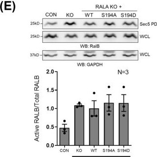
Detection of RalA/B by Western Blot
Role of RALA and its S194 phosphorylation in regulating RAL activation in MCF7 and MiaPaCa2 cells. (A,B) Western blot detection of RALA (WB: RALA) and actin (WB: actin) in cell lysates of MCF7 and MiaPaCa2 control (CON), RALA KO (KO) and RALA KO cells reconstituted with RALA mutants (WT/S194A/S194D). MCF7 Lysates were further probed for additional proteins of interest, with the results shown in Fig S3C. Blots were reused where required, and the RALA and actin blots from A are also shown for comparison in Fig. S3C. (C,D,E,F) Western blot detection (upper panel) and quantitation (lower panel) of active RALA and active RALB (in samples incubated with GST-Sec5, which pulls down active RALA and RALB from the same lysate), as well as total RALA and total RALB from lysates of control (CON), Cas9 control (Cas9), RALA KO (KO), WT-RALA (WT), S194A-RALA (S194A) and S194D-RALA (S194D) mutant reconstituted MCF7 (C,E) and MiaPaCa2 (D,F) cells. The same GAPDH blot is shown in C and E as the common loading control for RALA and RALB in the MCF lysates, and the same GAPDH blot is shown in D and F as the common loading control for RALA and RALB in the MiaPaCa2 lysates. (G,H) Western blot detection (upper panel) and quantitation (lower panel) of active RALA and total RALA from lysates of MCF7 and MiaPaCa2 cells treated with DMSO (CON), empty nano-vesicle scaffold (DEX), 0.02μM Free MLN and 0.02μM VMLN. The graphs represent the ratio of active to total protein (RALA and RALB) as mean±s.e. from at least three independent experiments. Statistical analysis was done using the Mann–Whitney test, and P-values, if significant, are represented in the graph (*P<0.05, **P<0.01, ***P<0.001). Image collected and cropped by CiteAb from the following open publication (https://pubmed.ncbi.nlm.nih.gov/40568758), licensed under a CC-BY license. Not internally tested by R&D Systems.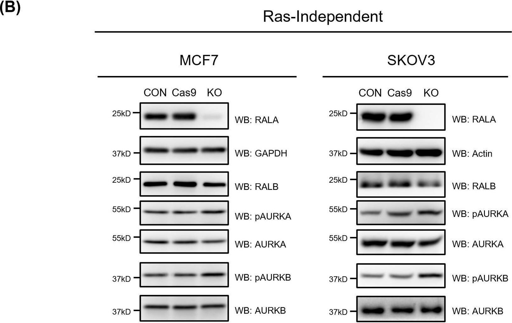
Detection of RalA/B by Western Blot
CRISPR-Cas9 mediated knockout of RALA in RAS-independent and RAS-dependent cancer cells. (A) The schematic shows an exon map (yellow boxes) for RALA, marking the location of sgRNAs designed against exon 2 (2.1 and 2.2) and exon 4 (4.0) to target RALA. The red box represents the functional RALA protein coded by these exons. (B) Western blot detection (WB) of RALA, RALB, AURKA, AURKB in untreated control (CON), Cas9 control (Cas9), RALA knockout (KO) clones of RAS-independent (MCF7 and SKOV3) (single clone each) and RAS-dependent (T24, UMUC3 and MiaPaCa2) (two clones for some) cells. Clone 2 selected based on these studies is marked by a box. This was accompanied by the western blot detection (WB) of active AURKA (pAURKA – pThr288 AURKA) and active AURKB (pAURKB – pThr232 AURKB). Lysates were further probed for additional proteins of interest, with the results shown in Fig. 2A and Fig. S1B. Blots were reused where required, and the RALA and GAPDH blots from B are also shown for comparison in Fig. 2A. Image collected and cropped by CiteAb from the following open publication (https://pubmed.ncbi.nlm.nih.gov/40568758), licensed under a CC-BY license. Not internally tested by R&D Systems.Applications for RalA/RalB Antibody
Application
Recommended Usage
Simple Western
10 µg/mL
Sample: Exosome Standards (HT-29) (Catalog # NBP3-11685) and U937 human histiocytic lymphoma cell line
Sample: Exosome Standards (HT-29) (Catalog # NBP3-11685) and U937 human histiocytic lymphoma cell line
Western Blot
1 µg/mL
Sample: U937 human histiocytic lymphoma cell line and NIH-3T3 mouse embryonic fibroblast cell line
Sample: U937 human histiocytic lymphoma cell line and NIH-3T3 mouse embryonic fibroblast cell line
Formulation, Preparation, and Storage
Purification
Antigen Affinity-purified
Reconstitution
Reconstitute at 0.2 mg/mL in sterile PBS. For liquid material, refer to CoA for concentration.
Loading...
Formulation
Lyophilized from a 0.2 μm filtered solution in PBS with Trehalose. See Certificate of Analysis for details.<br/>*Small pack size (-SP) is supplied either lyophilized or as a 0.2 µm filtered solution in PBS.
Shipping
Lyophilized product is shipped at ambient temperature. Liquid small pack size (-SP) is shipped with polar packs. Upon receipt, store immediately at the temperature recommended below.
Stability & Storage
Use a manual defrost freezer and avoid repeated freeze-thaw cycles.
- 12 months from date of receipt, -20 to -70 °C as supplied.
- 1 month, 2 to 8 °C under sterile conditions after reconstitution.
- 6 months, -20 to -70 °C under sterile conditions after reconstitution.
Calculators
Background: RalA/RalB
Long Name
Ras-like Protein A/B
Alternate Names
MGC48949, RAL, Ras family small GTP binding protein RALA, RAS-like protein A, ras-related protein Ral-A, v-ral simian leukemia viral oncogene homolog A (ras related)
UniProt
Additional RalA/RalB Products
Product Documents for RalA/RalB Antibody
Certificate of Analysis
To download a Certificate of Analysis, please enter a lot or batch number in the search box below.
Note: Certificate of Analysis not available for kit components.
Product Specific Notices for RalA/RalB Antibody
For research use only
Citations for RalA/RalB Antibody
 Powered by Bioz
Customer Reviews for RalA/RalB Antibody
There are currently no reviews for this product. Be the first to review RalA/RalB Antibody and earn rewards!
Have you used RalA/RalB Antibody?
Submit a review and receive an Amazon gift card!
$25/€18/£15/$25CAN/¥2500 for a review with an image
$10/€7/£6/$10CAN/¥1110 for a review without an image
Submit a review
Protocols
Find general support by application which include: protocols, troubleshooting, illustrated assays, videos and webinars.
- Cellular Response to Hypoxia Protocols
- R&D Systems Quality Control Western Blot Protocol
- Troubleshooting Guide: Western Blot Figures
- Western Blot Conditions
- Western Blot Protocol
- Western Blot Protocol for Cell Lysates
- Western Blot Troubleshooting
- Western Blot Troubleshooting Guide
- View all Protocols, Troubleshooting, Illustrated assays and Webinars
Loading...