
The cyclin-dependent kinase (cdk) inhibitor p27/Kip1 is a tumor suppressor protein that acts in the nucleus to enforce cell cycle checkpoints. p27/Kip1 binds to and inhibits many cyclin/cdk complexes, including cyclin D/cdk4, cyclin E/cdk2, and cyclin A/cdk2. This inhibition can subsequently block progression through different phases of the cell cycle. p27/Kip1 is phosphorylated at T198 in cells treated with the AMP-dependent protein kinase (AMPK) activator AICAR (5-amino-4‑imidazolecarboxamide riboside) or glucose withdrawal.
Human phospho-p27/Kip1 (T198) Antibody
R&D Systems | Catalog # AF3994


Scientific images may be resized, cropped, or adjusted for brightness or contrast for presentation purposes. These updates are limited to presentation and do not affect the underlying scientific interpretation of the data. Where available, additional source data may be provided upon request.
Key Product Details
Species Reactivity
Validated:
Human
Cited:
Human, Mouse, Rat
Applications
Validated:
Western Blot
Cited:
Western Blot, Immunocytochemistry, Immunoprecipitation
Label
Unconjugated
Antibody Source
Polyclonal Rabbit IgG
Loading...
Product Specifications
Immunogen
Phosphopeptide containing human p27/Kip1 T198 site
Specificity
Detects human p27/Kip1 when phosphorylated at T198. Does not recognize p27/Kip1 when unphosphorylated at T198.
Clonality
Polyclonal
Host
Rabbit
Isotype
IgG
Scientific Data Images for Human phospho-p27/Kip1 (T198) Antibody
Detection of Human Phospho-p27/Kip1 (T198) by Western Blot.
Western blot shows lysates of MCF-7 human breast cancer cell line, non-transfected, transfected with plasmid expressing YFP-p27/Kip1 wild type (WT), or transfected with YFP-p27/Kip1 T198A mutant (T198A). PVDF membrane was probed with 0.5 µg/mL Rabbit Anti-Human Phospho-p27/Kip1 (T198) Antigen Affinity-purified Polyclonal Antibody (Catalog # AF3994) followed by HRP-conjugated Anti-Rabbit IgG Secondary Antibody (Catalog # HAF008). A specific band for p27/Kip1 was detected at approximately 60 kDa (as indicated). This experiment was conducted under reducing conditions and using Immunoblot Buffer Group 1.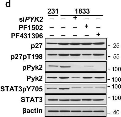
Detection of Phospho-p27/Kip1 (T198) by Western Blot
STAT3/cJun/p27 co-repress PTPN12 in complex with YY1, SIN3A, and HDAC1.a Volcano plot of genes differentially expressed in 231DD vs. 231. bPTPN12 mRNA expression by qPCR in the indicated lines. c Western shows effects of p27CK-DD on total and activated STAT3 and Pyk2 and effects of PYK2 loss on STAT3 levels and STAT3pY705. d Western shows effects of p27pT198 (reduced by PI3K inhibitor, PF1502) and Pyk2 loss (siPYK2) and Pyk2 inhibition by PF431396 on STAT3 levels and activation (STAT3pY705) e p27, STAT3, and cJun co-occupied peaks at +2 kb of the PTPN12 TSS. f–k ChIP-qPCR shows p27 (f); STAT3 (g); cJun (h); SIN3A (i); YY1 (j) and HDAC1 (k) binding at +2 kb of the PTPN12 TSS where p27 peaks overlap with binding peaks of STAT3 and cJun. l cJun ChIP-Western blot (right) shows interaction on chromatin of the indicated proteins. Input is shown on the left. m Schematic representation for p27/STAT3/cJun in activator (left) and repressor (right) complexes. All graphs show mean ± SEM from N = 3 biological replicate assays. p-values were calculated by paired one-tailed Student’s T Test and are indicated in graphs. Source data are provided as a Source data file. Image collected and cropped by CiteAb from the following open publication (https://www.nature.com/articles/s41467-024-48742-y), licensed under a CC-BY license. Not internally tested by R&D Systems.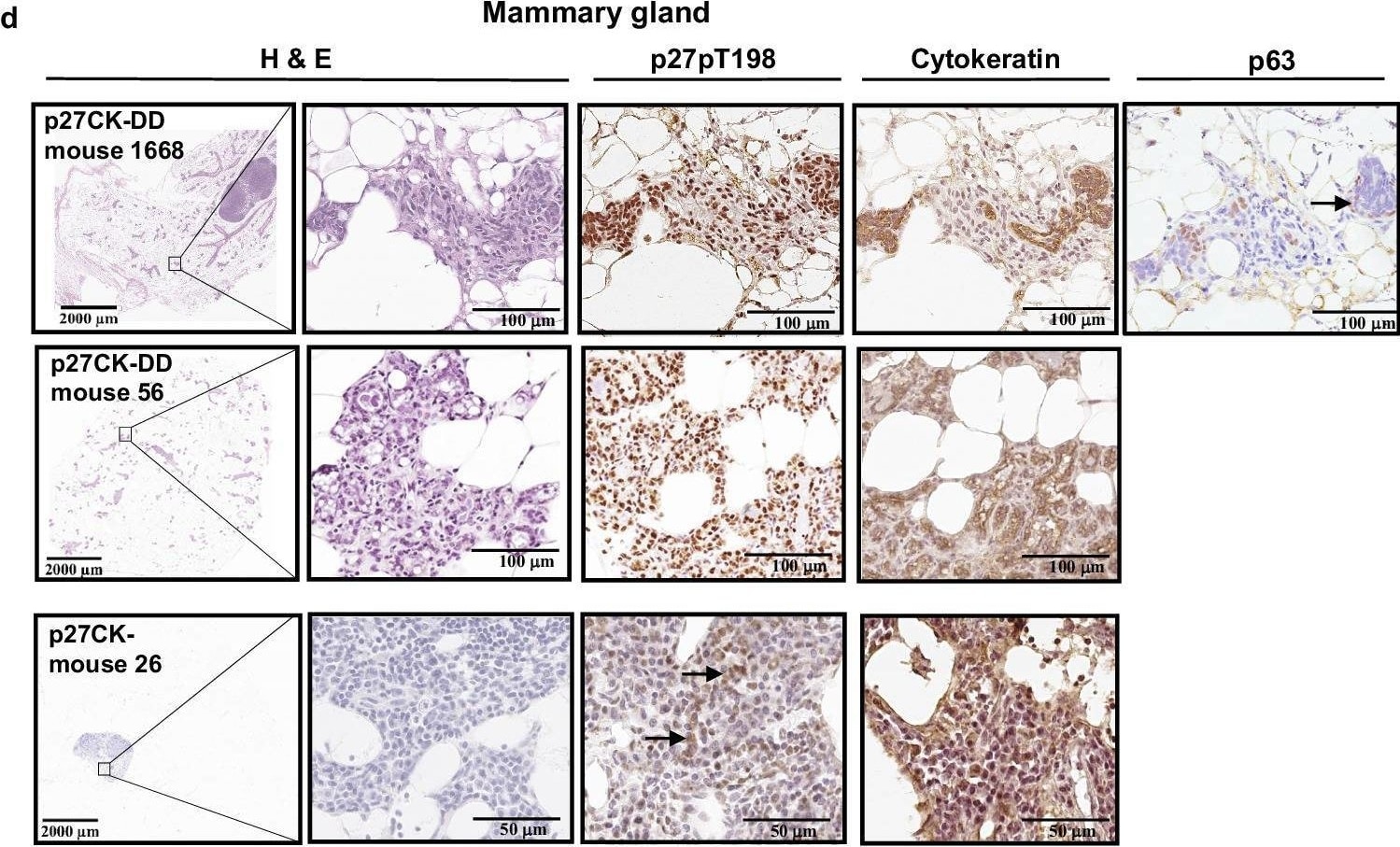
Detection of Phospho-p27/Kip1 (T198) by Immunohistochemistry
Mammary transgenic expression of p27CK-DD increases mammary duct branching, mammary hyperplasia and yields mammary cancers that generate liver metastases.a Representative mammary gland whole mounts stained with Carmine red from MMTVCre controls and MMTV-Cre X TGp27CK- (p27CK-) and MMTV-Cre X TGp27CK-DD (p27CK-DD) mice show greater duct branching (top 2 panels) and hyperplasia (bottom panels) in the p27CK-DD bigenics. b Duct hyperplasia was quantitated from whole mounts as described and graphed as mean %+/−SEM. p values were calculated by one-way ANOVA with post hoc 2 by 2 comparisons using Tukey correction. N = 8 mice for MMTVCre controls, N = 7 mice for TGp27CK-, N = 6 mice for TGp27CK-DD (See also SFig. 6b for quantitation of duct branching). c The number of mice with mammary cancers over the total number evaluated for each genotype is graphed as %. p values calculated by Fisher’s Exact Test are shown. N = 14 mice for MMTVCre controls, N = 14 mice for TGp27CK-, N = 14 mice for TGp27CK-DD. d FFPE mammary glands from the indicated genotypes were stained with hematoxylin and eosin (H&E) or with antibodies to p27pT198, to cytokeratin or to myoepithelial marker, p63, as indicated and representative photomicrographs shown. e Livers show representative micrometastases from mammary cancer-bearing MMTV Cre X p27CK-DD mouse 1668 (see also Fig. S6). Source data are provided as a Source data file. Image collected and cropped by CiteAb from the following open publication (https://www.nature.com/articles/s41467-024-48742-y), licensed under a CC-BY license. Not internally tested by R&D Systems.Applications for Human phospho-p27/Kip1 (T198) Antibody
Application
Recommended Usage
Western Blot
0.5 µg/mL
Sample: MCF‑7 human breast cancer cell line transfected with plasmid expressing YFP-p27/Kip1 wild type (WT), or transfected with YFP-p27/Kip1 T198A mutant (T198A)
Sample: MCF‑7 human breast cancer cell line transfected with plasmid expressing YFP-p27/Kip1 wild type (WT), or transfected with YFP-p27/Kip1 T198A mutant (T198A)
Formulation, Preparation, and Storage
Purification
Antigen Affinity-purified
Reconstitution
Reconstitute at 0.2 mg/mL in sterile PBS. For liquid material, refer to CoA for concentration.
Loading...
Formulation
Lyophilized from a 0.2 μm filtered solution in PBS with Trehalose. *Small pack size (SP) is supplied either lyophilized or as a 0.2 µm filtered solution in PBS.
Shipping
Lyophilized product is shipped at ambient temperature. Liquid small pack size (-SP) is shipped with polar packs. Upon receipt, store immediately at the temperature recommended below.
Stability & Storage
Use a manual defrost freezer and avoid repeated freeze-thaw cycles.
- 12 months from date of receipt, -20 to -70 °C as supplied.
- 1 month, 2 to 8 °C under sterile conditions after reconstitution.
- 6 months, -20 to -70 °C under sterile conditions after reconstitution.
Calculators
Background: p27/Kip1
Long Name
p27 Cyclin Dependent Kinase 4 Inhibitor 1B
Alternate Names
CDKN1B, Kip1
Gene Symbol
CDKN1B
Additional p27/Kip1 Products
Product Documents for Human phospho-p27/Kip1 (T198) Antibody
Certificate of Analysis
To download a Certificate of Analysis, please enter a lot or batch number in the search box below.
Note: Certificate of Analysis not available for kit components.
Product Specific Notices for Human phospho-p27/Kip1 (T198) Antibody
For research use only
Related Research Areas
Citations for Human phospho-p27/Kip1 (T198) Antibody
 Powered by Bioz
Customer Reviews for Human phospho-p27/Kip1 (T198) Antibody
There are currently no reviews for this product. Be the first to review Human phospho-p27/Kip1 (T198) Antibody and earn rewards!
Have you used Human phospho-p27/Kip1 (T198) Antibody?
Submit a review and receive an Amazon gift card!
$25/€18/£15/$25CAN/¥2500 for a review with an image
$10/€7/£6/$10CAN/¥1110 for a review without an image
Submit a review
Protocols
Find general support by application which include: protocols, troubleshooting, illustrated assays, videos and webinars.
- Cellular Response to Hypoxia Protocols
- R&D Systems Quality Control Western Blot Protocol
- Troubleshooting Guide: Western Blot Figures
- Western Blot Conditions
- Western Blot Protocol
- Western Blot Protocol for Cell Lysates
- Western Blot Troubleshooting
- Western Blot Troubleshooting Guide
- View all Protocols, Troubleshooting, Illustrated assays and Webinars
Loading...
Associated Pathways
