The human polymeric immunoglobulin receptor (pIgR; also known as membrane secretory component) is a 100 kDa type I transmembrane glycoprotein that is synthesized as a 764 amino acid (aa) precursor. It includes a signal sequence (aa 1-18), an extracellular region (aa 19-638), a transmembrane segment (aa 639-661), and a cytoplasmic domain (aa 662-764) (1-3). The extracellular region consists of five Ig-like domains and a sixth non-Ig domain that connects to the membrane region. pIgR is expressed on secretory epithelial cells of exocrine tissues. Immunoglobulin isotypes consist of two heavy (H) and two light (L) chains. For IgA and IgM, this H2L2 monomer can form larger polymers through association with a joining chain (J chain). The Fc regions of IgA and IgM have a carboxy-terminal extension called a secretory tailpiece that binds the J chain (4). pIgR functions as a carrier that transports IgA and IgM across epithelium (5). On the basolateral surface of epithelial cells, the receptor initially binds non-covalently to IgA via a docking site on the J chain. This initiates a rearrangement in which a disulfide bond forms between pIgR and an IgA heavy chain (2). The complexes are then internalized and transcytosed to the apical surface. A soluble covalent complex called secretory IgA (SIgA) is now generated by proteolytic cleavage of the sixth extracellular domain of pIgR and released into the lumen (6). This IgA-bound and proteolytically generated pIgR fragment is referred to as secretory component (SC). Notably, human pIgR transcytoses constitutively, with or without ligand, creating both bound and free, 78 kDa SC following cleavage (3). The extracellular region of pIgR is 64%, 65%, and 70% aa identical to the equivalent region in rat, mouse and porcine, respectively. The receptor component of the complex anchors the SIgA molecule to mucous (7). SIgA is a crucial component of the mucosal immune system serving to protect the large expanse of mucous membranes that form a barrier between the interior of the body and the external environment (8).

Key Product Details
Species Reactivity
Validated:
Human
Cited:
Human
Applications
Validated:
Immunohistochemistry
Cited:
Immunohistochemistry
Label
Unconjugated
Antibody Source
Monoclonal Mouse IgG1 Clone # 825732
Loading...
Product Specifications
Immunogen
Mouse myeloma cell line NS0-derived recombinant human pIgR
Lys19-Arg638
Accession # P01833
Lys19-Arg638
Accession # P01833
Specificity
Detects human pIgR in ELISAs.
Clonality
Monoclonal
Host
Mouse
Isotype
IgG1
Scientific Data Images for Human pIgR Antibody (825732)
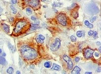
pIgR in Human Liver Cancer Tissue.
pIgR was detected in formalin fixed paraffin-embedded sections of human liver cancer tissue using Mouse Anti-Human pIgR Monoclonal Antibody (Catalog # MAB27171) at 15 µg/mL overnight at 4 °C. Tissue was stained using the Anti-Mouse HRP-DAB Cell & Tissue Staining Kit (brown; Catalog # CTS002) and counterstained with hematoxylin (blue). Specific staining was localized in the cytoplasm. View our protocol for Chromogenic IHC Staining of Paraffin-embedded Tissue Sections.Applications for Human pIgR Antibody (825732)
Application
Recommended Usage
Immunohistochemistry
8-25 µg/mL
Sample: Formalin fixed paraffin-embedded sections of human liver cancer tissue
Sample: Formalin fixed paraffin-embedded sections of human liver cancer tissue
Reviewed Applications
Read 1 review rated 5 using MAB27171 in the following applications:
Formulation, Preparation, and Storage
Purification
Protein A or G purified from hybridoma culture supernatant
Reconstitution
Reconstitute at 0.5 mg/mL in sterile PBS. For liquid material, refer to CoA for concentration.
Loading...
Formulation
Lyophilized from a 0.2 μm filtered solution in PBS and NaCl with Trehalose. *Small pack size (SP) is supplied either lyophilized or as a 0.2 µm filtered solution in PBS.
Shipping
Lyophilized product is shipped at ambient temperature. Liquid small pack size (-SP) is shipped with polar packs. Upon receipt, store immediately at the temperature recommended below.
Stability & Storage
Use a manual defrost freezer and avoid repeated freeze-thaw cycles.
- 12 months from date of receipt, -20 to -70 °C as supplied.
- 1 month, 2 to 8 °C under sterile conditions after reconstitution.
- 6 months, -20 to -70 °C under sterile conditions after reconstitution.
Calculators
Background: pIgR
References
- Krajci, P. et al. (1989) Biochem. Biophys. Res. Commun. 158:783.
- Piskurich, J. et al. (1995) J. Immunol. 154:1735.
- Brandtzaeg, P. and F-E. Johansen (2001) Trends Immunol. 22:545.
- Braathen, R. et al. (2002) J. Biol. Chem. 277:42755.
- Ben-Hur, H. et al. (2004) Int. J. Mol. Med. 14:35.
- Asano, M. et al. (2004) Immunology 112:583.
- Phalipon, A. and B. Corthesy (2003) Trends Immunol. 24:55.
- Uren, T. et al. (2003) J. Immunol. 170:2531.
Long Name
Polymeric Immunoglobulin Receptor
Alternate Names
FLJ22667, hepatocellular carcinoma associated protein TB6, Hepatocellular carcinoma-associated protein TB6, MGC125361, MGC125362, PIgR, Poly-Ig receptor, polymeric immunoglobulin receptor
Gene Symbol
PIGR
UniProt
Additional pIgR Products
Product Documents for Human pIgR Antibody (825732)
Certificate of Analysis
To download a Certificate of Analysis, please enter a lot or batch number in the search box below.
Note: Certificate of Analysis not available for kit components.
Product Specific Notices for Human pIgR Antibody (825732)
For research use only
Citations for Human pIgR Antibody (825732)
 Powered by Bioz
Customer Reviews for Human pIgR Antibody (825732) (1)
5 out of 5
1 Customer Rating
Have you used Human pIgR Antibody (825732)?
Submit a review and receive an Amazon gift card!
$25/€18/£15/$25CAN/¥2500 Yen for a review with an image
$10/€7/£6/$10CAN/¥1110 Yen for a review without an image
Submit a review
Customer Images
Showing
1
-
1 of
1 review
Showing All
Filter By:
-
Application: ImmunohistochemistrySample Tested: Tumor tissue and Kidney cancer tissueSpecies: HumanVerified Customer | Posted 02/03/2022

There are no reviews that match your criteria.
Protocols
Find general support by application which include: protocols, troubleshooting, illustrated assays, videos and webinars.
- Antigen Retrieval Protocol (PIER)
- Antigen Retrieval for Frozen Sections Protocol
- Appropriate Fixation of IHC/ICC Samples
- Cellular Response to Hypoxia Protocols
- Chromogenic IHC Staining of Formalin-Fixed Paraffin-Embedded (FFPE) Tissue Protocol
- Chromogenic Immunohistochemistry Staining of Frozen Tissue
- ClariTSA™ Fluorophore Kits
- Detection & Visualization of Antibody Binding
- Fluorescent IHC Staining of Frozen Tissue Protocol
- Graphic Protocol for Heat-induced Epitope Retrieval
- Graphic Protocol for the Preparation and Fluorescent IHC Staining of Frozen Tissue Sections
- Graphic Protocol for the Preparation and Fluorescent IHC Staining of Paraffin-embedded Tissue Sections
- Graphic Protocol for the Preparation of Gelatin-coated Slides for Histological Tissue Sections
- IHC Sample Preparation (Frozen sections vs Paraffin)
- Immunofluorescent IHC Staining of Formalin-Fixed Paraffin-Embedded (FFPE) Tissue Protocol
- Immunohistochemistry (IHC) and Immunocytochemistry (ICC) Protocols
- Immunohistochemistry Frozen Troubleshooting
- Immunohistochemistry Paraffin Troubleshooting
- Preparing Samples for IHC/ICC Experiments
- Preventing Non-Specific Staining (Non-Specific Binding)
- Primary Antibody Selection & Optimization
- Protocol for Heat-Induced Epitope Retrieval (HIER)
- Protocol for Making a 4% Formaldehyde Solution in PBS
- Protocol for VisUCyte™ HRP Polymer Detection Reagent
- Protocol for the Preparation & Fixation of Cells on Coverslips
- Protocol for the Preparation and Chromogenic IHC Staining of Frozen Tissue Sections
- Protocol for the Preparation and Chromogenic IHC Staining of Frozen Tissue Sections - Graphic
- Protocol for the Preparation and Chromogenic IHC Staining of Paraffin-embedded Tissue Sections
- Protocol for the Preparation and Chromogenic IHC Staining of Paraffin-embedded Tissue Sections - Graphic
- Protocol for the Preparation and Fluorescent IHC Staining of Frozen Tissue Sections
- Protocol for the Preparation and Fluorescent IHC Staining of Paraffin-embedded Tissue Sections
- Protocol for the Preparation of Gelatin-coated Slides for Histological Tissue Sections
- TUNEL and Active Caspase-3 Detection by IHC/ICC Protocol
- The Importance of IHC/ICC Controls
- Troubleshooting Guide: Immunohistochemistry
- View all Protocols, Troubleshooting, Illustrated assays and Webinars
Loading...