Human S100A8/S100A9 Heterodimer Antibody (1099F)
R&D Systems | Catalog # MAB45701

Key Product Details
Species Reactivity
Validated:
Cited:
Applications
Validated:
Cited:
Label
Antibody Source
Product Specifications
Immunogen
Met1-Glu93 (S100A8) & Thr2-Pro114 (S100A9)
Accession # P05109 (S100A8) and P06702 (S100A9)
Specificity
Clonality
Host
Isotype
Scientific Data Images for Human S100A8/S100A9 Heterodimer Antibody (1099F)
S100A8/S100A9 Heterodimer in HL60 Human Cell Line.
S100A8/S100A9 Heterodimer was detected in immersion fixed HL60 human promyelocytic leukemia cell line using Rabbit Anti-Human S100A8/S100A9 Heterodimer Monoclonal Antibody (Catalog # MAB45701) at 3 µg/mL for 3 hours at room temperature. Cells were stained using the NorthernLights™ 557-conjugated Anti-Rabbit IgG Secondary Antibody (red; Catalog # NL004) and counterstained with DAPI (blue). Specific staining was localized to cytoplasm (punctate). View our protocol for Fluorescent ICC Staining of Non-adherent Cells.Applications for Human S100A8/S100A9 Heterodimer Antibody (1099F)
Immunocytochemistry
Sample: Immersion fixed HL60 human promyelocytic leukemia cell line
Reviewed Applications
Read 1 review rated 5 using MAB45701 in the following applications:
Formulation, Preparation, and Storage
Purification
Reconstitution
Reconstitute at 0.5 mg/mL in sterile PBS. For liquid material, refer to CoA for concentration.
Formulation
*Small pack size (-SP) is supplied either lyophilized or as a 0.2 µm filtered solution in PBS.
Shipping
Stability & Storage
- 12 months from date of receipt, -20 to -70 °C as supplied.
- 1 month, 2 to 8 °C under sterile conditions after reconstitution.
- 6 months, -20 to -70 °C under sterile conditions after reconstitution.
Calculators
Background: S100A8/S100A9 Heterodimer
References
- Averill, M.M. et al. (2012) Arterioscler. Thromb. Vasc. Biol. 32:223.
- Vogl, T. et al. (2012) Int. J. Mol. Sci. 13:2893.
- Siegenthaler, G. et al. (1997) J. Biol. Chem. 272:9371.
- Sunahori, K. et al. (2006) Arthritis Res. Ther. 8:R69.
- Volz, H.C. et al. (2012) Basic Res. Cardiol. 107:250.
- Odink, K. et al. (1987) Nature 330:80.
- Dorin, J.R. et al. (1987) Nature 326:614.
- Teigelkamp, S. et al. (1991) J. Biol. Chem. 266:13462.
- Ryckman, C. et al. (2003) J. Immunol. 170:3233.
- Vogl, T. et al. (2006) Biochim. Biophys. Acta 1763:1298.
- Damo, S.M. et al. (2013) Proc. Natl. Acad. Sci. USA 110:3841.
- Ryu, M-J. et al. (2012) J. Biol. Chem. 287:22948.
Long Name
Alternate Names
Entrez Gene IDs
Gene Symbol
Additional S100A8/S100A9 Heterodimer Products
Product Documents for Human S100A8/S100A9 Heterodimer Antibody (1099F)
Certificate of Analysis
To download a Certificate of Analysis, please enter a lot or batch number in the search box below.
Note: Certificate of Analysis not available for kit components.
Product Specific Notices for Human S100A8/S100A9 Heterodimer Antibody (1099F)
* Contains <0.1% Sodium Azide, which is not hazardous at this concentration according to GHS classifications. Refer to SDS for additional information and handling instructions.
For research use only
Citations for Human S100A8/S100A9 Heterodimer Antibody (1099F)
 Powered by Bioz
Customer Reviews for Human S100A8/S100A9 Heterodimer Antibody (1099F) (1)
Have you used Human S100A8/S100A9 Heterodimer Antibody (1099F)?
Submit a review and receive an Amazon gift card!
$25/€18/£15/$25CAN/¥2500 Yen for a review with an image
$10/€7/£6/$10CAN/¥1110 Yen for a review without an image
Submit a review
Customer Images
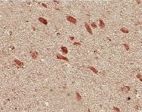
-
Application: ImmunohistochemistrySample Tested: Brain tissueSpecies: HumanVerified Customer | Posted 10/23/2022

There are no reviews that match your criteria.
Protocols
Find general support by application which include: protocols, troubleshooting, illustrated assays, videos and webinars.
- Appropriate Fixation of IHC/ICC Samples
- Cellular Response to Hypoxia Protocols
- ClariTSA™ Fluorophore Kits
- Detection & Visualization of Antibody Binding
- ICC Cell Smear Protocol for Suspension Cells
- ICC Immunocytochemistry Protocol Videos
- ICC for Adherent Cells
- Immunocytochemistry (ICC) Protocol
- Immunocytochemistry Troubleshooting
- Immunofluorescence of Organoids Embedded in Cultrex Basement Membrane Extract
- Immunohistochemistry (IHC) and Immunocytochemistry (ICC) Protocols
- Preparing Samples for IHC/ICC Experiments
- Preventing Non-Specific Staining (Non-Specific Binding)
- Primary Antibody Selection & Optimization
- Protocol for VisUCyte™ HRP Polymer Detection Reagent
- Protocol for the Fluorescent ICC Staining of Cell Smears - Graphic
- Protocol for the Fluorescent ICC Staining of Cultured Cells on Coverslips - Graphic
- Protocol for the Preparation and Fluorescent ICC Staining of Cells on Coverslips
- Protocol for the Preparation and Fluorescent ICC Staining of Non-adherent Cells
- Protocol for the Preparation and Fluorescent ICC Staining of Stem Cells on Coverslips
- Protocol for the Preparation of a Cell Smear for Non-adherent Cell ICC - Graphic
- TUNEL and Active Caspase-3 Detection by IHC/ICC Protocol
- The Importance of IHC/ICC Controls
- View all Protocols, Troubleshooting, Illustrated assays and Webinars