Human S100A9 (also MRP-14, Calgranulin-B, and p14) is a 14 kDa member of the S100 family of EF-hand calcium-binding proteins. It is 114 amino acids (aa) in length and contains short sequential modules. There is an N-terminal helical region, followed by a calcium-binding EF-hand domain, two more helical regions, a second EF-hand domain, and three additional helical regions. S100A9 will noncovalently heterodimerize with S100A8. In the presence of calcium, this heterodimer will form a heterotetramer. S100A9 is expressed in granulocytes, monocytes, and macrophages during acute and chronic inflammation. Human S100A9 shares 62% and 57% aa identity with rat and mouse S100A9, respectively.

Key Product Details
Validated by
Knockout/Knockdown
Species Reactivity
Human
Applications
Knockout Validated, Immunohistochemistry, Western Blot, Intracellular Staining by Flow Cytometry, Immunocytochemistry
Label
Unconjugated
Antibody Source
Polyclonal Sheep IgG
Loading...
Product Specifications
Immunogen
E. coli-derived recombinant human S100A9
The2-Pro114
Accession # P06702
The2-Pro114
Accession # P06702
Specificity
Detects human S100A9 in direct ELISAs and Western blots. In direct ELISAs, less than 5% cross-reactivity with recombinant mouse S100A9 is observed.
Clonality
Polyclonal
Host
Sheep
Isotype
IgG
Scientific Data Images for Human S100A9 Antibody
Detection of Human S100A9 by Western Blot.
Western blot shows lysates of human peripheral blood mononuclear cells (PBMC), human spleen tissue, human tonsil tissue, and human cartilage tissue. PVDF membrane was probed with 0.2 µg/mL of Sheep Anti-Human S100A9 Antigen Affinity-purified Polyclonal Antibody (Catalog # AF5578) followed by HRP-conjugated Anti-Sheep IgG Secondary Antibody (Catalog # HAF016). A specific band was detected for S100A9 at approximately 14 kDa (as indicated). This experiment was conducted under reducing conditions and using Immunoblot Buffer Group 1.
S100A9 in MDA‑MB‑468 Human Cell Line.
S100A9 was detected in immersion fixed MDA-MB-468 human breast cancer cell line using Sheep Anti-Human S100A9 Antigen Affinity-purified Polyclonal Antibody (Catalog # AF5578) at 1.7 µg/mL for 3 hours at room temperature. Cells were stained using the NorthernLights™ 557-conjugated Anti-Sheep IgG Secondary Antibody (red; Catalog # NL010) and counterstained with DAPI (blue). Specific staining was localized to cytoplasm. View our protocol for Fluorescent ICC Staining of Cells on Coverslips.
S100A9 in Human Cartilage.
S100A9 was detected in immersion fixed paraffin-embedded sections of human cartilage using Sheep Anti-Human S100A9 Antigen Affinity-purified Polyclonal Antibody (Catalog # AF5578) at 1 µg/mL overnight at 4 °C. Tissue was stained using the Anti-Sheep HRP-DAB Cell & Tissue Staining Kit (brown; Catalog # CTS019) and counterstained with hematoxylin (blue). Specific staining was localized to cytoplasm of chondrocytes. View our protocol for Chromogenic IHC Staining of Paraffin-embedded Tissue Sections.
Western Blot Shows Human S100A9 Specificity by Using Knockout Cell Line.
Western blot shows lysates of MDA-MB-468 human breast cancer parental cell line and S100A9 knock out MDA-MB-468 cell line (KO). PVDF membrane was probed with 0.2 µg/mL of Sheep Anti-Human S100A9 Antigen Affinity-purified Polyclonal Antibody (Catalog # AF5578) followed by HRP-conjugated Anti-Sheep IgG Secondary Antibody (Catalog # HAF016). A specific band was detected for S100A9 at approximately 14 kDa (as indicated) in the parental MDA-MB-468 cell line, but is not detectable in knockout MDA-MB-468 cell line. This experiment was conducted under reducing conditions and using Immunoblot Buffer Group 1.
Detection of S100A9 in Human PBMCs by Flow Cytometry.
Human peripheral blood monocytes (PBMC) were stained with Mouse Anti-Human CD14 PE-conjugated Monoclonal Antibody (Catalog # FAB3832P) and either (A) Sheep Anti-Human S100A9 Polyclonal Antibody (Catalog # AF5578) or (B) Sheep IgG Isotype Control (Catalog # 5-001-A) followed by anti-Sheep IgG APC-conjugated Secondary Antibody (Catalog # F0127). To facilitate intracellular staining, cells were fixed with Flow Cytometry Fixation Buffer (Catalog # FC004) and permeabilized with Flow Cytometry Permeabilization/Wash Buffer (Catalog # FC005). View our protocol for Staining Membrane-associated Proteins.
S100A9 Specificity is Shown by Immunocytochemistry in Knockout Cell Line.
S100A9 was detected in immersion fixed MDA-MB-468 human breast cancer cell line but is not detected in S100A9 knockout (KO) MDA-MB-468 cell line using Sheep Anti-Human S100A9 Antigen Affinity-purified Polyclonal Antibody (Catalog # AF5578) at 1.7 µg/mL for 3 hours at room temperature. Cells were stained using the NorthernLights™ 557-conjugated Anti-Sheep IgG Secondary Antibody (red; Catalog # NL010) and counterstained with DAPI (blue). Specific staining was localized to cytoplasm. View our protocol for Fluorescent ICC Staining of Cells on Coverslips.Applications for Human S100A9 Antibody
Application
Recommended Usage
Immunocytochemistry
1-25 µg/mL
Sample: Immersion fixed MDA‑MB‑468 human breast cancer cell line
Sample: Immersion fixed MDA‑MB‑468 human breast cancer cell line
Immunohistochemistry
5-15 µg/mL
Sample: Immersion fixed paraffin-embedded sections of human cartilage
Sample: Immersion fixed paraffin-embedded sections of human cartilage
Intracellular Staining by Flow Cytometry
0.25 µg/106 cells
Sample: Human peripheral blood monocytes (PBMC) fixed with Flow Cytometry Fixation Buffer (Catalog # FC004) and permeabilized with Flow Cytometry Permeabilization/Wash Buffer (Catalog # FC005)
Sample: Human peripheral blood monocytes (PBMC) fixed with Flow Cytometry Fixation Buffer (Catalog # FC004) and permeabilized with Flow Cytometry Permeabilization/Wash Buffer (Catalog # FC005)
Knockout Validated
S100A9 is specifically detected in MDA‑MB‑468 human breast cancer parental cell line but is not detectable in S100A9 knockout MDA‑MB‑468 cell line.
Western Blot
0.2 µg/mL
Sample: Human peripheral blood mononuclear cells (PBMC), human spleen tissue, human tonsil tissue, and human cartilage tissue
Sample: Human peripheral blood mononuclear cells (PBMC), human spleen tissue, human tonsil tissue, and human cartilage tissue
Reviewed Applications
Read 1 review rated 4 using AF5578 in the following applications:
Flow Cytometry Panel Builder
Bio-Techne Knows Flow Cytometry
Save time and reduce costly mistakes by quickly finding compatible reagents using the Panel Builder Tool.
Advanced Features
- Spectra Viewer - Custom analysis of spectra from multiple fluorochromes
- Spillover Popups - Visualize the spectra of individual fluorochromes
- Antigen Density Selector - Match fluorochrome brightness with antigen density

Formulation, Preparation, and Storage
Purification
Antigen Affinity-purified
Reconstitution
Reconstitute at 0.2 mg/mL in sterile PBS. For liquid material, refer to CoA for concentration.
Loading...
Formulation
Lyophilized from a 0.2 μm filtered solution in PBS with Trehalose. *Small pack size (SP) is supplied either lyophilized or as a 0.2 µm filtered solution in PBS.
Shipping
Lyophilized product is shipped at ambient temperature. Liquid small pack size (-SP) is shipped with polar packs. Upon receipt, store immediately at the temperature recommended below.
Stability & Storage
Use a manual defrost freezer and avoid repeated freeze-thaw cycles.
- 12 months from date of receipt, -20 to -70 °C as supplied.
- 1 month, 2 to 8 °C under sterile conditions after reconstitution.
- 6 months, -20 to -70 °C under sterile conditions after reconstitution.
Calculators
Background: S100A9
Long Name
S100 Calcium Binding Protein A9
Alternate Names
Calgranulin B, MRP-14
Gene Symbol
S100A9
UniProt
Additional S100A9 Products
Product Documents for Human S100A9 Antibody
Certificate of Analysis
To download a Certificate of Analysis, please enter a lot or batch number in the search box below.
Note: Certificate of Analysis not available for kit components.
Product Specific Notices for Human S100A9 Antibody
For research use only
Customer Reviews for Human S100A9 Antibody (1)
4 out of 5
1 Customer Rating
Have you used Human S100A9 Antibody?
Submit a review and receive an Amazon gift card!
$25/€18/£15/$25CAN/¥2500 Yen for a review with an image
$10/€7/£6/$10CAN/¥1110 Yen for a review without an image
Submit a review
Customer Images
Showing
1
-
1 of
1 review
Showing All
Filter By:
-
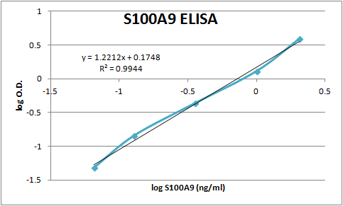
Application: ELISASample Tested: Serum and PlasmaSpecies: HumanVerified Customer | Posted 01/05/2018This antibody was used as both capture and detection in an ELISA reaction measuring S100A9 in human samples.

There are no reviews that match your criteria.
Protocols
Find general support by application which include: protocols, troubleshooting, illustrated assays, videos and webinars.
- 7-Amino Actinomycin D (7-AAD) Cell Viability Flow Cytometry Protocol
- Antigen Retrieval Protocol (PIER)
- Antigen Retrieval for Frozen Sections Protocol
- Appropriate Fixation of IHC/ICC Samples
- Cellular Response to Hypoxia Protocols
- Chromogenic IHC Staining of Formalin-Fixed Paraffin-Embedded (FFPE) Tissue Protocol
- Chromogenic Immunohistochemistry Staining of Frozen Tissue
- ClariTSA™ Fluorophore Kits
- Detection & Visualization of Antibody Binding
- Extracellular Membrane Flow Cytometry Protocol
- Flow Cytometry Protocol for Cell Surface Markers
- Flow Cytometry Protocol for Staining Membrane Associated Proteins
- Flow Cytometry Staining Protocols
- Flow Cytometry Troubleshooting Guide
- Fluorescent IHC Staining of Frozen Tissue Protocol
- Graphic Protocol for Heat-induced Epitope Retrieval
- Graphic Protocol for the Preparation and Fluorescent IHC Staining of Frozen Tissue Sections
- Graphic Protocol for the Preparation and Fluorescent IHC Staining of Paraffin-embedded Tissue Sections
- Graphic Protocol for the Preparation of Gelatin-coated Slides for Histological Tissue Sections
- ICC Cell Smear Protocol for Suspension Cells
- ICC Immunocytochemistry Protocol Videos
- ICC for Adherent Cells
- IHC Sample Preparation (Frozen sections vs Paraffin)
- Immunocytochemistry (ICC) Protocol
- Immunocytochemistry Troubleshooting
- Immunofluorescence of Organoids Embedded in Cultrex Basement Membrane Extract
- Immunofluorescent IHC Staining of Formalin-Fixed Paraffin-Embedded (FFPE) Tissue Protocol
- Immunohistochemistry (IHC) and Immunocytochemistry (ICC) Protocols
- Immunohistochemistry Frozen Troubleshooting
- Immunohistochemistry Paraffin Troubleshooting
- Intracellular Flow Cytometry Protocol Using Alcohol (Methanol)
- Intracellular Flow Cytometry Protocol Using Detergents
- Intracellular Nuclear Staining Flow Cytometry Protocol Using Detergents
- Intracellular Staining Flow Cytometry Protocol Using Alcohol Permeabilization
- Intracellular Staining Flow Cytometry Protocol Using Detergents to Permeabilize Cells
- Preparing Samples for IHC/ICC Experiments
- Preventing Non-Specific Staining (Non-Specific Binding)
- Primary Antibody Selection & Optimization
- Propidium Iodide Cell Viability Flow Cytometry Protocol
- Protocol for Heat-Induced Epitope Retrieval (HIER)
- Protocol for Liperfluo
- Protocol for Making a 4% Formaldehyde Solution in PBS
- Protocol for VisUCyte™ HRP Polymer Detection Reagent
- Protocol for the Characterization of Human Th22 Cells
- Protocol for the Characterization of Human Th9 Cells
- Protocol for the Fluorescent ICC Staining of Cell Smears - Graphic
- Protocol for the Fluorescent ICC Staining of Cultured Cells on Coverslips - Graphic
- Protocol for the Preparation & Fixation of Cells on Coverslips
- Protocol for the Preparation and Chromogenic IHC Staining of Frozen Tissue Sections
- Protocol for the Preparation and Chromogenic IHC Staining of Frozen Tissue Sections - Graphic
- Protocol for the Preparation and Chromogenic IHC Staining of Paraffin-embedded Tissue Sections
- Protocol for the Preparation and Chromogenic IHC Staining of Paraffin-embedded Tissue Sections - Graphic
- Protocol for the Preparation and Fluorescent ICC Staining of Cells on Coverslips
- Protocol for the Preparation and Fluorescent ICC Staining of Non-adherent Cells
- Protocol for the Preparation and Fluorescent ICC Staining of Stem Cells on Coverslips
- Protocol for the Preparation and Fluorescent IHC Staining of Frozen Tissue Sections
- Protocol for the Preparation and Fluorescent IHC Staining of Paraffin-embedded Tissue Sections
- Protocol for the Preparation of Gelatin-coated Slides for Histological Tissue Sections
- Protocol for the Preparation of a Cell Smear for Non-adherent Cell ICC - Graphic
- Protocol: Annexin V and PI Staining by Flow Cytometry
- Protocol: Annexin V and PI Staining for Apoptosis by Flow Cytometry
- R&D Systems Quality Control Western Blot Protocol
- TUNEL and Active Caspase-3 Detection by IHC/ICC Protocol
- The Importance of IHC/ICC Controls
- Troubleshooting Guide: Fluorokine Flow Cytometry Kits
- Troubleshooting Guide: Immunohistochemistry
- Troubleshooting Guide: Western Blot Figures
- Western Blot Conditions
- Western Blot Protocol
- Western Blot Protocol for Cell Lysates
- Western Blot Troubleshooting
- Western Blot Troubleshooting Guide
- View all Protocols, Troubleshooting, Illustrated assays and Webinars
Loading...