SIRT1 (SIR2-like protein 1; also NAD-dependent protein deacetylase sirtuin-1 and hSIR2) is a class I member of the sirtuin family of enzymes. Although its predicted MW is 81 kDa, it runs anomalously at 110-120 kDa in SDS-PAGE. It is a widely expressed nuclear protein that participates in the deacetylation of multiple proteins, including p300, p53, LKB1 and histone H1. Functionally, this has the effect of promoting heterochromatin formation, cell survival and resistance to oxidative stress. Metabolically, SIRT1 induces insulin secretion, inhibits glycolysis and suppresses fatty acid synthesis. Human SIRT1 is 747 amino acids (aa) in length. It possesses two NLS's (aa 32-39 and 223-230), an NES (aa 138-145), and a sertuin-type deacetylase domain (aa 241-495) that contains an NAD and Zn binding motif. There are at least 12 utilized Ser/Thr phosphorylation sites, plus two nitrosylated Cys and one acetylated Ala. There are also four potential isoform variants. One is 95 kDa in size and shows a deletion of aa 454-639, a second is 17 kDa in size and contains a 16 aa substitution for aa 149-747, and a third contains an alternative start site at Met296. SIRT1 is also known to undergo proteolysis by cathepsin B at Val533Ser534, generating a fourth, C-terminally truncated 75 kDa isoform. Full-length SIRT1 is suggested to form trimers, while the 17 kDa isoform appears to form dimers. Over aa 2-747, human and mouse SIRT1 share 86% aa sequence identity.

Key Product Details
Validated by
Knockout/Knockdown
Species Reactivity
Validated:
Human
Cited:
Human
Applications
Validated:
Knockout Validated, Western Blot, Flow Cytometry, CyTOF-ready
Cited:
Western Blot
Label
Unconjugated
Antibody Source
Polyclonal Sheep IgG
Loading...
Product Specifications
Immunogen
E. coli-derived recombinant human Sirtuin 1/SIRT1
Ala2-Ser747
Accession # Q96EB6
Ala2-Ser747
Accession # Q96EB6
Specificity
Detects human Sirtuin 1/SIRT1 in direct ELISAs and Western blots. In direct ELISAs, less than 1% cross-reactivity with recombinant human SIRT2 is observed.
Clonality
Polyclonal
Host
Sheep
Isotype
IgG
Scientific Data Images for Human Sirtuin 1/SIRT1 Antibody
Detection of Human Sirtuin 1/SIRT1 by Western Blot.
Western blot shows lysates of A172 human glioblastoma cell line, A549 human lung carcinoma cell line, and HeLa human cervical epithelial carcinoma cell line. PVDF membrane was probed with 0.2 µg/mL of Sheep Anti-Human Sirtuin 1/SIRT1 Antigen Affinity-purified Polyclonal Antibody (Catalog # AF7714) followed by HRP-conjugated Anti-Sheep IgG Secondary Antibody (Catalog # HAF016). A specific band was detected for Sirtuin 1/SIRT1 at approximately 120 kDa (as indicated). This experiment was conducted under reducing conditions and using Immunoblot Buffer Group 1.
Detection of Sirtuin 1/SIRT1 in HepG2 Human Cell Line by Flow Cytometry.
HepG2 human hepatocellular carcinoma cell line was stained with Sheep Anti-Human Sirtuin 1/SIRT1 Antigen Affinity-purified Polyclonal Antibody (Catalog # AF7714, filled histogram) or isotype control antibody (Catalog # 5-001-A, open histogram), followed by Allophycocyanin-conjugated Anti-Sheep IgG Secondary Antibody (Catalog # F0127). To facilitate intracellular staining, cells were fixed and permeabilized with FlowX FoxP3 Fixation & Permeabilization Buffer Kit (Catalog # FC012). View our protocol for Staining Intracellular Molecules.
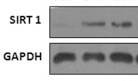
Western Blot Shows Human Sirtuin 1/SIRT1 Specificity by Using Knockout Cell Line.
Western blot shows lysates of HeLa human cervical epithelial carcinoma parental cell line and SIRT1 knockout HeLa cell line (KO). PVDF membrane was probed with 0.2 µg/mL of Sheep Anti-Human Sirtuin 1/SIRT1 Antigen Affinity-purified Polyclonal Antibody (Catalog # AF7714) followed by HRP-conjugated Anti-Sheep IgG Secondary Antibody (Catalog # HAF016). A specific band was detected for Sirtuin 1/SIRT1 at approximately 120 kDa (as indicated) in the parental HeLa cell line, but is not detectable in knockout HeLa cell line. GAPDH (Catalog # AF5718) is shown as a loading control. This experiment was conducted under reducing conditions and using Immunoblot Buffer Group 1.Applications for Human Sirtuin 1/SIRT1 Antibody
Application
Recommended Usage
CyTOF-ready
Ready to be labeled using established conjugation methods. No BSA or other carrier proteins that could interfere with conjugation.
Flow Cytometry
0.25 µg/106 cells
Sample: HepG2 human hepatocellular carcinoma cell line fixed and permeabilized with FlowX FoxP3 Fixation & Permeabilization Buffer Kit
Sample: HepG2 human hepatocellular carcinoma cell line fixed and permeabilized with FlowX FoxP3 Fixation & Permeabilization Buffer Kit
Knockout Validated
Sirtuin
1/SIRT1 is specifically detected in HeLa human cervical epithelial carcinoma parental cell line but is not
detectable in Sirtuin 1/SIRT1 knockout HeLa cell line.
Western Blot
0.2 µg/mL
Sample: A172 human glioblastoma cell line, A549 human lung carcinoma cell line, and HeLa human cervical epithelial carcinoma cell line
Sample: A172 human glioblastoma cell line, A549 human lung carcinoma cell line, and HeLa human cervical epithelial carcinoma cell line
Reviewed Applications
Read 1 review rated 5 using AF7714 in the following applications:
Flow Cytometry Panel Builder
Bio-Techne Knows Flow Cytometry
Save time and reduce costly mistakes by quickly finding compatible reagents using the Panel Builder Tool.
Advanced Features
- Spectra Viewer - Custom analysis of spectra from multiple fluorochromes
- Spillover Popups - Visualize the spectra of individual fluorochromes
- Antigen Density Selector - Match fluorochrome brightness with antigen density

Formulation, Preparation, and Storage
Purification
Antigen Affinity-purified
Reconstitution
Sterile PBS to a final concentration of 0.2 mg/mL. For liquid material, refer to CoA for concentration.
Loading...
Formulation
Lyophilized from a 0.2 μm filtered solution in PBS with Trehalose. *Small pack size (SP) is supplied either lyophilized or as a 0.2 µm filtered solution in PBS.
Shipping
Lyophilized product is shipped at ambient temperature. Liquid small pack size (-SP) is shipped with polar packs. Upon receipt, store immediately at the temperature recommended below.
Stability & Storage
Use a manual defrost freezer and avoid repeated freeze-thaw cycles.
- 12 months from date of receipt, -20 to -70 °C as supplied.
- 1 month, 2 to 8 °C under sterile conditions after reconstitution.
- 6 months, -20 to -70 °C under sterile conditions after reconstitution.
Calculators
Background: Sirtuin 1/SIRT1
Long Name
Silent Mating Type Information Regulation 2 Homolog
Alternate Names
SIR2-like protein 1, SIR2alpha, SIR2L1, SIRT1
Gene Symbol
SIRT1
UniProt
Additional Sirtuin 1/SIRT1 Products
Product Documents for Human Sirtuin 1/SIRT1 Antibody
Certificate of Analysis
To download a Certificate of Analysis, please enter a lot or batch number in the search box below.
Note: Certificate of Analysis not available for kit components.
Product Specific Notices for Human Sirtuin 1/SIRT1 Antibody
For research use only
Related Research Areas
Citations for Human Sirtuin 1/SIRT1 Antibody
 Powered by Bioz
Customer Reviews for Human Sirtuin 1/SIRT1 Antibody (1)
5 out of 5
1 Customer Rating
Have you used Human Sirtuin 1/SIRT1 Antibody?
Submit a review and receive an Amazon gift card!
$25/€18/£15/$25CAN/¥2500 Yen for a review with an image
$10/€7/£6/$10CAN/¥1110 Yen for a review without an image
Submit a review
Customer Images
Showing
1
-
1 of
1 review
Showing All
Filter By:
-
Application: Western BlotSample Tested: HEK293 human embryonic kidney cell lineSpecies: HumanVerified Customer | Posted 01/06/2018

There are no reviews that match your criteria.
Protocols
Find general support by application which include: protocols, troubleshooting, illustrated assays, videos and webinars.
- 7-Amino Actinomycin D (7-AAD) Cell Viability Flow Cytometry Protocol
- Cellular Response to Hypoxia Protocols
- Extracellular Membrane Flow Cytometry Protocol
- Flow Cytometry Protocol for Cell Surface Markers
- Flow Cytometry Protocol for Staining Membrane Associated Proteins
- Flow Cytometry Staining Protocols
- Flow Cytometry Troubleshooting Guide
- Intracellular Flow Cytometry Protocol Using Alcohol (Methanol)
- Intracellular Flow Cytometry Protocol Using Detergents
- Intracellular Nuclear Staining Flow Cytometry Protocol Using Detergents
- Intracellular Staining Flow Cytometry Protocol Using Alcohol Permeabilization
- Intracellular Staining Flow Cytometry Protocol Using Detergents to Permeabilize Cells
- Propidium Iodide Cell Viability Flow Cytometry Protocol
- Protocol for Liperfluo
- Protocol for the Characterization of Human Th22 Cells
- Protocol for the Characterization of Human Th9 Cells
- Protocol: Annexin V and PI Staining by Flow Cytometry
- Protocol: Annexin V and PI Staining for Apoptosis by Flow Cytometry
- R&D Systems Quality Control Western Blot Protocol
- Troubleshooting Guide: Fluorokine Flow Cytometry Kits
- Troubleshooting Guide: Western Blot Figures
- Western Blot Conditions
- Western Blot Protocol
- Western Blot Protocol for Cell Lysates
- Western Blot Troubleshooting
- Western Blot Troubleshooting Guide
- View all Protocols, Troubleshooting, Illustrated assays and Webinars
Loading...
Associated Pathways

