Syndecan-1, designated CD138, is a dimeric type I transmembrane (TM) protein that belongs to the syndecan family of Type 1 transmembrane proteins (1, 2). The four syndecan family members are major carriers of heparan sulfate (HS) and chondroitin sulfate glycosaminoglycans (GAGs) that have different expression patterns and extracellular sequences. Syndecan-1 forms weak non-covalent homodimers, or heterodimers with Syndecan-2 or -3, through interactions of the transmembrane domain (3). It is synthesized as a 310 amino acid (aa) precursor with a 17 aa signal sequence, a 234 aa extracellular domain (ECD) that includes three closely-spaced consensus Ser-Gly HS attachment sites near the N-terminus, a 25 aa TM segment, and a 34 aa cytoplasmic region that includes a PDZ binding motif with a tyrosine phosphorylation site. The ECD is variably modified by GAGs, producing molecular weights of 120-200 kDa for native Syndecan-1. Soluble forms are shed via proteolytic cleavage. Human Syndecan-1 ECD shares 65-71% aa identity with the ECD of rat, mouse, canine, equine and bovine Syndecan-1. Syndecan-1 shows highest expression on epithelial cells such as keratinocytes, and terminally differentiated B cells such as plasma cells (4, 5). It aids wound healing in skin, cornea, and heart following myocardial infarction by promoting re-epithelialization, migration, and collagen deposition (4-8). It binds chemokines, creating chemotactic gradients when shed, but also binds and modulates integrins to control the influx of leukocytes (5, 7, 9). The net effect is to allow, but limit, inflammation. In myeloma and other cancers, shedding of Syndecan-1 can facilitate growth, angiogenesis and metastasis (10-12). Growth factors, such as FGFs and HGF, bind GAG chains and use Syndecan-1 as a coreceptor (12, 13). The GAG chains may also be used by a variety of viruses and bacteria for cell adhesion and uptake (4).

Key Product Details
Validated by
Orthogonal Validation
Species Reactivity
Validated:
Human
Cited:
Human, Mouse, Xenograft
Applications
Validated:
Immunohistochemistry, Flow Cytometry, Dual RNAscope ISH-IHC Compatible, Immunocytochemistry, CyTOF-ready
Cited:
Immunohistochemistry
Label
Unconjugated
Antibody Source
Monoclonal Rat IgG1 Clone # 359103
Loading...
Product Specifications
Immunogen
Mouse myeloma cell line NS0-derived recombinant human Syndecan-1/CD138
Gln18-Glu251
Accession # NP_002988
Gln18-Glu251
Accession # NP_002988
Specificity
Detects human Syndecan-1 in direct ELISAs. In direct ELISAs, this antibody shows approximately 60% cross-reactivity with recombinant mouse (rm) Syndecan‑1 and no cross-reactivity with recombinant human (rh) Syndecan-2, rhSyndecan-3, or rmSyndecan-4.
Clonality
Monoclonal
Host
Rat
Isotype
IgG1
Scientific Data Images for Human Syndecan‑1/CD138 Antibody
Detection of Syndecan‑1/CD138 in RPMI 8226 Human Cell Line by Flow Cytometry.
RPMI 8226 human multiple myeloma cell line was stained with Rat Anti-Human Syndecan-1/CD138 Monoclonal Antibody (Catalog # MAB2780, filled histogram) or isotype control antibody (Catalog # MAB005, open histogram), followed by Phycoerythrin-conjugated Anti-Rat IgG Secondary Antibody (Catalog # F0105B).
Syndecan‑1/CD138 in U266 Human Cell Line.
Syndecan-1/CD138 was detected in immersion fixed U266 human myeloma cell line using Rat Anti-Human Syndecan-1/CD138 Monoclonal Antibody (Catalog # MAB2780) at 3 µg/mL for 3 hours at room temperature. Cells were stained using the NorthernLights™ 557-conjugated Anti-Rat IgG Secondary Antibody (red; Catalog # NL013) and counterstained with DAPI (blue). Specific staining was localized to plasma membrane. View our protocol for Fluorescent ICC Staining of Non-adherent Cells.
Syndecan‑1/CD138 in Human Cervical Cancer Tissue.
Syndecan-1/CD138 was detected in immersion fixed paraffin-embedded sections of human cervical cancer tissue using Rat Anti-Human Syndecan-1/CD138 Monoclonal Antibody (Catalog # MAB2780) at 1.7 µg/mL overnight at 4 °C. Tissue was stained using the Anti-Rat HRP-DAB Cell & Tissue Staining Kit (brown; Catalog # CTS017) and counterstained with hematoxylin (blue). Specific staining was localized to plasma membrane. View our protocol for Chromogenic IHC Staining of Paraffin-embedded Tissue Sections.
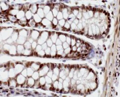
Syndecan-1/CD138 in Human Colon Using Dual RNAscope®ISH and IHC.
Syndecan-1/CD138 mRNA was detected in formalin-fixed paraffin-embedded tissue sections of human colon probed with ACD RNAScope®Probe (Catalog # 416961) and stained using ACD RNAscope®2.5 HD Duplex Detection Reagents (red, Catalog # 322500). Adjacent tissue section was processed for immunohistochemistry using R&D Systems Rat Anti-Human Syndecan-1/CD138 Monoclonal Antibody (Catalog# MAB2780) at 0.5 ug/mL for 1 hour at room temperature followed by incubation with the Anti-Rat IgG VisUCyte HRP Polymer Antibody (R&D Systems, Catalog # VC005) and DAB chromogen (brown). Tissues were counterstained with hematoxylin (blue).Applications for Human Syndecan‑1/CD138 Antibody
Application
Recommended Usage
CyTOF-ready
Ready to be labeled using established conjugation methods. No BSA or other carrier proteins that could interfere with conjugation.
Dual RNAscope ISH-IHC Compatible
0.5-25 µg/mL
Sample: Immersion fixed paraffin-embedded tissue sections of human colon
Sample: Immersion fixed paraffin-embedded tissue sections of human colon
Flow Cytometry
0.25 µg/106 cells
Sample: RPMI 8226 human multiple myeloma cell line
Sample: RPMI 8226 human multiple myeloma cell line
Immunocytochemistry
3-25 µg/mL
Sample: Immersion fixed U266 human myeloma cell line
Sample: Immersion fixed U266 human myeloma cell line
Immunohistochemistry
8-25 µg/mL
Sample: Immersion fixed paraffin-embedded sections of human intestine, and immersion fixed paraffin-embedded sections of human cervical cancer tissue
Sample: Immersion fixed paraffin-embedded sections of human intestine, and immersion fixed paraffin-embedded sections of human cervical cancer tissue
Reviewed Applications
Read 3 reviews rated 4.3 using MAB2780 in the following applications:
Flow Cytometry Panel Builder
Bio-Techne Knows Flow Cytometry
Save time and reduce costly mistakes by quickly finding compatible reagents using the Panel Builder Tool.
Advanced Features
- Spectra Viewer - Custom analysis of spectra from multiple fluorochromes
- Spillover Popups - Visualize the spectra of individual fluorochromes
- Antigen Density Selector - Match fluorochrome brightness with antigen density

Formulation, Preparation, and Storage
Purification
Protein A or G purified from hybridoma culture supernatant
Reconstitution
Reconstitute at 0.5 mg/mL in sterile PBS. For liquid material, refer to CoA for concentration.
Loading...
Formulation
Lyophilized from a 0.2 μm filtered solution in PBS with Trehalose. *Small pack size (SP) is supplied either lyophilized or as a 0.2 µm filtered solution in PBS.
Shipping
Lyophilized product is shipped at ambient temperature. Liquid small pack size (-SP) is shipped with polar packs. Upon receipt, store immediately at the temperature recommended below.
Stability & Storage
Use a manual defrost freezer and avoid repeated freeze-thaw cycles.
- 12 months from date of receipt, -20 to -70 °C as supplied.
- 1 month, 2 to 8 °C under sterile conditions after reconstitution.
- 6 months, -20 to -70 °C under sterile conditions after reconstitution.
Calculators
Background: Syndecan-1/CD138
References
- Tkachenko, E. et al. (2005) Circ. Res. 96:488.
- Mali, M. et al. (1990) J. Biol. Chem. 265:6884.
- Dews, I.C. and K.R. MacKenzie (2007) Proc. Natl. Acad. Sci. USA 104:20782.
- Fears, C.Y. and A. Woods (2006) Matrix Biol. 25:443.
- Stepp, M.A. et al. (2002) J. Cell Sci. 115:4517.
- Ojeh, N. et al. (2008) J. Invest. Dermatol. 128:26.
- Stepp, M.A. et al. (2007) J. Cell Sci. 120:2851.
- Vanhoutte, D. et al. (2007) Circulation 115:475.
- Li, Q. et al. (2002) Cell 111:635.
- Beauvais, D.M. et al. (2009) J. Exp. Med. 206:691.
- Yang, Y. et al. (2007) J. Biol. Chem. 282:13326.
- Derksen, P.W.B. et al. (2002) Blood 99:1405.
- Su, G. et al. (2007) J. Biol. Chem. 282:14906.
Alternate Names
CD138, SDC1, Syndecan1
Gene Symbol
SDC1
UniProt
Additional Syndecan-1/CD138 Products
Product Documents for Human Syndecan‑1/CD138 Antibody
Certificate of Analysis
To download a Certificate of Analysis, please enter a lot or batch number in the search box below.
Note: Certificate of Analysis not available for kit components.
Product Specific Notices for Human Syndecan‑1/CD138 Antibody
For research use only
Related Research Areas
Citations for Human Syndecan‑1/CD138 Antibody
 Powered by Bioz
Customer Reviews for Human Syndecan‑1/CD138 Antibody (3)
4.3 out of 5
3 Customer Ratings
Have you used Human Syndecan‑1/CD138 Antibody?
Submit a review and receive an Amazon gift card!
$25/€18/£15/$25CAN/¥2500 Yen for a review with an image
$10/€7/£6/$10CAN/¥1110 Yen for a review without an image
Submit a review
Customer Images
Showing
1
-
3 of
3 reviews
Showing All
Filter By:
-
Application: ImmunohistochemistrySample Tested: Colon tissueSpecies: HumanVerified Customer | Posted 02/10/2022

-
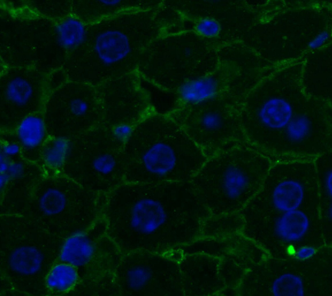
Application: Immunocytochemistry/ImmunofluorescenceSample Tested: Liver tissueSpecies: MouseVerified Customer | Posted 07/14/2021
-
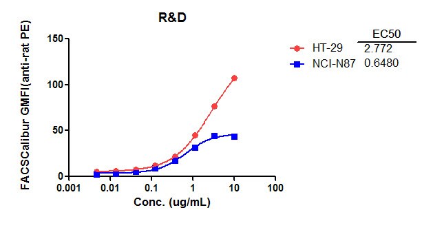
Application: Flow CytometrySample Tested: HT-29 human colon adenocarcinoma cell line and NCI-N87 human gastric carcinoma cell lineSpecies: HumanVerified Customer | Posted 02/22/2016
There are no reviews that match your criteria.
Protocols
Find general support by application which include: protocols, troubleshooting, illustrated assays, videos and webinars.
- 7-Amino Actinomycin D (7-AAD) Cell Viability Flow Cytometry Protocol
- Antigen Retrieval Protocol (PIER)
- Antigen Retrieval for Frozen Sections Protocol
- Appropriate Fixation of IHC/ICC Samples
- Cellular Response to Hypoxia Protocols
- Chromogenic IHC Staining of Formalin-Fixed Paraffin-Embedded (FFPE) Tissue Protocol
- Chromogenic Immunohistochemistry Staining of Frozen Tissue
- ClariTSA™ Fluorophore Kits
- Detection & Visualization of Antibody Binding
- Extracellular Membrane Flow Cytometry Protocol
- Flow Cytometry Protocol for Cell Surface Markers
- Flow Cytometry Protocol for Staining Membrane Associated Proteins
- Flow Cytometry Staining Protocols
- Flow Cytometry Troubleshooting Guide
- Fluorescent IHC Staining of Frozen Tissue Protocol
- Graphic Protocol for Heat-induced Epitope Retrieval
- Graphic Protocol for the Preparation and Fluorescent IHC Staining of Frozen Tissue Sections
- Graphic Protocol for the Preparation and Fluorescent IHC Staining of Paraffin-embedded Tissue Sections
- Graphic Protocol for the Preparation of Gelatin-coated Slides for Histological Tissue Sections
- ICC Cell Smear Protocol for Suspension Cells
- ICC Immunocytochemistry Protocol Videos
- ICC for Adherent Cells
- IHC Sample Preparation (Frozen sections vs Paraffin)
- ISH-IHC Protocol for Chromogenic Detection on Formalin Fixed Paraffin Embedded (FFPE) Tissue
- Immunocytochemistry (ICC) Protocol
- Immunocytochemistry Troubleshooting
- Immunofluorescence of Organoids Embedded in Cultrex Basement Membrane Extract
- Immunofluorescent IHC Staining of Formalin-Fixed Paraffin-Embedded (FFPE) Tissue Protocol
- Immunohistochemistry (IHC) and Immunocytochemistry (ICC) Protocols
- Immunohistochemistry Frozen Troubleshooting
- Immunohistochemistry Paraffin Troubleshooting
- Intracellular Flow Cytometry Protocol Using Alcohol (Methanol)
- Intracellular Flow Cytometry Protocol Using Detergents
- Intracellular Nuclear Staining Flow Cytometry Protocol Using Detergents
- Intracellular Staining Flow Cytometry Protocol Using Alcohol Permeabilization
- Intracellular Staining Flow Cytometry Protocol Using Detergents to Permeabilize Cells
- Preparing Samples for IHC/ICC Experiments
- Preventing Non-Specific Staining (Non-Specific Binding)
- Primary Antibody Selection & Optimization
- Propidium Iodide Cell Viability Flow Cytometry Protocol
- Protocol for Heat-Induced Epitope Retrieval (HIER)
- Protocol for Liperfluo
- Protocol for Making a 4% Formaldehyde Solution in PBS
- Protocol for VisUCyte™ HRP Polymer Detection Reagent
- Protocol for the Characterization of Human Th22 Cells
- Protocol for the Characterization of Human Th9 Cells
- Protocol for the Fluorescent ICC Staining of Cell Smears - Graphic
- Protocol for the Fluorescent ICC Staining of Cultured Cells on Coverslips - Graphic
- Protocol for the Preparation & Fixation of Cells on Coverslips
- Protocol for the Preparation and Chromogenic IHC Staining of Frozen Tissue Sections
- Protocol for the Preparation and Chromogenic IHC Staining of Frozen Tissue Sections - Graphic
- Protocol for the Preparation and Chromogenic IHC Staining of Paraffin-embedded Tissue Sections
- Protocol for the Preparation and Chromogenic IHC Staining of Paraffin-embedded Tissue Sections - Graphic
- Protocol for the Preparation and Fluorescent ICC Staining of Cells on Coverslips
- Protocol for the Preparation and Fluorescent ICC Staining of Non-adherent Cells
- Protocol for the Preparation and Fluorescent ICC Staining of Stem Cells on Coverslips
- Protocol for the Preparation and Fluorescent IHC Staining of Frozen Tissue Sections
- Protocol for the Preparation and Fluorescent IHC Staining of Paraffin-embedded Tissue Sections
- Protocol for the Preparation of Gelatin-coated Slides for Histological Tissue Sections
- Protocol for the Preparation of a Cell Smear for Non-adherent Cell ICC - Graphic
- Protocol: Annexin V and PI Staining by Flow Cytometry
- Protocol: Annexin V and PI Staining for Apoptosis by Flow Cytometry
- TUNEL and Active Caspase-3 Detection by IHC/ICC Protocol
- The Importance of IHC/ICC Controls
- Troubleshooting Guide: Fluorokine Flow Cytometry Kits
- Troubleshooting Guide: Immunohistochemistry
- View all Protocols, Troubleshooting, Illustrated assays and Webinars


