![Western Blot: IKK beta Antibody [NB600-477]](https://resources.rndsystems.com/images/products/IKK-beta-Antibody-Western-Blot-NB600-477-img0006.jpg "Western Blot: IKK beta Antibody [NB600-477]")
Loading...
Key Product Details
Species Reactivity
Human, Mouse, Rat
Applications
Immunohistochemistry, Immunohistochemistry-Paraffin, Western Blot, ELISA, Simple Western, Dot Blot
Label
Unconjugated
Antibody Source
Polyclonal Rabbit Serum
Loading...
Product Specifications
Immunogen
IKK beta peptide corresponding to the highly conserved C-terminus region of the human protein conjugated to Keyhole Limpet Hemocyanin (KLH). (Uniprot: O14920)
Reactivity Notes
This product may react non-specifically with other proteins. Control peptide will compete only with the specific reaction of antiserum with the IKKb subunit.
Localization
Cytoplasmic
Specificity
Anti-IKKb may react non-specifically with other proteins. Control peptide will compete only with the specific reaction of antiserum with the IKKb subunit.
Clonality
Polyclonal
Host
Rabbit
Isotype
Serum
Description
This product was prepared from monospecific antiserum by delipidation and defibrination
Store vial at -20C prior to opening. Aliquot contents and freeze at -20C or below for extended storage. Avoid cycles of freezing and thawing. Centrifuge product if not completely clear after standing at room temperature. This product is stable for several weeks at 4C as an undiluted liquid. Dilute only prior to immediate use.
Store vial at -20C prior to opening. Aliquot contents and freeze at -20C or below for extended storage. Avoid cycles of freezing and thawing. Centrifuge product if not completely clear after standing at room temperature. This product is stable for several weeks at 4C as an undiluted liquid. Dilute only prior to immediate use.
Scientific Data Images for IKK beta Antibody
Western Blot: IKK beta Antibody [NB600-477]
Western Blot: IKK beta Antibody [NB600-477] - Western blot of anti-IKKBeta antibody reacted with HeLa cell extract. All incubations except color development were performed using TBS supplemented with 0.1% Tween-20 at room temperature. The membrane was blocked in 5% dry milk for 2 h. After washing, a 1:500 dilution of the primary antibody was added to the membrane and incubated for 2 h. Washes with buffer were performed 4 times for 5' each. The western blot was incubated with secondary antibody (HRP Goat-a-Rabbit IgG [H&L]) diluted 1:2,000 for 1h. Washes with TBS preceded color development.![Immunohistochemistry-Paraffin: IKK beta Antibody [NB600-477]](https://resources.rndsystems.com/images/products/IKK-beta-Antibody-Immunohistochemistry-Paraffin-NB600-477-img0003.jpg "Immunohistochemistry-Paraffin: IKK beta Antibody [NB600-477]")
Immunohistochemistry-Paraffin: IKK beta Antibody [NB600-477]
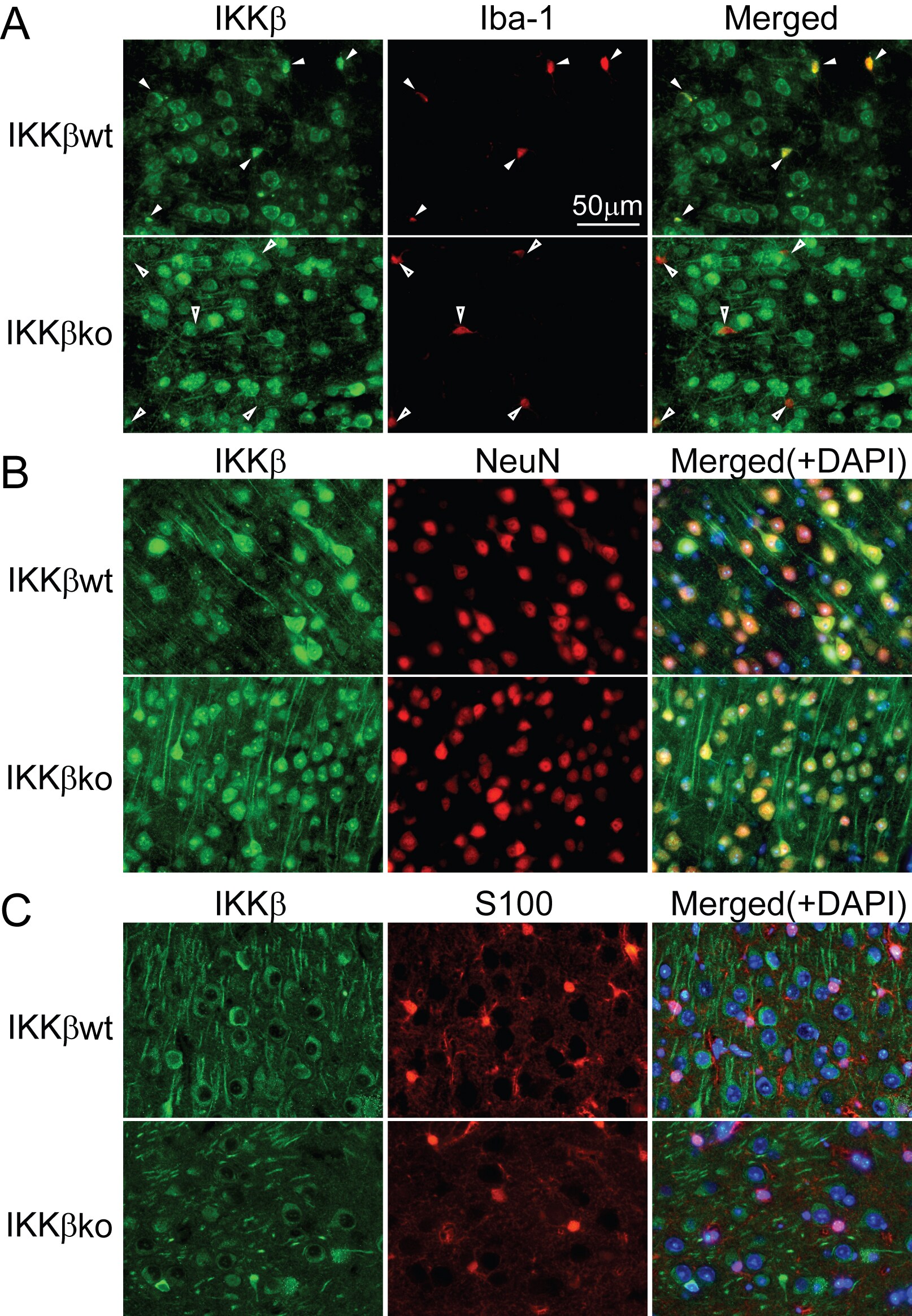
Immunohistochemistry-Paraffin: IKK beta Antibody [NB600-477] - Paraffin-embedded mouse brain sections (IKK beta KO or IKK beta WT) were stained with IKK beta (green). Note: during staining, remember to add 0.1-0.3% Triton-100 in the antibody incubation buffer. See PMID 25253847 for further details regarding the experiemental conditions. Image from verified customer review.![Immunohistochemistry-Paraffin: IKK beta Antibody [NB600-477]](https://resources.rndsystems.com/images/products/IKK-beta-Antibody-Immunohistochemistry-Paraffin-NB600-477-img0004.jpg "Immunohistochemistry-Paraffin: IKK beta Antibody [NB600-477]")
Immunohistochemistry-Paraffin: IKK beta Antibody [NB600-477]
Immunohistochemistry-Paraffin: IKK beta Antibody [NB600-477] - Human placenta tissue.Applications for IKK beta Antibody
Application
Recommended Usage
ELISA
1:1000-1:5000
Immunohistochemistry
1:200-1:1000
Immunohistochemistry-Paraffin
1:10-1:500
Western Blot
1:200-1:1000
Application Notes
This product was tested by immunoblot and found to be reactive against IKK-b at a dilution of 1:1000 followed by reaction with Peroxidase conjugated Affinity Purified anti-Rabbit IgG [H&L] (Goat). Anti-IKKb is suitable for the detection by immunoblot of human, mouse and rat IKKb showing an 87 kDa band. Anti-IKKb has been tested in IHC using human placenta tissue.
Simple Western Antibody Database for Simple Western validation: tested in HeLa-/+EGF, Jurkat/TNF; separated by charge, antibody dilution of 1:50.
Simple Western Antibody Database for Simple Western validation: tested in HeLa-/+EGF, Jurkat/TNF; separated by charge, antibody dilution of 1:50.
Reviewed Applications
Read 1 review rated 5 using NB600-477 in the following applications:
Formulation, Preparation, and Storage
Purification
Delipidation and Defibrination
Formulation
Antiserum
Preservative
0.01% Sodium Azide
Concentration
Please see the vial label for concentration. If unlisted please contact technical services.
Shipping
The product is shipped with polar packs. Upon receipt, store it immediately at the temperature recommended below.
Stability & Storage
Store at -20C short term. Aliquot and store at -80C long term. Avoid freeze-thaw cycles.
Background: IKK beta
Long Name
IkB Kinase beta
Alternate Names
IkBKB, IKK2, NFKBIKB
Gene Symbol
IKBKB
UniProt
Additional IKK beta Products
Product Documents for IKK beta Antibody
Certificate of Analysis
To download a Certificate of Analysis, please enter a lot or batch number in the search box below.
Product Specific Notices for IKK beta Antibody
This product is for research use only and is not approved for use in humans or in clinical diagnosis. Primary Antibodies are guaranteed for 1 year from date of receipt.
Citations for IKK beta Antibody
 Powered by Bioz
Customer Reviews for IKK beta Antibody (1)
5 out of 5
1 Customer Rating
Have you used IKK beta Antibody?
Submit a review and receive an Amazon gift card!
$25/€18/£15/$25CAN/¥2500 Yen for a review with an image
$10/€7/£6/$10CAN/¥1110 Yen for a review without an image
Submit a review
Customer Images
Showing
1
-
1 of
1 review
Showing All
Filter By:
-
Application: ImmunofluorescenceSample Tested:Species: MouseVerified Customer | Posted 12/19/2014Figure 1. LysM-Cre efficiently excises the floxed ikbkb gene in microglia and brain macrophages.

There are no reviews that match your criteria.
Protocols
Find general support by application which include: protocols, troubleshooting, illustrated assays, videos and webinars.
- Antigen Retrieval Protocol (PIER)
- Antigen Retrieval for Frozen Sections Protocol
- Appropriate Fixation of IHC/ICC Samples
- Cellular Response to Hypoxia Protocols
- Chromogenic IHC Staining of Formalin-Fixed Paraffin-Embedded (FFPE) Tissue Protocol
- Chromogenic Immunohistochemistry Staining of Frozen Tissue
- ClariTSA™ Fluorophore Kits
- Detection & Visualization of Antibody Binding
- ELISA Sample Preparation & Collection Guide
- ELISA Troubleshooting Guide
- Fluorescent IHC Staining of Frozen Tissue Protocol
- Graphic Protocol for Heat-induced Epitope Retrieval
- Graphic Protocol for the Preparation and Fluorescent IHC Staining of Frozen Tissue Sections
- Graphic Protocol for the Preparation and Fluorescent IHC Staining of Paraffin-embedded Tissue Sections
- Graphic Protocol for the Preparation of Gelatin-coated Slides for Histological Tissue Sections
- How to Run an R&D Systems DuoSet ELISA
- How to Run an R&D Systems Quantikine ELISA
- How to Run an R&D Systems Quantikine™ QuicKit™ ELISA
- IHC Sample Preparation (Frozen sections vs Paraffin)
- Immunofluorescent IHC Staining of Formalin-Fixed Paraffin-Embedded (FFPE) Tissue Protocol
- Immunohistochemistry (IHC) and Immunocytochemistry (ICC) Protocols
- Immunohistochemistry Frozen Troubleshooting
- Immunohistochemistry Paraffin Troubleshooting
- Preparing Samples for IHC/ICC Experiments
- Preventing Non-Specific Staining (Non-Specific Binding)
- Primary Antibody Selection & Optimization
- Protocol for Heat-Induced Epitope Retrieval (HIER)
- Protocol for Making a 4% Formaldehyde Solution in PBS
- Protocol for VisUCyte™ HRP Polymer Detection Reagent
- Protocol for the Preparation & Fixation of Cells on Coverslips
- Protocol for the Preparation and Chromogenic IHC Staining of Frozen Tissue Sections
- Protocol for the Preparation and Chromogenic IHC Staining of Frozen Tissue Sections - Graphic
- Protocol for the Preparation and Chromogenic IHC Staining of Paraffin-embedded Tissue Sections
- Protocol for the Preparation and Chromogenic IHC Staining of Paraffin-embedded Tissue Sections - Graphic
- Protocol for the Preparation and Fluorescent IHC Staining of Frozen Tissue Sections
- Protocol for the Preparation and Fluorescent IHC Staining of Paraffin-embedded Tissue Sections
- Protocol for the Preparation of Gelatin-coated Slides for Histological Tissue Sections
- Quantikine HS ELISA Kit Assay Principle, Alkaline Phosphatase
- Quantikine HS ELISA Kit Principle, Streptavidin-HRP Polymer
- R&D Systems Quality Control Western Blot Protocol
- Sandwich ELISA (Colorimetric) – Biotin/Streptavidin Detection Protocol
- Sandwich ELISA (Colorimetric) – Direct Detection Protocol
- TUNEL and Active Caspase-3 Detection by IHC/ICC Protocol
- The Importance of IHC/ICC Controls
- Troubleshooting Guide: ELISA
- Troubleshooting Guide: Immunohistochemistry
- Troubleshooting Guide: Western Blot Figures
- Western Blot Conditions
- Western Blot Protocol
- Western Blot Protocol for Cell Lysates
- Western Blot Troubleshooting
- Western Blot Troubleshooting Guide
- View all Protocols, Troubleshooting, Illustrated assays and Webinars
Loading...
Associated Pathways


Notch Signaling Pathways
 Pathogen or Damage-activated C-Type Lectin Receptor Signaling Pathways
 Toll-Like Receptor Signaling Pathways
