Integrin beta 1/CD29 Antibody (P4C10) - BSA Free
Novus Biologicals | Catalog # NBP2-36561
Key Product Details
Species Reactivity
Validated:
Cited:
Applications
Validated:
Cited:
Label
Antibody Source
Format
Product Specifications
Immunogen
Reactivity Notes
Specificity
Clonality
Host
Isotype
Theoretical MW
Disclaimer note: The observed molecular weight of the protein may vary from the listed predicted molecular weight due to post translational modifications, post translation cleavages, relative charges, and other experimental factors.
Scientific Data Images for Integrin beta 1/CD29 Antibody (P4C10) - BSA Free
![Immunocytochemistry/ Immunofluorescence: Integrin beta 1/CD29 Antibody (P4C10) - BSA Free [NBP2-36561]](https://resources.rndsystems.com/images/products/Integrin-beta-1-CD29-Antibody-P4C10-Immunocytochemistry-Immunofluorescence-NBP2-36561-img0001.jpg "Immunocytochemistry/ Immunofluorescence: Integrin beta 1/CD29 Antibody (P4C10) - BSA Free [NBP2-36561]")
Immunocytochemistry/ Immunofluorescence: Integrin beta 1/CD29 Antibody (P4C10) - BSA Free [NBP2-36561]
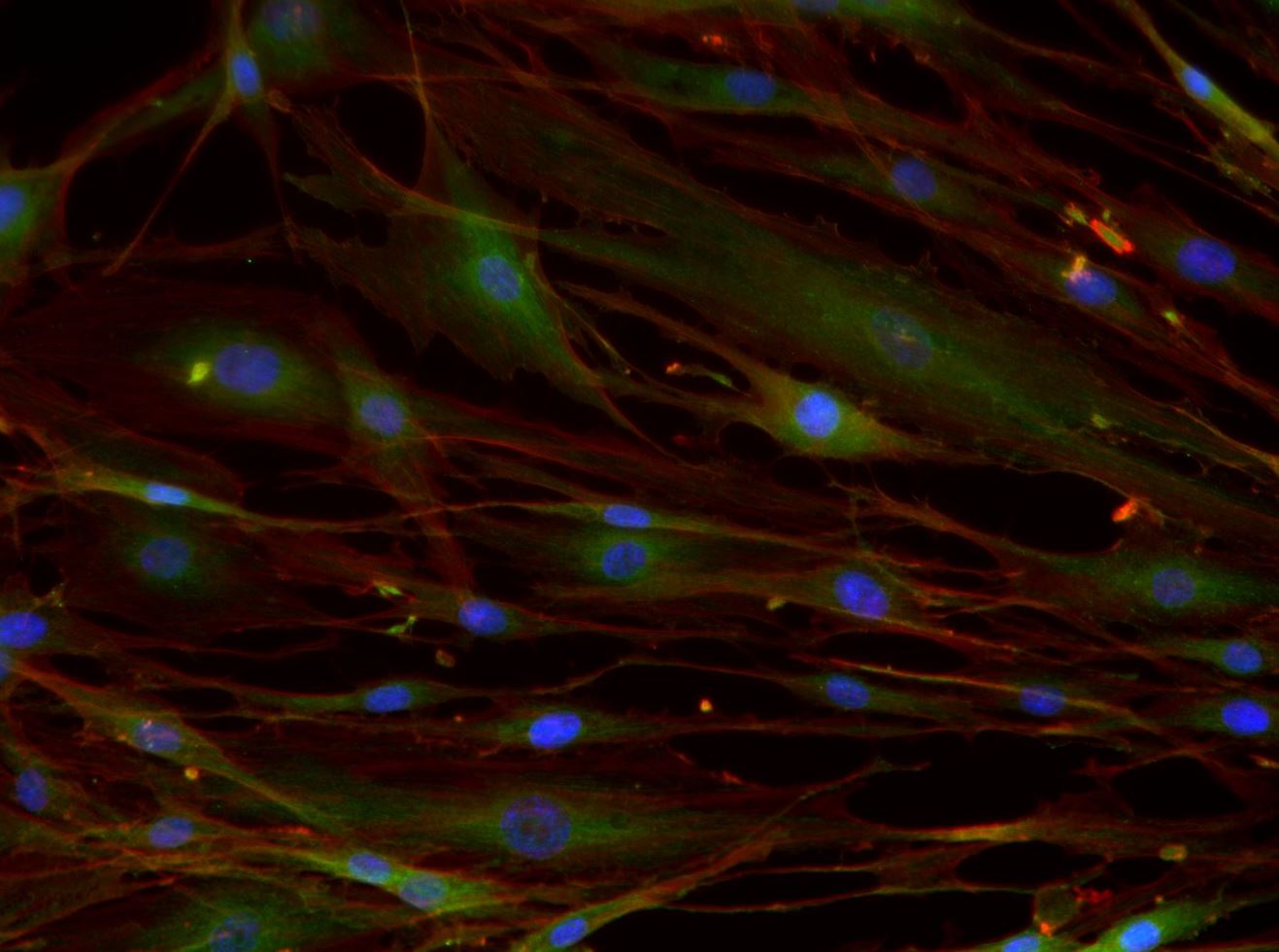
Immunocytochemistry/Immunofluorescence: Integrin beta 1/CD29 Antibody (P4C10) [NBP2-36561] - Integrin beta 1 (P4C10) antibody was tested in -20 degree MeOH fixed HeLa cells at a 1:200 dilution against DyLight 488 (green). Actin and nuclei were counterstained against Phalloidin 568 (red) and DAPI (blue), respectively.![Flow Cytometry: Integrin beta 1/CD29 Antibody (P4C10) - BSA Free [NBP2-36561]](https://resources.rndsystems.com/images/products/Integrin-beta-1-CD29-Antibody-P4C10-Flow-Cytometry-NBP2-36561-img0005.jpg "Flow Cytometry: Integrin beta 1/CD29 Antibody (P4C10) - BSA Free [NBP2-36561]")
Flow Cytometry: Integrin beta 1/CD29 Antibody (P4C10) - BSA Free [NBP2-36561]
Flow Cytometry: Integrin beta 1/CD29 Antibody (P4C10) [NBP2-36561] - An intracellular stain was performed on A431 cells with Integrin beta 1/CD29 [P4C10] Antibody NBP2-36561B (blue) and a matched isotype control (orange). Both antibodies were conjugated to Biotin. Cells were fixed with 4% PFA and then permeabilized with 0.1% saponin. Cells were incubated in an antibody dilution of 2.5 ug/mL for 30 minutes at room temperature, followed by Streptavidin - R-Phycoerythrin Protein (2012-1000, Novus Biologicals).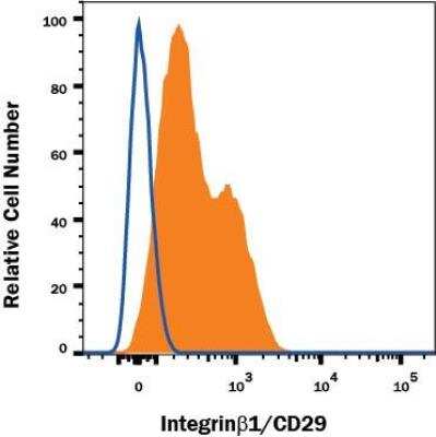
![Flow Cytometry: Integrin beta 1/CD29 Antibody (P4C10) - BSA Free [NBP2-36561]](https://resources.rndsystems.com/images/products/Integrin-beta-1-CD29-Antibody-P4C10-Flow-Cytometry-NBP2-36561-img0003.jpg "Flow Cytometry: Integrin beta 1/CD29 Antibody (P4C10) - BSA Free [NBP2-36561]")
Flow Cytometry: Integrin beta 1/CD29 Antibody (P4C10) - BSA Free [NBP2-36561]
Flow Cytometry: Integrin beta 1/CD29 Antibody (P4C10) [NBP2-36561] - A surface stain was performed on HeLa cells with Integrin beta 1/CD29 [P4C10] Antibody NBP2-36561PE (blue) and a matched isotype control (orange). Cells were incubated in an antibody dilution of 2.5 ug/mL for 20 minutes at room temperature. Both antibodies were conjugated to Phycoerythrin.![Flow Cytometry: Integrin beta 1/CD29 Antibody (P4C10) - BSA Free [NBP2-36561]](https://resources.rndsystems.com/images/products/Integrin-beta-1-CD29-Antibody-P4C10-Flow-Cytometry-NBP2-36561-img0004.jpg "Flow Cytometry: Integrin beta 1/CD29 Antibody (P4C10) - BSA Free [NBP2-36561]")
Flow Cytometry: Integrin beta 1/CD29 Antibody (P4C10) - BSA Free [NBP2-36561]
Flow Cytometry: Integrin beta 1/CD29 Antibody (P4C10) [NBP2-36561] - A surface stain was performed on HeLa cells with Integrin beta 1/CD29 [P4C10] Antibody NBP2-36561AF488 (blue) and a matched isotype control (orange). Cells were incubated in an antibody dilution of 5 ug/mL for 20 minutes at room temperature. Both antibodies were conjugated to Alexa Fluor 488. [NBP2-36561] -")
Immunocytochemistry/Immunofluorescence: Integrin beta 1/CD29 Antibody (P4C10) [NBP2-36561] -
Immunocytochemistry/Immunofluorescence: Integrin beta 1/CD29 Antibody (P4C10) [NBP2-36561] - Overnight incubation with 1:50 Integrin beta 1/CD29 primary antibody in PBS/BSA 1%, at 4ºC. Green: Integrin beta 1/CD29 Antibody (P4C10), Red: Phalloidin, Blue: DAPI. Image from verified customer review. - BSA Free [NBP2-36561] -")
Western Blot: Integrin beta 1/CD29 Antibody (P4C10) - BSA Free [NBP2-36561] -
Generation and characterization of ADSC-EVs and ADSC-NVs. A TEM images of ADSC-EVs and ADSC-NVs. Scale bar: 100 nm. B NTA analysis of ADSC-EVs and ADSC-NVs. The peak diameter of both ranged between 100 and 150 nm. C, D The yields of EVs and NVs from equivalent ADSCs were examined by particle number (C) and the protein amount (D) (n = 3 per group). E Protein expression of TSG101, CD81, and CD29 was assessed by western blot analysis for ADSC-EVs (EVs), ADSC-NVs (NVs), and ADSC. F-H The internalization of PKH26-labeled ADSC-EVs and ADSC-NVs in equivalent amount particles by HUVECs was evaluated at 1, 2, 3 and 4 h respectively. The internalization rate (F) was measured by calculating area ratio between ADSC-EVs/ ADSC-NVs and HUVECs (n = 3 per group). G, H Representative images of internalization of ADSC-EVs or ADSC-NVs at 1, 2, 3, 4 h. Scale bar: 100 μm. Red: ADSC-EVs/NVs, Green: HUVECs, blue: Nucleus of HUVECs. ns, no significant difference, ***p < 0.001, ****p < 0.0001 Image collected and cropped by CiteAb from the following open publication (https://pubmed.ncbi.nlm.nih.gov/36528594), licensed under a CC-BY license. Not internally tested by Novus Biologicals.Applications for Integrin beta 1/CD29 Antibody (P4C10) - BSA Free
Flow Cytometry
Immunocytochemistry/ Immunofluorescence
Immunohistochemistry-Frozen
Western Blot
Reviewed Applications
Read 1 review rated 4 using NBP2-36561 in the following applications:
Flow Cytometry Panel Builder
Bio-Techne Knows Flow Cytometry
Save time and reduce costly mistakes by quickly finding compatible reagents using the Panel Builder Tool.
Advanced Features
- Spectra Viewer - Custom analysis of spectra from multiple fluorochromes
- Spillover Popups - Visualize the spectra of individual fluorochromes
- Antigen Density Selector - Match fluorochrome brightness with antigen density

Formulation, Preparation, and Storage
Purification
Formulation
Format
Preservative
Concentration
Shipping
Stability & Storage
Background: Integrin beta 1/CD29
Additional Integrin beta 1/CD29 Products
Product Documents for Integrin beta 1/CD29 Antibody (P4C10) - BSA Free
Certificate of Analysis
To download a Certificate of Analysis, please enter a lot or batch number in the search box below.
Product Specific Notices for Integrin beta 1/CD29 Antibody (P4C10) - BSA Free
This product is for research use only and is not approved for use in humans or in clinical diagnosis. Primary Antibodies are guaranteed for 1 year from date of receipt.
Citations for Integrin beta 1/CD29 Antibody (P4C10) - BSA Free
 Powered by Bioz
Customer Reviews for Integrin beta 1/CD29 Antibody (P4C10) - BSA Free (1)
Have you used Integrin beta 1/CD29 Antibody (P4C10) - BSA Free?
Submit a review and receive an Amazon gift card!
$25/€18/£15/$25CAN/¥2500 Yen for a review with an image
$10/€7/£6/$10CAN/¥1110 Yen for a review without an image
Submit a review
Customer Images
-
Application: ImmunocytochemistrySample Tested: fibroblast cellsSpecies: HumanVerified Customer | Posted 06/13/2023Overnight incubation with 1:50 anti-ITGB1 primary antibody in PBS/BSA 1%, at 4ºC. Green: Integrin beta 1/CD29 Antibody (P4C10) Red: Phalloidin Blue: DAPI

There are no reviews that match your criteria.
Protocols
View specific protocols for Integrin beta 1/CD29 Antibody (P4C10) - BSA Free (NBP2-36561):
Sample Preparation.
1. Grow cells to 60-85% confluency. Flow cytometry requires between 2 x 10⁵ and 1 x 10⁶ cells for optimal performance.
2. If cells are adherent, harvest gently by washing once with staining buffer and then scraping. Avoid using trypsin as this can disrupt certain epitopes of interest. If enzymatic harvest is required, use Accutase, Collagenase, or TrypLE Express for a less damaging option.
3. Reserve 100 uL for counting, then transfer cell volume into a 15 mL conical tube and centrifuge for 4 minutes at 400 RCF.
a. Count cells using a hemocytometer and a 1:1 trypan blue exclusion stain to determine cell viability before starting the flow protocol. If cells appear blue, do not proceed.
4. Re-suspend cells to a concentration of 1 x 10⁶ cells/mL in staining buffer (NBP2-26247).
5. Aliquot out 100 uL samples in accordance with your experimental samples.
Tip: When cell surface and intracellular staining are required in the same sample, it is advisable that the cell surface staining be performed first since the fixation and permeablization steps might reduce the availability of surface antigens.
Cell surface staining
1. Recommended: Block non-specific interactions using 0.5-1 ug of a species specific Fc-blocking reagent such as an anti-mouse CD16/CD32 antibody (NBP1-27946).
2. Add appropriate amount of each antibody (eg. 1 test or 1 ug per sample, as experimentally determined) to 100 uL of staining buffer (NBP2-26247) per sample (eg. use 1 mL of staining buffer for 10 samples).
3. Mix well and incubate at room temperature in dark for 20 minutes.
4. Add 1-2 mL of staining buffer and centrifuge at 400 RCF for 1 minute and discard supernatant.
5. Wash twice by re-suspending cells in staining buffer (2 mL for tubes or 200 uL for wells) and centrifuging at 400 RCF for 5 minutes. Discard supernatant.
6. Add appropriate amount of secondary antibody (as experimentally determined) to each sample.
7. Incubate at room temperature in dark for 20 minutes.
8. Add 1-2 mL of staining buffer and centrifuge at 400 RCF for 1 minute and discard supernatant.
9. Wash twice by re-suspending cells in staining buffer (2 mL for tubes or 200 uL for wells) and centrifuging at 400 RCF for 5 minutes. Discard supernatant.
10. Resuspend in an appropriate volume of staining buffer (usually 500 uL per sample) and proceed with analysis on your flow cytometer.
Culture cells to appropriate density in 35 mm culture dishes or 6-well plates.
1. Remove culture medium and wash the cells briefly in PBS. Add 10% formalin to the dish and fix at room temperature for 10 minutes.
2. Remove the formalin and wash the cells in PBS.
3. Permeablize the cells with 0.1% Triton X100 or other suitable detergent for 10 min.
4. Remove the permeablization buffer and wash three times for 10 minutes each in PBS. Be sure to not let the specimen dry out.
5. To block nonspecific antibody binding, incubate in 10% normal goat serum from 1 hour to overnight at room temperature.
6. Add primary antibody at appropriate dilution and incubate overnight at 4C.
7. Remove primary antibody and replace with PBS. Wash three times for 10 minutes each.
8. Add secondary antibody at appropriate dilution. Incubate for 1 hour at room temperature.
9. Remove secondary antibody and replace with PBS. Wash three times for 10 minutes each.
10. Counter stain DNA with DAPi if required.
Find general support by application which include: protocols, troubleshooting, illustrated assays, videos and webinars.
- 7-Amino Actinomycin D (7-AAD) Cell Viability Flow Cytometry Protocol
- Antigen Retrieval Protocol (PIER)
- Antigen Retrieval for Frozen Sections Protocol
- Appropriate Fixation of IHC/ICC Samples
- Cellular Response to Hypoxia Protocols
- Chromogenic IHC Staining of Formalin-Fixed Paraffin-Embedded (FFPE) Tissue Protocol
- Chromogenic Immunohistochemistry Staining of Frozen Tissue
- ClariTSA™ Fluorophore Kits
- Detection & Visualization of Antibody Binding
- ELISA Sample Preparation & Collection Guide
- ELISA Troubleshooting Guide
- Extracellular Membrane Flow Cytometry Protocol
- Flow Cytometry Protocol for Cell Surface Markers
- Flow Cytometry Protocol for Staining Membrane Associated Proteins
- Flow Cytometry Staining Protocols
- Flow Cytometry Troubleshooting Guide
- Fluorescent IHC Staining of Frozen Tissue Protocol
- Graphic Protocol for Heat-induced Epitope Retrieval
- Graphic Protocol for the Preparation and Fluorescent IHC Staining of Frozen Tissue Sections
- Graphic Protocol for the Preparation and Fluorescent IHC Staining of Paraffin-embedded Tissue Sections
- Graphic Protocol for the Preparation of Gelatin-coated Slides for Histological Tissue Sections
- How to Run an R&D Systems DuoSet ELISA
- How to Run an R&D Systems Quantikine ELISA
- How to Run an R&D Systems Quantikine™ QuicKit™ ELISA
- ICC Cell Smear Protocol for Suspension Cells
- ICC Immunocytochemistry Protocol Videos
- ICC for Adherent Cells
- IHC Sample Preparation (Frozen sections vs Paraffin)
- Immunocytochemistry (ICC) Protocol
- Immunocytochemistry Troubleshooting
- Immunofluorescence of Organoids Embedded in Cultrex Basement Membrane Extract
- Immunofluorescent IHC Staining of Formalin-Fixed Paraffin-Embedded (FFPE) Tissue Protocol
- Immunohistochemistry (IHC) and Immunocytochemistry (ICC) Protocols
- Immunohistochemistry Frozen Troubleshooting
- Immunohistochemistry Paraffin Troubleshooting
- Immunoprecipitation Protocol
- Intracellular Flow Cytometry Protocol Using Alcohol (Methanol)
- Intracellular Flow Cytometry Protocol Using Detergents
- Intracellular Nuclear Staining Flow Cytometry Protocol Using Detergents
- Intracellular Staining Flow Cytometry Protocol Using Alcohol Permeabilization
- Intracellular Staining Flow Cytometry Protocol Using Detergents to Permeabilize Cells
- Preparing Samples for IHC/ICC Experiments
- Preventing Non-Specific Staining (Non-Specific Binding)
- Primary Antibody Selection & Optimization
- Propidium Iodide Cell Viability Flow Cytometry Protocol
- Protocol for Heat-Induced Epitope Retrieval (HIER)
- Protocol for Liperfluo
- Protocol for Making a 4% Formaldehyde Solution in PBS
- Protocol for VisUCyte™ HRP Polymer Detection Reagent
- Protocol for the Characterization of Human Th22 Cells
- Protocol for the Characterization of Human Th9 Cells
- Protocol for the Fluorescent ICC Staining of Cell Smears - Graphic
- Protocol for the Fluorescent ICC Staining of Cultured Cells on Coverslips - Graphic
- Protocol for the Preparation & Fixation of Cells on Coverslips
- Protocol for the Preparation and Chromogenic IHC Staining of Frozen Tissue Sections
- Protocol for the Preparation and Chromogenic IHC Staining of Frozen Tissue Sections - Graphic
- Protocol for the Preparation and Chromogenic IHC Staining of Paraffin-embedded Tissue Sections
- Protocol for the Preparation and Chromogenic IHC Staining of Paraffin-embedded Tissue Sections - Graphic
- Protocol for the Preparation and Fluorescent ICC Staining of Cells on Coverslips
- Protocol for the Preparation and Fluorescent ICC Staining of Non-adherent Cells
- Protocol for the Preparation and Fluorescent ICC Staining of Stem Cells on Coverslips
- Protocol for the Preparation and Fluorescent IHC Staining of Frozen Tissue Sections
- Protocol for the Preparation and Fluorescent IHC Staining of Paraffin-embedded Tissue Sections
- Protocol for the Preparation of Gelatin-coated Slides for Histological Tissue Sections
- Protocol for the Preparation of a Cell Smear for Non-adherent Cell ICC - Graphic
- Protocol: Annexin V and PI Staining by Flow Cytometry
- Protocol: Annexin V and PI Staining for Apoptosis by Flow Cytometry
- Quantikine HS ELISA Kit Assay Principle, Alkaline Phosphatase
- Quantikine HS ELISA Kit Principle, Streptavidin-HRP Polymer
- R&D Systems Quality Control Western Blot Protocol
- Sandwich ELISA (Colorimetric) – Biotin/Streptavidin Detection Protocol
- Sandwich ELISA (Colorimetric) – Direct Detection Protocol
- TUNEL and Active Caspase-3 Detection by IHC/ICC Protocol
- The Importance of IHC/ICC Controls
- Troubleshooting Guide: ELISA
- Troubleshooting Guide: Fluorokine Flow Cytometry Kits
- Troubleshooting Guide: Immunohistochemistry
- Troubleshooting Guide: Western Blot Figures
- Western Blot Conditions
- Western Blot Protocol
- Western Blot Protocol for Cell Lysates
- Western Blot Troubleshooting
- Western Blot Troubleshooting Guide
- View all Protocols, Troubleshooting, Illustrated assays and Webinars
FAQs for Integrin beta 1/CD29 Antibody (P4C10) - BSA Free
-
Q: I'm looking for a good antibody for integrins that works in rats. I would need it for Western Blot, Immunocyto- as well as Immunohistochemistry. I'm especially interested in beta1.
A: I would recommend NB110-57123 as it has been validated using rat samples in WB, IHC-P, and FACS. Although we do not list ICC on the datasheet for this antibody I believe it will work in ICC because it works in Flow Cytometry and has been validated to stain tissue. We have several other Integrin targets. To search for what you are looking for I recommend typing Integrin into the search area at the top of our website, then review the list of suggested targets that poplulate below the search. Once you click on a particular Integrin you can narrow the results with our filter. I recommend filtering to primary antibodies, by target name, species of interest you can filter to rat. If you so choose you can also filter further by application, host, clonality, etc.
Associated Pathways


