Artemin is a member of the Glia Cell-Derived Neurotrophic factor (GDNF) family ligands, which include GDNF, Persephin, Artemin, and Neurturin. GDNF family ligands are distant members of the Transforming Growth Factor beta (TGF-beta ) superfamily (1‑4). Similar to other TGF-beta family proteins, Artemin is synthesized as a large precursor protein that is cleaved at the dibasic cleavage site (RXXR) to release the carboxy-terminal domain. The carboxy-terminal domain of Artemin contains the characteristic seven conserved cysteine residues necessary for the formation of the cysteine-knot and the single interchain disulfide bond. Biologically active Artemin is a disulfide-linked homodimer of the carboxy-terminal 113 amino acid residues. Mature mouse Artemin shares 88.5% amino acid sequence similarity with human Artemin. Mature Artemin also shares approximately 40% amino acid sequence identity with the other three members of the GDNF family ligands (5). Bioactivities of all GDNF family ligands are mediated through a receptor complex composed of a high affinity ligand binding component (GFR alpha 1‑GFR alpha 4) and a common signaling component, cRET (receptor tyrosine kinase) (5‑8). Artemin prefers to bind to GFR alpha 3 and activites the GFR alpha 3‑RET. However, in the presence of RET, it can bind to GFR alpha 1 as well (4, 5, 9). Artemin has been shown to promote the survival and growth of various peripheral and central neurons, including sympathetic and dopaminergic neurons. It may also play an important role in the development of sympathetic neurons and several organs (5, 10, 11).
Mouse Artemin Antibody (185234)
R&D Systems | Catalog # MAB10851

Key Product Details
Species Reactivity
Validated:
Mouse
Cited:
Mouse, Transgenic Mouse
Applications
Validated:
Immunohistochemistry, Western Blot
Cited:
Immunohistochemistry, Neutralization, Immunocytochemistry, In vivo assay
Label
Unconjugated
Antibody Source
Monoclonal Rat IgG2A Clone # 185234
Loading...
Product Specifications
Immunogen
E. coli-derived recombinant mouse Artemin
Ala112-Gly224
Accession # Q9Z0L2.1
Ala112-Gly224
Accession # Q9Z0L2.1
Specificity
Detects mouse Artemin in direct ELISAs and Western blots. In direct ELISAs, no cross-reactivity with recombinant mouse (rm) AgRP, rmCripto, recombinant human (rh) Cripto-1, recombinant drosophila Dpp, recombinant rat GDNF, rhLAP, rhLatent TGF-beta 1, rhLefty A, rmLefty‑1, rmMIS, rhNeurturin, rmNodal, rhTGF-alpha, rhTGF-beta 1, rhTGF-beta 1.2, rhTGF-beta 2, rhTGF-beta 3, or recombinant Amphibian TGF-beta 5 is observed.
Clonality
Monoclonal
Host
Rat
Isotype
IgG2A
Scientific Data Images for Mouse Artemin Antibody (185234)
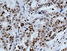
Artemin in Human tonsil.
Artemin was detected in immersion fixed paraffin-embedded sections of human tonsil using Rat Anti-Mouse Artemin Monoclonal Antibody (Catalog # MAB10851) at 3 µg/mL for 1 hour at room temperature followed by incubation with the Anti-Rabbit IgG VisUCyte™ HRP Polymer Antibody (VC003). Before incubation with the primary antibody, tissue was subjected to heat-induced epitope retrieval using Antigen Retrieval Reagent-Basic (CTS013). Tissue was stained using DAB (brown) and counterstained with hematoxylin (blue). Specific staining was localized to lymphocytes in germinal centers. Staining was performed using our protocol for IHC Staining with VisUCyte HRP Polymer Detection Reagents.Applications for Mouse Artemin Antibody (185234)
Application
Recommended Usage
Immunohistochemistry
3-25 µg/mL
Sample: Immersion fixed paraffin-embedded sections of human tonsil
Sample: Immersion fixed paraffin-embedded sections of human tonsil
Western Blot
1 µg/mL
Sample: Recombinant Mouse Artemin (Catalog # 1085-AR)
under non-reducing conditions only
Sample: Recombinant Mouse Artemin (Catalog # 1085-AR)
under non-reducing conditions only
Reviewed Applications
Read 2 reviews rated 5 using MAB10851 in the following applications:
Formulation, Preparation, and Storage
Purification
Protein A or G purified from hybridoma culture supernatant
Reconstitution
Reconstitute at 0.5 mg/mL in sterile PBS. For liquid material, refer to CoA for concentration.
Loading...
Formulation
Lyophilized from a 0.2 μm filtered solution in PBS with Trehalose. *Small pack size (SP) is supplied either lyophilized or as a 0.2 µm filtered solution in PBS.
Shipping
Lyophilized product is shipped at ambient temperature. Liquid small pack size (-SP) is shipped with polar packs. Upon receipt, store immediately at the temperature recommended below.
Stability & Storage
Use a manual defrost freezer and avoid repeated freeze-thaw cycles.
- 12 months from date of receipt, -20 to -70 °C as supplied.
- 1 month, 2 to 8 °C under sterile conditions after reconstitution.
- 6 months, -20 to -70 °C under sterile conditions after reconstitution.
Calculators
Background: Artemin
References
- Lin, L-F.H. et al. (1993) Science 260:1130.
- Milbrandt, J. et al. (1998) Neuron 20:245.
- Kotzbauer, P.T. et al. (1996) Nature 384:467.
- Baloh, R.H. et al. (1998) Neuron 21:1291.
- Takahashi, M. (2001) Cytokine and Growth Factor Reviews 12:361.
- Baloh, R.H. et al. (1997) Neuron 18:793.
- Jing, S. et al. (1996) Cell 85:1113.
- Jing, S. et al. (1997) J Biol Chem 272:33111.
- Nishino, J. et al. (1999) Neuron 23:725.
- Enomoto, H. et al. (2001) Development 128:3963.
- Andres, R. et al. (2001) Development 128:3685.
Alternate Names
ARTN, Enovin, EVN, Neublastin
Gene Symbol
ARTN
UniProt
Additional Artemin Products
Product Documents for Mouse Artemin Antibody (185234)
Certificate of Analysis
To download a Certificate of Analysis, please enter a lot or batch number in the search box below.
Note: Certificate of Analysis not available for kit components.
Product Specific Notices for Mouse Artemin Antibody (185234)
For research use only
Related Research Areas
Citations for Mouse Artemin Antibody (185234)
 Powered by Bioz
Customer Reviews for Mouse Artemin Antibody (185234) (2)
5 out of 5
2 Customer Ratings
Have you used Mouse Artemin Antibody (185234)?
Submit a review and receive an Amazon gift card!
$25/€18/£15/$25CAN/¥2500 Yen for a review with an image
$10/€7/£6/$10CAN/¥1110 Yen for a review without an image
Submit a review
Customer Images
Showing
1
-
2 of
2 reviews
Showing All
Filter By:
-
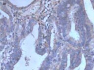
Application: ImmunohistochemistrySample Tested: Lung cancer tissueSpecies: MouseVerified Customer | Posted 07/08/2022

-
Application: ImmunohistochemistrySample Tested: Endometrial carcinomaSpecies: MouseVerified Customer | Posted 10/22/2021
There are no reviews that match your criteria.
Protocols
Find general support by application which include: protocols, troubleshooting, illustrated assays, videos and webinars.
- Antigen Retrieval Protocol (PIER)
- Antigen Retrieval for Frozen Sections Protocol
- Appropriate Fixation of IHC/ICC Samples
- Cellular Response to Hypoxia Protocols
- Chromogenic IHC Staining of Formalin-Fixed Paraffin-Embedded (FFPE) Tissue Protocol
- Chromogenic Immunohistochemistry Staining of Frozen Tissue
- ClariTSA™ Fluorophore Kits
- Detection & Visualization of Antibody Binding
- Fluorescent IHC Staining of Frozen Tissue Protocol
- Graphic Protocol for Heat-induced Epitope Retrieval
- Graphic Protocol for the Preparation and Fluorescent IHC Staining of Frozen Tissue Sections
- Graphic Protocol for the Preparation and Fluorescent IHC Staining of Paraffin-embedded Tissue Sections
- Graphic Protocol for the Preparation of Gelatin-coated Slides for Histological Tissue Sections
- IHC Sample Preparation (Frozen sections vs Paraffin)
- Immunofluorescent IHC Staining of Formalin-Fixed Paraffin-Embedded (FFPE) Tissue Protocol
- Immunohistochemistry (IHC) and Immunocytochemistry (ICC) Protocols
- Immunohistochemistry Frozen Troubleshooting
- Immunohistochemistry Paraffin Troubleshooting
- Preparing Samples for IHC/ICC Experiments
- Preventing Non-Specific Staining (Non-Specific Binding)
- Primary Antibody Selection & Optimization
- Protocol for Heat-Induced Epitope Retrieval (HIER)
- Protocol for Making a 4% Formaldehyde Solution in PBS
- Protocol for VisUCyte™ HRP Polymer Detection Reagent
- Protocol for the Preparation & Fixation of Cells on Coverslips
- Protocol for the Preparation and Chromogenic IHC Staining of Frozen Tissue Sections
- Protocol for the Preparation and Chromogenic IHC Staining of Frozen Tissue Sections - Graphic
- Protocol for the Preparation and Chromogenic IHC Staining of Paraffin-embedded Tissue Sections
- Protocol for the Preparation and Chromogenic IHC Staining of Paraffin-embedded Tissue Sections - Graphic
- Protocol for the Preparation and Fluorescent IHC Staining of Frozen Tissue Sections
- Protocol for the Preparation and Fluorescent IHC Staining of Paraffin-embedded Tissue Sections
- Protocol for the Preparation of Gelatin-coated Slides for Histological Tissue Sections
- R&D Systems Quality Control Western Blot Protocol
- TUNEL and Active Caspase-3 Detection by IHC/ICC Protocol
- The Importance of IHC/ICC Controls
- Troubleshooting Guide: Immunohistochemistry
- Troubleshooting Guide: Western Blot Figures
- Western Blot Conditions
- Western Blot Protocol
- Western Blot Protocol for Cell Lysates
- Western Blot Troubleshooting
- Western Blot Troubleshooting Guide
- View all Protocols, Troubleshooting, Illustrated assays and Webinars
Loading...