CD27 is a lymphocyte-specific member of the tumor necrosis factor receptor superfamily (TNFRSF) and is designated TNFRSF7 (1, 2). Mouse CD27 cDNA encodes a 250 amino acid (aa) residue type I transmembrane protein with a 20 aa putative signal peptide, a 162 aa extracellular region containing three TNFR cysteine-rich repeats, a 21 aa transmembrane domain and a 47 aa cytoplasmic region (3). Mouse and human CD27 share approximately 65% amino acid identity. CD27 exists as homodimers on the cell surface via an extracellular disulfide bond in the membrane-proximal region. A soluble form of CD27 is also produced during the immune response and is found in various body fluids (4). CD27 is expressed on subsets of T and B cells. The expression of CD27 is upregulated upon T-cell activation. Although CD27 appears to be a marker for human memory B cells, it is only expressed in a small population of mouse B cells in germinal centers and at sites of B cell stimulation, suggesting that mouse CD27 may be a marker for activated B cells (5). CD27 interacts with CD27 ligand (also named CD70 and TNFSF7), which is a member of the TNF ligand superfamily. Ligation of CD27 on T cells provides costimulatory signals that are required for T cell proliferation, clonal expansion and the promotion of effector T cell formation (1, 2). Ligation of CD27 on B cells has been shown to inhibit terminal differentiation of activated mouse B cells into plasma cells and enhances commitment to memory B cell responses (5).
Mouse CD27/TNFRSF7 Antibody (137910)
R&D Systems | Catalog # MAB574

Key Product Details
Species Reactivity
Validated:
Mouse
Cited:
Human
Applications
Validated:
Western Blot, Immunocytochemistry
Cited:
Western Blot
Label
Unconjugated
Antibody Source
Monoclonal Rat IgG2A Clone # 137910
Loading...
Product Specifications
Immunogen
Mouse myeloma cell line NS0-derived recombinant mouse CD27/TNFRSF7
Thr21-Arg182
Accession # P41272
Thr21-Arg182
Accession # P41272
Specificity
Detects mouse CD27 in direct ELISAs and Western blots. In Western blots, does not cross-react with recombinant human (rh) CD27, rhCD30, rhCD40, rhDR3, rhDR6, rhFas Ligand, rhTRAIL R3, rhTRAIL R4, rhLT beta R, rhNGF-R1, rh4-1BB, rhGITR, rhTNFR1, rhTNF RII, rhBAFF, rhTAJ, rhEDA, rhOPG, rhRELT, rhRANK, recombinant mouse (rm) CD30, rmCD40, rmGITR, rmHVEM, rmRANK, rmFas, rmTRAIL R2, rm4-1BB, rmLT beta R, rmOPG, rmNGFR, rmDR3, rmBAFF R, rmEDAR, rmOX-40, rmTWEAK R, rmTNF R1, recombinant rat Fas, or recombinant feline Fas.
Clonality
Monoclonal
Host
Rat
Isotype
IgG2A
Scientific Data Images for Mouse CD27/TNFRSF7 Antibody (137910)
Detection of Mouse CD27/TNFRSF7 by Western Blot.
Western blot shows lysates of mouse spleen tissue. PVDF Membrane was probed with 2 µg/mL of Mouse CD27/TNFRSF7 Monoclonal Antibody (Catalog # MAB574) followed by HRP-conjugated Anti-Rat IgG Secondary Antibody (Catalog # HAF005). A specific band was detected for CD27/TNFRSF7 at approximately 45 kDa (as indicated). This experiment was conducted under reducing conditions and using Immunoblot Buffer Group 7.
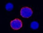
CD27/TNFRSF7 in Mouse Splenocytes.
CD27/TNFRSF7 was detected in immersion fixed mouse splenocytes using Rat Anti-Mouse CD27/TNFRSF7 Monoclonal Antibody (Catalog # MAB574) at 8 µg/mL for 3 hours at room temperature. Cells were stained using the NorthernLights™ 557-conjugated Anti-Rat IgG Secondary Antibody (red; Catalog # NL013) and counterstained with DAPI (blue). Specific staining was localized to plasma membrane. View our protocol for Fluorescent ICC Staining of Non-adherent Cells.
Detection of CD27/TNFRSF7 by Western Blot
KMT9 is expressed in lung cancer tissue and cell lines. a Dynamics of KMT9 alpha expression in matched normal and stage 1a lung adenocarcinoma tissue from eleven patients that underwent curative lobectomy. Normal samples were taken at 6 cm distance from macroscopic tumor sites. Data were extracted from (GSE83213). Red lines indicate increased expression of KMT9 alpha in tumor (n = 8), green lines indicate decreased expression of KMT9 alpha in tumor (n = 3). b TCGA data comparing KMT9 alpha expression in n = 515 lung adenocarcinoma with non-matched normal lung tissue (n = 59). Data represent interquartile range including minimum, 25th percentile, median, 75th percentile and maximum values. Significance was accessed by t test. c Kaplan–Meier survival analysis of patients with adenocarcinoma expressing high (n = 58) and low (n = 57) KMT9 alpha. Data were extracted from GSE26939. HR = hazard ratio. d Western blots of matched tissue from normal and tumor samples from patients with adenocarcinoma (#1 and #2) or SCLC (#3 and #4). Western blots were performed with the indicated antibodies. e Expression levels of KMT9 alpha and KMT9 beta in human cell lines from SCLC (GLC-2 and NCI-H82), adenocarcinoma (A549, PC-9 and NCI-H2087) and human immortalized normal lung fibroblasts (CRL-7000 and IMR-90) were analyzed by western blot using the indicated antibodies. f In A549 cells, KMT9 alpha and KMT9 beta are present in both nuclear and cytoplasmic compartments. Western blots were performed with the indicated antibodies. g Levels of H4K12me1 in SCLC (GLC-2 and NCI-H82) and adenocarcinoma (A549, PC-9 and NCI-H2087) cells were analyzed by western blotting using the indicated antibodies Image collected and cropped by CiteAb from the following open publication (https://pubmed.ncbi.nlm.nih.gov/32095117), licensed under a CC-BY license. Not internally tested by R&D Systems.
Detection of CD27/TNFRSF7 by Western Blot
KMT9 controls expression of genes involved in the organization of organelles, cells death and cell proliferation. a Venn diagram showing overlap and number of genes/proteins in A549 cells that are differentially expressed upon RNAi mediated knock-down of KMT9 alpha (log2(fold-change) > ± 0.26). In total, 460 targets are concomitantly up- or down-regulated on RNA and protein level upon knock-down of KMT9 alpha. Enriched GO_cellular components b and GO_biological processes c gene sets obtained for the indicated 460 KMT9 alpha -regulated target genes. d Heat map displaying mRNA levels of the 460 KMT9 alpha -regulated genes involved in cell proliferation (GO:0042127) in A549 cells treated with siControl or siKMT9 alpha #1. e RNA sequencing reads (left panel) and mass spectrometry volcano plot (right panel) for the indicated genes and proteins are represented exemplarily. f Western blot displaying expression of the target proteins indicated in e upon knock-down of KMT9 alpha in A549 cells. The indicated antibodies were used. g Quantitative real-time PCR analysis of the mRNA expression of the target genes displayed in e after knock-down of KMT9 alpha. Data represent means + standard deviation. Significance was accessed by two-tailed t test, n = 3 (TIMP2 n = 6) Image collected and cropped by CiteAb from the following open publication (https://pubmed.ncbi.nlm.nih.gov/32095117), licensed under a CC-BY license. Not internally tested by R&D Systems.Applications for Mouse CD27/TNFRSF7 Antibody (137910)
Application
Recommended Usage
Immunocytochemistry
8-25 µg/mL
Sample: Immersion fixed mouse splenocytes
Sample: Immersion fixed mouse splenocytes
Western Blot
2 µg/mL
Sample: Mouse spleen tissue
Sample: Mouse spleen tissue
Reviewed Applications
Read 1 review rated 5 using MAB574 in the following applications:
Formulation, Preparation, and Storage
Purification
Protein A or G purified from hybridoma culture supernatant
Reconstitution
Reconstitute at 0.5 mg/mL in sterile PBS. For liquid material, refer to CoA for concentration.
Loading...
Formulation
Lyophilized from a 0.2 μm filtered solution in PBS with Trehalose. *Small pack size (SP) is supplied either lyophilized or as a 0.2 µm filtered solution in PBS.
Shipping
Lyophilized product is shipped at ambient temperature. Liquid small pack size (-SP) is shipped with polar packs. Upon receipt, store immediately at the temperature recommended below.
Stability & Storage
Use a manual defrost freezer and avoid repeated freeze-thaw cycles.
- 12 months from date of receipt, -20 to -70 °C as supplied.
- 1 month, 2 to 8 °C under sterile conditions after reconstitution.
- 6 months, -20 to -70 °C under sterile conditions after reconstitution.
Calculators
Background: CD27/TNFRSF7
References
- Croft, M. (2003) Nature Reviews Immunol. 3:609.
- Croft, M. (2003) Cytokine and Growth Factor Reviews 14:265.
- Gravestein, L.A. et al. (1993) Eur. J. Immunol. 23:943.
- Lens, S.M. et al. (1998) Semin. Immunol. 10:491.
- Raman, V.S. et al. (2003) J. Immunol. 171:5876.
Alternate Names
CD27, TNFRSF7
Gene Symbol
CD27
UniProt
Additional CD27/TNFRSF7 Products
Product Documents for Mouse CD27/TNFRSF7 Antibody (137910)
Certificate of Analysis
To download a Certificate of Analysis, please enter a lot or batch number in the search box below.
Note: Certificate of Analysis not available for kit components.
Product Specific Notices for Mouse CD27/TNFRSF7 Antibody (137910)
For research use only
Related Research Areas
Citations for Mouse CD27/TNFRSF7 Antibody (137910)
 Powered by Bioz
Customer Reviews for Mouse CD27/TNFRSF7 Antibody (137910) (1)
5 out of 5
1 Customer Rating
Have you used Mouse CD27/TNFRSF7 Antibody (137910)?
Submit a review and receive an Amazon gift card!
$25/€18/£15/$25CAN/¥2500 Yen for a review with an image
$10/€7/£6/$10CAN/¥1110 Yen for a review without an image
Submit a review
Customer Images
Showing
1
-
1 of
1 review
Showing All
Filter By:
-
Application: Immunocytochemistry/ImmunofluorescenceSample Tested: SplenocytesSpecies: MouseVerified Customer | Posted 01/25/2022

There are no reviews that match your criteria.
Protocols
Find general support by application which include: protocols, troubleshooting, illustrated assays, videos and webinars.
- Appropriate Fixation of IHC/ICC Samples
- Cellular Response to Hypoxia Protocols
- ClariTSA™ Fluorophore Kits
- Detection & Visualization of Antibody Binding
- ICC Cell Smear Protocol for Suspension Cells
- ICC Immunocytochemistry Protocol Videos
- ICC for Adherent Cells
- Immunocytochemistry (ICC) Protocol
- Immunocytochemistry Troubleshooting
- Immunofluorescence of Organoids Embedded in Cultrex Basement Membrane Extract
- Immunohistochemistry (IHC) and Immunocytochemistry (ICC) Protocols
- Preparing Samples for IHC/ICC Experiments
- Preventing Non-Specific Staining (Non-Specific Binding)
- Primary Antibody Selection & Optimization
- Protocol for VisUCyte™ HRP Polymer Detection Reagent
- Protocol for the Fluorescent ICC Staining of Cell Smears - Graphic
- Protocol for the Fluorescent ICC Staining of Cultured Cells on Coverslips - Graphic
- Protocol for the Preparation and Fluorescent ICC Staining of Cells on Coverslips
- Protocol for the Preparation and Fluorescent ICC Staining of Non-adherent Cells
- Protocol for the Preparation and Fluorescent ICC Staining of Stem Cells on Coverslips
- Protocol for the Preparation of a Cell Smear for Non-adherent Cell ICC - Graphic
- R&D Systems Quality Control Western Blot Protocol
- TUNEL and Active Caspase-3 Detection by IHC/ICC Protocol
- The Importance of IHC/ICC Controls
- Troubleshooting Guide: Western Blot Figures
- Western Blot Conditions
- Western Blot Protocol
- Western Blot Protocol for Cell Lysates
- Western Blot Troubleshooting
- Western Blot Troubleshooting Guide
- View all Protocols, Troubleshooting, Illustrated assays and Webinars
Loading...
Associated Pathways
