CEACAM-1 (Carcinoembryonic antigen-related cell adhesion molecule 1; also BGP-1, CD66a and MHVR1) is a 110-120 kDa member of the CEACAM subfamily, CEA family of proteins. It has a wide expression pattern, being found on neutrophils, dendritic cells, endothelial cells, colonic epithelium and hepatocytes. It mediates cell adhesion, and appears to regulate insulin levels and signaling by interacting with the insulin receptor. It also demonstrates proangiogenic effects by inducing endothelial cells to proliferate and form capillary-like tubules. Finally, CEACAM-1 is a known receptor for mouse hepatitis virus. Mature mouse CEACAM-1 is a 487 amino acid (aa) type I transmembrane glycoprotein. Its contains a 394 aa extracellular region (aa 35-428) that shows one V-type (aa 35-142) and three C2-type (aa 147-411) Ig-like domains, plus a 74 aa cytoplasmic domain. Three alternate splice forms exist. One contains a four aa substitution for aa 455-521, a second shows a Gln substitution for aa 142-322, and a third possesses a combination of the first two splice patterns. CEACAM-1 forms homodimers. Over aa 35-428, mouse CEACAM-1 shares 56% and 70% aa identity with human and rat CEACAM-1, respectively.
Mouse CEACAM-1/CD66a Antibody (723629)
R&D Systems | Catalog # MAB6480

Key Product Details
Species Reactivity
Mouse
Applications
Immunohistochemistry, Western Blot, Flow Cytometry, CyTOF-ready
Label
Unconjugated
Antibody Source
Monoclonal Rat IgG1 Clone # 723629
Loading...
Product Specifications
Immunogen
Mouse myeloma cell line NS0-derived recombinant mouse CEACAM-1/CD66a
Glu35-Gly428
Accession # P31809
Glu35-Gly428
Accession # P31809
Specificity
Detects mouse CEACAM-1/CD66a in direct ELISAs and Western blots.
In direct ELISAs, no cross-reactivity
with recombinant human CECAM-1, -3, -5, or -6 is observed. In Western blots, no
cross-reactivity with rhCEACAM-1, -3, -4, -5, -6, or -7 is observed.
Clonality
Monoclonal
Host
Rat
Isotype
IgG1
Scientific Data Images for Mouse CEACAM-1/CD66a Antibody (723629)
Detection of Mouse CEACAM‑1/CD66a by Western Blot.
Western blot shows lysates of mouse liver tissue. PVDF membrane was probed with 0.1 µg/mL of Rat Anti-Mouse CEACAM-1/CD66a Monoclonal Antibody (Catalog # MAB6480) followed by HRP-conjugated Anti-Rat IgG Secondary Antibody (Catalog # HAF005). A specific band was detected for CEACAM-1/CD66a at approximately 110-120 kDa (as indicated). This experiment was conducted under reducing conditions and using Immunoblot Buffer Group 1.
Detection of CEACAM-1 in Mouse Splenocytes by Flow Cytometry.
Mouse splenocytes were stained with Rat Anti-Mouse CEACAM-1/CD66a Monoclonal Antibody (Catalog # MAB6480) followed by Fluorescein-conjugated Anti-Rat IgG Secondary Antibody (Catalog # F0104B) and Rat Anti-Mouse B220/CD45R Allophycocyanin-conjugated Monoclonal Antibody (Catalog # FAB1217A). Quadrant markers were set based on control antibody staining (Catalog # MAB005).
CEACAM‑1/CD66a in Mouse Liver.
CEACAM-1/CD66a was detected in perfusion fixed frozen sections of mouse liver using Rat Anti-Mouse CEACAM-1/CD66a Monoclonal Antibody (Catalog # MAB6480) at 25 µg/mL overnight at 4 °C. Tissue was stained using the NorthernLights™ 557-conjugated Anti-Rat IgG Secondary Antibody (red; Catalog # NL013) and counterstained with DAPI (blue). Specific staining was localized to bile canaliculi. View our protocol for Fluorescent IHC Staining of Frozen Tissue Sections.Applications for Mouse CEACAM-1/CD66a Antibody (723629)
Application
Recommended Usage
CyTOF-ready
Ready to be labeled using established conjugation methods. No BSA or other carrier proteins that could interfere with conjugation.
Flow Cytometry
2.5 µg/106 cells
Sample: Mouse splenocytes
Sample: Mouse splenocytes
Immunohistochemistry
8-25 µg/mL
Sample: Perfusion fixed frozen sections of mouse liver
Sample: Perfusion fixed frozen sections of mouse liver
Western Blot
0.1 µg/mL
Sample: Mouse liver tissue
Sample: Mouse liver tissue
Reviewed Applications
Read 2 reviews rated 5 using MAB6480 in the following applications:
Flow Cytometry Panel Builder
Bio-Techne Knows Flow Cytometry
Save time and reduce costly mistakes by quickly finding compatible reagents using the Panel Builder Tool.
Advanced Features
- Spectra Viewer - Custom analysis of spectra from multiple fluorochromes
- Spillover Popups - Visualize the spectra of individual fluorochromes
- Antigen Density Selector - Match fluorochrome brightness with antigen density

Formulation, Preparation, and Storage
Purification
Protein A or G purified from hybridoma culture supernatant
Reconstitution
Sterile PBS to a final concentration of 0.5 mg/mL. For liquid material, refer to CoA for concentration.
Loading...
Formulation
Lyophilized from a 0.2 μm filtered solution in PBS with Trehalose. *Small pack size (SP) is supplied either lyophilized or as a 0.2 µm filtered solution in PBS.
Shipping
Lyophilized product is shipped at ambient temperature. Liquid small pack size (-SP) is shipped with polar packs. Upon receipt, store immediately at the temperature recommended below.
Stability & Storage
Use a manual defrost freezer and avoid repeated freeze-thaw cycles.
- 12 months from date of receipt, -20 to -70 °C as supplied.
- 1 month, 2 to 8 °C under sterile conditions after reconstitution.
- 6 months, -20 to -70 °C under sterile conditions after reconstitution.
Calculators
Background: CEACAM-1/CD66a
Long Name
Carcinoembryonic Antigen-related Cell Adhesion Molecule 1
Alternate Names
BGP1, Biliary Glycoprotein 1, CD66a, Cea-1, CEACAM1, Hv-1, Hv-2, MHVR
Gene Symbol
CEACAM1
UniProt
Additional CEACAM-1/CD66a Products
Product Documents for Mouse CEACAM-1/CD66a Antibody (723629)
Certificate of Analysis
To download a Certificate of Analysis, please enter a lot or batch number in the search box below.
Note: Certificate of Analysis not available for kit components.
Product Specific Notices for Mouse CEACAM-1/CD66a Antibody (723629)
For research use only
Citations for Mouse CEACAM-1/CD66a Antibody (723629)
 Powered by Bioz
Customer Reviews for Mouse CEACAM-1/CD66a Antibody (723629) (2)
5 out of 5
2 Customer Ratings
Have you used Mouse CEACAM-1/CD66a Antibody (723629)?
Submit a review and receive an Amazon gift card!
$25/€18/£15/$25CAN/¥2500 Yen for a review with an image
$10/€7/£6/$10CAN/¥1110 Yen for a review without an image
Submit a review
Customer Images
Showing
1
-
2 of
2 reviews
Showing All
Filter By:
-
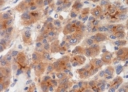
Application: ImmunohistochemistrySample Tested: Liver tissueSpecies: MouseVerified Customer | Posted 10/30/2021

-
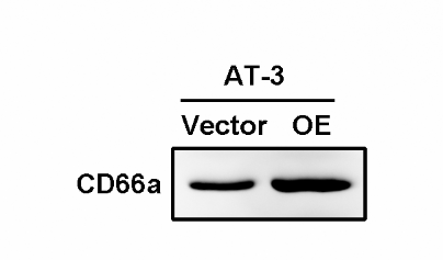
Application: Western BlotSample Tested: Cancer CellsSpecies: MouseVerified Customer | Posted 02/16/2020
There are no reviews that match your criteria.
Protocols
Find general support by application which include: protocols, troubleshooting, illustrated assays, videos and webinars.
- 7-Amino Actinomycin D (7-AAD) Cell Viability Flow Cytometry Protocol
- Antigen Retrieval Protocol (PIER)
- Antigen Retrieval for Frozen Sections Protocol
- Appropriate Fixation of IHC/ICC Samples
- Cellular Response to Hypoxia Protocols
- Chromogenic IHC Staining of Formalin-Fixed Paraffin-Embedded (FFPE) Tissue Protocol
- Chromogenic Immunohistochemistry Staining of Frozen Tissue
- ClariTSA™ Fluorophore Kits
- Detection & Visualization of Antibody Binding
- Extracellular Membrane Flow Cytometry Protocol
- Flow Cytometry Protocol for Cell Surface Markers
- Flow Cytometry Protocol for Staining Membrane Associated Proteins
- Flow Cytometry Staining Protocols
- Flow Cytometry Troubleshooting Guide
- Fluorescent IHC Staining of Frozen Tissue Protocol
- Graphic Protocol for Heat-induced Epitope Retrieval
- Graphic Protocol for the Preparation and Fluorescent IHC Staining of Frozen Tissue Sections
- Graphic Protocol for the Preparation and Fluorescent IHC Staining of Paraffin-embedded Tissue Sections
- Graphic Protocol for the Preparation of Gelatin-coated Slides for Histological Tissue Sections
- IHC Sample Preparation (Frozen sections vs Paraffin)
- Immunofluorescent IHC Staining of Formalin-Fixed Paraffin-Embedded (FFPE) Tissue Protocol
- Immunohistochemistry (IHC) and Immunocytochemistry (ICC) Protocols
- Immunohistochemistry Frozen Troubleshooting
- Immunohistochemistry Paraffin Troubleshooting
- Intracellular Flow Cytometry Protocol Using Alcohol (Methanol)
- Intracellular Flow Cytometry Protocol Using Detergents
- Intracellular Nuclear Staining Flow Cytometry Protocol Using Detergents
- Intracellular Staining Flow Cytometry Protocol Using Alcohol Permeabilization
- Intracellular Staining Flow Cytometry Protocol Using Detergents to Permeabilize Cells
- Preparing Samples for IHC/ICC Experiments
- Preventing Non-Specific Staining (Non-Specific Binding)
- Primary Antibody Selection & Optimization
- Propidium Iodide Cell Viability Flow Cytometry Protocol
- Protocol for Heat-Induced Epitope Retrieval (HIER)
- Protocol for Liperfluo
- Protocol for Making a 4% Formaldehyde Solution in PBS
- Protocol for VisUCyte™ HRP Polymer Detection Reagent
- Protocol for the Characterization of Human Th22 Cells
- Protocol for the Characterization of Human Th9 Cells
- Protocol for the Preparation & Fixation of Cells on Coverslips
- Protocol for the Preparation and Chromogenic IHC Staining of Frozen Tissue Sections
- Protocol for the Preparation and Chromogenic IHC Staining of Frozen Tissue Sections - Graphic
- Protocol for the Preparation and Chromogenic IHC Staining of Paraffin-embedded Tissue Sections
- Protocol for the Preparation and Chromogenic IHC Staining of Paraffin-embedded Tissue Sections - Graphic
- Protocol for the Preparation and Fluorescent IHC Staining of Frozen Tissue Sections
- Protocol for the Preparation and Fluorescent IHC Staining of Paraffin-embedded Tissue Sections
- Protocol for the Preparation of Gelatin-coated Slides for Histological Tissue Sections
- Protocol: Annexin V and PI Staining by Flow Cytometry
- Protocol: Annexin V and PI Staining for Apoptosis by Flow Cytometry
- R&D Systems Quality Control Western Blot Protocol
- TUNEL and Active Caspase-3 Detection by IHC/ICC Protocol
- The Importance of IHC/ICC Controls
- Troubleshooting Guide: Fluorokine Flow Cytometry Kits
- Troubleshooting Guide: Immunohistochemistry
- Troubleshooting Guide: Western Blot Figures
- Western Blot Conditions
- Western Blot Protocol
- Western Blot Protocol for Cell Lysates
- Western Blot Troubleshooting
- Western Blot Troubleshooting Guide
- View all Protocols, Troubleshooting, Illustrated assays and Webinars
Loading...
Associated Pathways
