Mouse Type II Collagen is a 100 kDa fibrillar collagen found in cartilage, vitreous body and vertebral disk. It is synthesized as a 1459 amino acid (aa) preproprecursor that contains an N-terminal and a C-terminal proregion. Intracellularly, type II Collagen homotrimerizes into a fibril and is then secreted. The proregions are then cleaved by procollagenases and the processed homotrimer covalently crosslinks to other type II fibrils. Mature mouse Collagen II shares 95% and 98% aa sequence identity with human and rat Collagen II, respectively.

Key Product Details
Species Reactivity
Validated:
Mouse
Cited:
Mouse, Transgenic Mouse
Applications
Validated:
Western Blot, Immunocytochemistry
Cited:
Immunohistochemistry, Immunohistochemistry-Paraffin, Western Blot, Immunocytochemistry
Label
Unconjugated
Antibody Source
Polyclonal Sheep IgG
Loading...
Product Specifications
Immunogen
Mouse sternum-derived Collagen II
Specificity
Detects mouse Collagen II in direct ELISAs and Western blots.
Clonality
Polyclonal
Host
Sheep
Isotype
IgG
Scientific Data Images for Mouse Collagen II Antibody
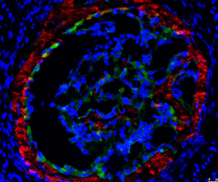
Collagen II in Mouse Chondrocytes.
Collagen II was detected in immersion fixed mouse mesenchymal stem cell-differentiated chondrocytes using 10 µg/mL Sheep Anti-Mouse Collagen II Antigen Affinity-purified Polyclonal Antibody (Catalog # AF3615) for 3 hours at room temperature. Cells were stained with the NorthernLights™ 557-conjugated Anti-Sheep IgG Secondary Antibody (red; Catalog # NL010) and counterstained with DAPI (blue). View our protocol for Fluorescent ICC Staining of Cells on Coverslips.Applications for Mouse Collagen II Antibody
Application
Recommended Usage
Immunocytochemistry
5-15 µg/mL
Sample: Immersion fixed mouse mesenchymal stem cell-differentiated chondrocytes
Sample: Immersion fixed mouse mesenchymal stem cell-differentiated chondrocytes
Western Blot
0.1 µg/mL
Sample: Mouse Collagen II
Sample: Mouse Collagen II
Reviewed Applications
Read 1 review rated 5 using AF3615 in the following applications:
Formulation, Preparation, and Storage
Purification
Antigen Affinity-purified
Reconstitution
Reconstitute at 0.2 mg/mL in sterile PBS. For liquid material, refer to CoA for concentration.
Loading...
Formulation
Lyophilized from a 0.2 μm filtered solution in PBS with Trehalose. *Small pack size (SP) is supplied either lyophilized or as a 0.2 µm filtered solution in PBS.
Shipping
Lyophilized product is shipped at ambient temperature. Liquid small pack size (-SP) is shipped with polar packs. Upon receipt, store immediately at the temperature recommended below.
Stability & Storage
Use a manual defrost freezer and avoid repeated freeze-thaw cycles.
- 12 months from date of receipt, -20 to -70 °C as supplied.
- 1 month, 2 to 8 °C under sterile conditions after reconstitution.
- 6 months, -20 to -70 °C under sterile conditions after reconstitution.
Calculators
Background: Collagen II
Alternate Names
Chondrocalcin, COL2A1, SEDC
Gene Symbol
COL2A1
Additional Collagen II Products
Product Documents for Mouse Collagen II Antibody
Certificate of Analysis
To download a Certificate of Analysis, please enter a lot or batch number in the search box below.
Note: Certificate of Analysis not available for kit components.
Product Specific Notices for Mouse Collagen II Antibody
For research use only
Related Research Areas
Citations for Mouse Collagen II Antibody
 Powered by Bioz
Customer Reviews for Mouse Collagen II Antibody (1)
5 out of 5
1 Customer Rating
Have you used Mouse Collagen II Antibody?
Submit a review and receive an Amazon gift card!
$25/€18/£15/$25CAN/¥2500 Yen for a review with an image
$10/€7/£6/$10CAN/¥1110 Yen for a review without an image
Submit a review
Customer Images
Showing
1
-
1 of
1 review
Showing All
Filter By:
-
Application: Immunocytochemistry/ImmunofluorescenceSample Tested: embryonic leg boneSpecies: MouseVerified Customer | Posted 12/14/2018mouse E17.5 embryonic leg frozen section. Primary antibody was incubated overnight at 4 °C. Secondary antibody was donkey anit-sheep 555.

There are no reviews that match your criteria.
Protocols
Find general support by application which include: protocols, troubleshooting, illustrated assays, videos and webinars.
- Appropriate Fixation of IHC/ICC Samples
- Cellular Response to Hypoxia Protocols
- ClariTSA™ Fluorophore Kits
- Detection & Visualization of Antibody Binding
- ICC Cell Smear Protocol for Suspension Cells
- ICC Immunocytochemistry Protocol Videos
- ICC for Adherent Cells
- Immunocytochemistry (ICC) Protocol
- Immunocytochemistry Troubleshooting
- Immunofluorescence of Organoids Embedded in Cultrex Basement Membrane Extract
- Immunohistochemistry (IHC) and Immunocytochemistry (ICC) Protocols
- Preparing Samples for IHC/ICC Experiments
- Preventing Non-Specific Staining (Non-Specific Binding)
- Primary Antibody Selection & Optimization
- Protocol for VisUCyte™ HRP Polymer Detection Reagent
- Protocol for the Fluorescent ICC Staining of Cell Smears - Graphic
- Protocol for the Fluorescent ICC Staining of Cultured Cells on Coverslips - Graphic
- Protocol for the Preparation and Fluorescent ICC Staining of Cells on Coverslips
- Protocol for the Preparation and Fluorescent ICC Staining of Non-adherent Cells
- Protocol for the Preparation and Fluorescent ICC Staining of Stem Cells on Coverslips
- Protocol for the Preparation of a Cell Smear for Non-adherent Cell ICC - Graphic
- R&D Systems Quality Control Western Blot Protocol
- TUNEL and Active Caspase-3 Detection by IHC/ICC Protocol
- The Importance of IHC/ICC Controls
- Troubleshooting Guide: Western Blot Figures
- Western Blot Conditions
- Western Blot Protocol
- Western Blot Protocol for Cell Lysates
- Western Blot Troubleshooting
- Western Blot Troubleshooting Guide
- View all Protocols, Troubleshooting, Illustrated assays and Webinars
Loading...
Associated Pathways

