CXCR2, also known as IL-8 RB, is a G protein-coupled chemokine receptor expressed on neutrophils. It binds IL-8, GRO alpha, GRO beta, GRO gamma, NAP-2, and ENA-78.
Mouse CXCR2/IL-8RB Antibody (242216)
R&D Systems | Catalog # MAB2164

Key Product Details
Species Reactivity
Validated:
Mouse
Cited:
Human, Mouse, Rat, Transgenic Mouse
Applications
Validated:
Immunohistochemistry, Neutralization, Flow Cytometry, CyTOF-reported
Cited:
Immunohistochemistry, Immunohistochemistry-Frozen, Western Blot, Neutralization, Flow Cytometry, Immunocytochemistry, Bioassay, In vivo assay, Functional Assay
Label
Unconjugated
Antibody Source
Monoclonal Rat IgG2A Clone # 242216
Loading...
Product Specifications
Immunogen
C6 rat glioma cell line transfected with mouse CXCR2
Met1-Leu359
Accession # P35343
Met1-Leu359
Accession # P35343
Specificity
Stains mouse CXCR2 transfected
cells but not CXCR1 transfectants.
Clonality
Monoclonal
Host
Rat
Isotype
IgG2A
Endotoxin Level
<0.10 EU per 1 μg of the antibody by the LAL method.
Scientific Data Images for Mouse CXCR2/IL-8RB Antibody (242216)
Detection of CXCR2/IL‑8 RB in HEK293 Human Cell Line Transfected with Mouse CXCR2/IL-8 RB and eGFP by Flow Cytometry.
HEK293 human embryonic kidney cell line transfected with either (A) mouse CXCR2/IL-8 RB or (B) mouse CXCR1/IL-8 RA and eGFP was stained with Rat Anti-Mouse CXCR2/IL-8 RB Monoclonal Antibody (Catalog # MAB2164) followed by Allophycocyanin-conjugated Anti-Rat IgG Secondary Antibody (Catalog # F0113). Quadrant markers were set based on control antibody staining (Catalog # MAB006).
CXCR2/IL‑8 RB in Mouse Spleen.
CXCR2/IL-8 RB was detected in perfusion fixed frozen sections of mouse spleen using Rat Anti-Mouse CXCR2/IL-8 RB Monoclonal Antibody (Catalog # MAB2164) at 5 µg/mL for 1 hour at room temperature followed by incubation with the Anti-Rat IgG VisUCyte™ HRP Polymer Antibody (Catalog # VC005). Tissue was stained using DAB (brown) and counterstained with hematoxylin (blue). Specific staining was localized to cell sufaces in splenocytes. View our protocol for IHC Staining with VisUCyte HRP Polymer Detection Reagents.
Chemotaxis Induced by CXCL2/MIP‑2 and Neutralization by Mouse CXCR2/IL‑8 RB Antibody.
Recombinant Mouse CXCL2/MIP-2 (Catalog # 452-M2) chemoattracts the BaF3 mouse pro-B cell line transfected with mouse CXCR2 in a dose-dependent manner (orange line). The amount of cells that migrated through to the lower chemotaxis chamber was measured by Resazurin (Catalog # AR002). Chemotaxis elicited by Recombinant Mouse CXCL2/MIP-2 (2 ng/mL) is neutralized (green line) by increasing concentrations of Rat Anti-Mouse CXCR2/IL-8 RB Monoclonal Antibody (Catalog # MAB2164). The ND50 is typically 15-50 µg/mL.Applications for Mouse CXCR2/IL-8RB Antibody (242216)
Application
Recommended Usage
CyTOF-reported
Wang, G. et al. (2016) Cancer Discov. 6: 80. Ready to be labeled using established conjugation methods. No BSA or other carrier proteins that could interfere with conjugation.
Flow Cytometry
0.25 µg/106 cells
Sample: HEK293 human embryonic kidney cell line transfected with mouse CXCR2/IL-8 RB and eGFP
Sample: HEK293 human embryonic kidney cell line transfected with mouse CXCR2/IL-8 RB and eGFP
Immunohistochemistry
5-25 µg/mL
Sample: Perfusion fixed frozen sections of mouse spleen
Sample: Perfusion fixed frozen sections of mouse spleen
Neutralization
Measured by its ability to neutralize CXCL2/GRO beta /MIP‑2/CINC‑3-induced chemotaxis in the BaF3 mouse pro‑B cell line transfected with mouse CXCR2. The Neutralization Dose (ND50) is typically 15-50 µg/mL in the presence of 2 ng/mL Recombinant Mouse CXCL2/GRO beta /MIP‑2/CINC‑3.
Reviewed Applications
Read 1 review rated 5 using MAB2164 in the following applications:
Flow Cytometry Panel Builder
Bio-Techne Knows Flow Cytometry
Save time and reduce costly mistakes by quickly finding compatible reagents using the Panel Builder Tool.
Advanced Features
- Spectra Viewer - Custom analysis of spectra from multiple fluorochromes
- Spillover Popups - Visualize the spectra of individual fluorochromes
- Antigen Density Selector - Match fluorochrome brightness with antigen density

Formulation, Preparation, and Storage
Purification
Protein A or G purified from hybridoma culture supernatant
Reconstitution
Reconstitute at 0.5 mg/mL in sterile PBS. For liquid material, refer to CoA for concentration.
Loading...
Formulation
Lyophilized from a 0.2 μm filtered solution in PBS with Trehalose. *Small pack size (SP) is supplied either lyophilized or as a 0.2 µm filtered solution in PBS.
Shipping
Lyophilized product is shipped at ambient temperature. Liquid small pack size (-SP) is shipped with polar packs. Upon receipt, store immediately at the temperature recommended below.
Stability & Storage
Use a manual defrost freezer and avoid repeated freeze-thaw cycles.
- 12 months from date of receipt, -20 to -70 °C as supplied.
- 1 month, 2 to 8 °C under sterile conditions after reconstitution.
- 6 months, -20 to -70 °C under sterile conditions after reconstitution.
Calculators
Background: CXCR2/IL-8RB
Long Name
Interleukin 8 Receptor B
Alternate Names
CD182, CDw128b, CMKAR2, CXCR-2, IL-8 RB, IL8RB
Gene Symbol
CXCR2
UniProt
Additional CXCR2/IL-8RB Products
Product Documents for Mouse CXCR2/IL-8RB Antibody (242216)
Certificate of Analysis
To download a Certificate of Analysis, please enter a lot or batch number in the search box below.
Note: Certificate of Analysis not available for kit components.
Product Specific Notices for Mouse CXCR2/IL-8RB Antibody (242216)
For research use only
Citations for Mouse CXCR2/IL-8RB Antibody (242216)
 Powered by Bioz
Customer Reviews for Mouse CXCR2/IL-8RB Antibody (242216) (1)
5 out of 5
1 Customer Rating
Have you used Mouse CXCR2/IL-8RB Antibody (242216)?
Submit a review and receive an Amazon gift card!
$25/€18/£15/$25CAN/¥2500 Yen for a review with an image
$10/€7/£6/$10CAN/¥1110 Yen for a review without an image
Submit a review
Customer Images
Showing
1
-
1 of
1 review
Showing All
Filter By:
-
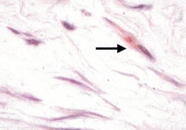
Application: Immunocytochemistry/ImmunofluorescenceSample Tested: fibroblastsSpecies: MouseVerified Customer | Posted 08/18/2021

There are no reviews that match your criteria.
Protocols
Find general support by application which include: protocols, troubleshooting, illustrated assays, videos and webinars.
- 7-Amino Actinomycin D (7-AAD) Cell Viability Flow Cytometry Protocol
- Antigen Retrieval Protocol (PIER)
- Antigen Retrieval for Frozen Sections Protocol
- Appropriate Fixation of IHC/ICC Samples
- Cellular Response to Hypoxia Protocols
- Chromogenic IHC Staining of Formalin-Fixed Paraffin-Embedded (FFPE) Tissue Protocol
- Chromogenic Immunohistochemistry Staining of Frozen Tissue
- ClariTSA™ Fluorophore Kits
- Detection & Visualization of Antibody Binding
- Extracellular Membrane Flow Cytometry Protocol
- Flow Cytometry Protocol for Cell Surface Markers
- Flow Cytometry Protocol for Staining Membrane Associated Proteins
- Flow Cytometry Staining Protocols
- Flow Cytometry Troubleshooting Guide
- Fluorescent IHC Staining of Frozen Tissue Protocol
- Graphic Protocol for Heat-induced Epitope Retrieval
- Graphic Protocol for the Preparation and Fluorescent IHC Staining of Frozen Tissue Sections
- Graphic Protocol for the Preparation and Fluorescent IHC Staining of Paraffin-embedded Tissue Sections
- Graphic Protocol for the Preparation of Gelatin-coated Slides for Histological Tissue Sections
- IHC Sample Preparation (Frozen sections vs Paraffin)
- Immunofluorescent IHC Staining of Formalin-Fixed Paraffin-Embedded (FFPE) Tissue Protocol
- Immunohistochemistry (IHC) and Immunocytochemistry (ICC) Protocols
- Immunohistochemistry Frozen Troubleshooting
- Immunohistochemistry Paraffin Troubleshooting
- Intracellular Flow Cytometry Protocol Using Alcohol (Methanol)
- Intracellular Flow Cytometry Protocol Using Detergents
- Intracellular Nuclear Staining Flow Cytometry Protocol Using Detergents
- Intracellular Staining Flow Cytometry Protocol Using Alcohol Permeabilization
- Intracellular Staining Flow Cytometry Protocol Using Detergents to Permeabilize Cells
- Preparing Samples for IHC/ICC Experiments
- Preventing Non-Specific Staining (Non-Specific Binding)
- Primary Antibody Selection & Optimization
- Propidium Iodide Cell Viability Flow Cytometry Protocol
- Protocol for Heat-Induced Epitope Retrieval (HIER)
- Protocol for Liperfluo
- Protocol for Making a 4% Formaldehyde Solution in PBS
- Protocol for VisUCyte™ HRP Polymer Detection Reagent
- Protocol for the Characterization of Human Th22 Cells
- Protocol for the Characterization of Human Th9 Cells
- Protocol for the Preparation & Fixation of Cells on Coverslips
- Protocol for the Preparation and Chromogenic IHC Staining of Frozen Tissue Sections
- Protocol for the Preparation and Chromogenic IHC Staining of Frozen Tissue Sections - Graphic
- Protocol for the Preparation and Chromogenic IHC Staining of Paraffin-embedded Tissue Sections
- Protocol for the Preparation and Chromogenic IHC Staining of Paraffin-embedded Tissue Sections - Graphic
- Protocol for the Preparation and Fluorescent IHC Staining of Frozen Tissue Sections
- Protocol for the Preparation and Fluorescent IHC Staining of Paraffin-embedded Tissue Sections
- Protocol for the Preparation of Gelatin-coated Slides for Histological Tissue Sections
- Protocol: Annexin V and PI Staining by Flow Cytometry
- Protocol: Annexin V and PI Staining for Apoptosis by Flow Cytometry
- TUNEL and Active Caspase-3 Detection by IHC/ICC Protocol
- The Importance of IHC/ICC Controls
- Troubleshooting Guide: Fluorokine Flow Cytometry Kits
- Troubleshooting Guide: Immunohistochemistry
- View all Protocols, Troubleshooting, Illustrated assays and Webinars
Loading...
Associated Pathways
