
EDAR is a type I transmembrane protein which is a member of the TNF Receptor Superfamily (TNFRSF). The extracellular domain contains 14 cysteine residues, six of which approximate the TNFRSF cysteine-rich region, the cytoplasmic domain contains a region with homology to the death domains found in other TNFRSF members. Mouse EDAR is a 488 amino acid (aa) protein with a predicted 30 aa signal, a 159 aa extracellular domain, a 22 aa transmembrane domain, and a 277 aa cytoplasmic domain. The human and mouse EDAR homologs share 91% identity. Within the TNFRSF, EDAR shares the highest homologies with XEDAR and TNFRSF19/TROY. EDA-A1 is the EDAR ligand. EDA and EDAR have been associated with hypohidrotic ectodermal dysplasia (HED). HED is characterized by abnormalities in hair, teeth and eccrine sweat gland morphogenesis. HED was initially found to associate with two gene loci, tabby and downless. Tabby was later identified as the gene for EDA and downless as the autosomal EDAR gene. EDA has two splice variants, EDA-A1 and EDA-A2 which differ by only two amino acids. Despite this minor difference, the EDA isoforms display strong receptor specificity. EDA-A1 only binds to EDAR, whereas EDA-A2 binds to XEDAR, an X-linked TNFRSF member with high homology to EDAR. Mutations in EDA, EDAR and XEDAR have been associated with HED.

Scientific images may be resized, cropped, or adjusted for brightness or contrast for presentation purposes. These updates are limited to presentation and do not affect the underlying scientific interpretation of the data. Where available, additional source data may be provided upon request.
Key Product Details
Species Reactivity
Validated:
Cited:
Applications
Validated:
Cited:
Label
Antibody Source
Product Specifications
Immunogen
Glu27-Ala187
Accession # Q9R187
Specificity
Clonality
Host
Isotype
Endotoxin Level
Scientific Data Images for Mouse EDAR Antibody
Detection of EDAR by Immunohistochemistry
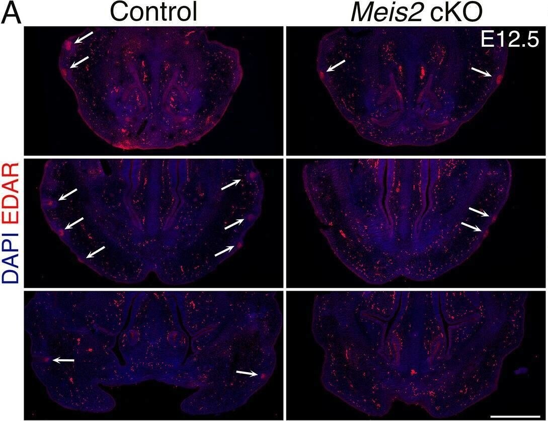
EDAR and LEF1 expression in Meis2 cKO mice.(A) Three examples of 10 μm FFPE sections stained with EDAR antibody showing affected expression of EDAR in Meis2 cKO mice. Arrows indicate EDAR-positive sites which are observed less frequently in the mutant snout. Scale bar: 500 μm. (B) Two representative images of normally developed escaper whisker follicles (WFs) in the mutants by LEF1 staining. LEF1 expression is similar in just forming placodes (top) and invaginated (bottom). Additionally, LEF1 expression shows a decline in the DC regions compared to peri-DC. Scale bar: 30 μm. (C) Three representative micrographs of LEF1-stained 10 μm FFPE snout sections showing normal expression of LEF1 in the dermis but the decreased number of LEF1+ placodes in the epithelium of the mutant snout. Scale bar: 500 μm. (D) Quantification of Lef1+ placode count and Lef1 fluorescence intensities in placodes, DCs, or interfollicular upper dermis. Data are presented as mean ± sem, ***p=0.0002. (E) In situ hybridization HCR-FISH using probes for Axin2 and Wnt10b in control snout at embryonic day 12.5 (E12.5) showing placodal expression of Wnt10b (green) and Axin2 (arrows, red) as well as widespread expression of Axin2 in the dermis. Scale bar: 60 μm. Image collected and cropped by CiteAb from the following open publication (https://elifesciences.org/articles/100854), licensed under a CC-BY license. Not internally tested by R&D Systems.Applications for Mouse EDAR Antibody
Blockade of Receptor-ligand Interaction
Western Blot
Sample: Recombinant Mouse EDAR Fc Chimera (Catalog # 745-ED)
Formulation, Preparation, and Storage
Purification
Reconstitution
Reconstitute at 0.2 mg/mL in sterile PBS. For liquid material, refer to CoA for concentration.
Formulation
Shipping
Stability & Storage
- 12 months from date of receipt, -20 to -70 °C as supplied.
- 1 month, 2 to 8 °C under sterile conditions after reconstitution.
- 6 months, -20 to -70 °C under sterile conditions after reconstitution.
Calculators
Background: EDAR
References
- Headon, D.J. and Overbeek, P.A. (1999) Nat. Genet. 22:370.
- Kumar, A. et al. (2000) J. Biol. Chem. 276:2668.
- Monreal, A.W. et al. (1999) Nat. Genet. 22:366.
- Schneider, P. et al. (2001) J. Biol. Chem. 276:18819.
- Srivastava, A.K. et al. (1997) Proc. Natl. Acad. Sci. USA 94:13069.
- Yan, M. et al. (2000) Science 209:523.
Long Name
Alternate Names
Gene Symbol
UniProt
Additional EDAR Products
Product Documents for Mouse EDAR Antibody
Certificate of Analysis
To download a Certificate of Analysis, please enter a lot or batch number in the search box below.
Note: Certificate of Analysis not available for kit components.
Product Specific Notices for Mouse EDAR Antibody
For research use only
Related Research Areas
Citations for Mouse EDAR Antibody
 Powered by Bioz
Customer Reviews for Mouse EDAR Antibody
There are currently no reviews for this product. Be the first to review Mouse EDAR Antibody and earn rewards!
Have you used Mouse EDAR Antibody?
Submit a review and receive an Amazon gift card!
$25/€18/£15/$25CAN/¥2500 for a review with an image
$10/€7/£6/$10CAN/¥1110 for a review without an image
Submit a review
Protocols
Find general support by application which include: protocols, troubleshooting, illustrated assays, videos and webinars.
- Cellular Response to Hypoxia Protocols
- R&D Systems Quality Control Western Blot Protocol
- Troubleshooting Guide: Western Blot Figures
- Western Blot Conditions
- Western Blot Protocol
- Western Blot Protocol for Cell Lysates
- Western Blot Troubleshooting
- Western Blot Troubleshooting Guide
- View all Protocols, Troubleshooting, Illustrated assays and Webinars
Associated Pathways
