
Mouse OCAM/NCAM2 Antibody
R&D Systems | Catalog # AF778

Scientific images may be resized, cropped, or adjusted for brightness or contrast for presentation purposes. These updates are limited to presentation and do not affect the underlying scientific interpretation of the data. Where available, additional source data may be provided upon request.
Key Product Details
Species Reactivity
Validated:
Cited:
Applications
Validated:
Cited:
Label
Antibody Source
Product Specifications
Immunogen
Leu20-Gly700
Accession # O35136
Specificity
Clonality
Host
Isotype
Scientific Data Images for Mouse OCAM/NCAM2 Antibody
Detection of OCAM/NCAM2 by Immunohistochemistry
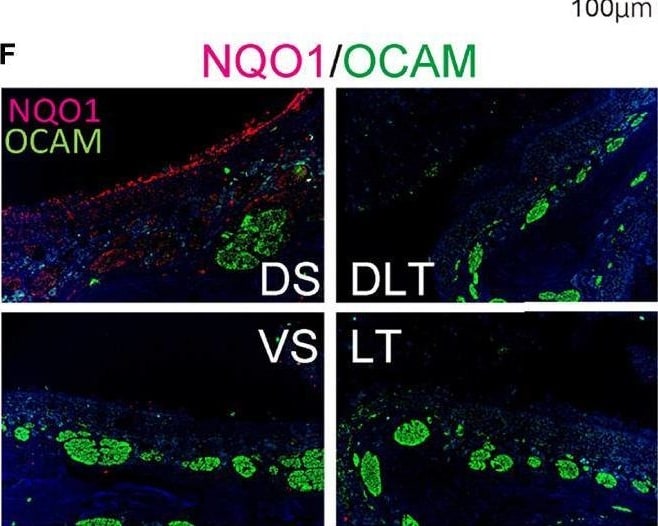
Infectious findings in the lungs and nasal cavity. (A) Severe acute respiratory syndrome coronavirus 2 (SARS-CoV-2) gene detection with RT- quantitative polymerase chain reaction (*P < 0.05). (B) Immunohistochemistry staining of SARS-CoV-2 in the lung (shown in brown, arrow heads). (C, D) Representative images of the olfactory epithelium from control hamsters (C: hematoxylin and eosin stain, D: immunohistological staining of NQO1 and OCAM). The boxes in (C) indicate the regions of the olfactory epithelium shown in D; the dorsal nasal septum (DS) area, ventral nasal septum (VS) area, dorsal lateral turbinate (DLT) area, and lateral turbinate (LT) area. Only the DS area appears positive for NQO1. (E, F) Representative images of the olfactory epithelium from SARS-CoV-2 hamsters. (G) Immunohistochemistry staining of the olfactory epithelium in SARS-CoV-2 hamsters. The virus is mainly present in the lateral region of the nasal cavity. (H) Double stained images of SARS-CoV-2 and OMP in each region of the olfactory epithelium. No double positive cells for SARS-CoV-2 and OMP are present in the olfactory epithelium. The arrow heads show SARS-CoV-2 positive cells. Image collected and cropped by CiteAb from the following open publication (https://www.frontiersin.org/articles/10.3389/fcimb.2022.924725/full), licensed under a CC-BY license. Not internally tested by R&D Systems.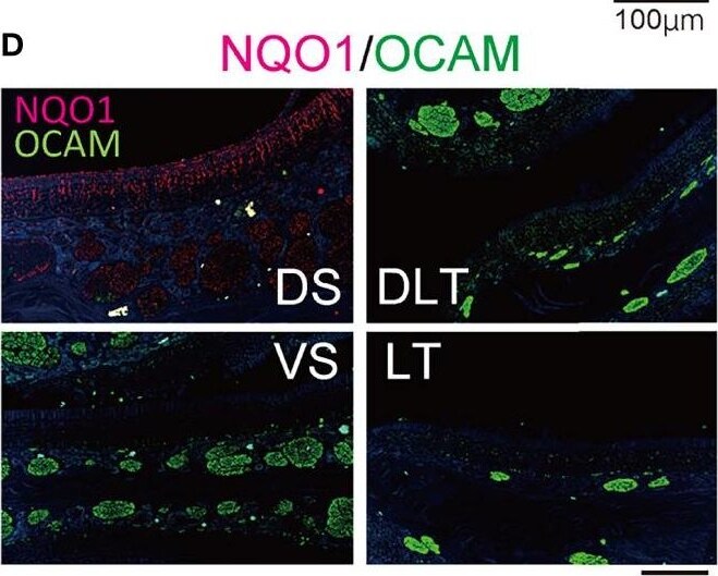
Detection of OCAM/NCAM2 by Immunohistochemistry
Infectious findings in the lungs and nasal cavity. (A) Severe acute respiratory syndrome coronavirus 2 (SARS-CoV-2) gene detection with RT- quantitative polymerase chain reaction (*P < 0.05). (B) Immunohistochemistry staining of SARS-CoV-2 in the lung (shown in brown, arrow heads). (C, D) Representative images of the olfactory epithelium from control hamsters (C: hematoxylin and eosin stain, D: immunohistological staining of NQO1 and OCAM). The boxes in (C) indicate the regions of the olfactory epithelium shown in D; the dorsal nasal septum (DS) area, ventral nasal septum (VS) area, dorsal lateral turbinate (DLT) area, and lateral turbinate (LT) area. Only the DS area appears positive for NQO1. (E, F) Representative images of the olfactory epithelium from SARS-CoV-2 hamsters. (G) Immunohistochemistry staining of the olfactory epithelium in SARS-CoV-2 hamsters. The virus is mainly present in the lateral region of the nasal cavity. (H) Double stained images of SARS-CoV-2 and OMP in each region of the olfactory epithelium. No double positive cells for SARS-CoV-2 and OMP are present in the olfactory epithelium. The arrow heads show SARS-CoV-2 positive cells. Image collected and cropped by CiteAb from the following open publication (https://www.frontiersin.org/articles/10.3389/fcimb.2022.924725/full), licensed under a CC-BY license. Not internally tested by R&D Systems.Applications for Mouse OCAM/NCAM2 Antibody
Western Blot
Sample: Recombinant Mouse OCAM
Formulation, Preparation, and Storage
Purification
Reconstitution
Reconstitute at 0.2 mg/mL in sterile PBS. For liquid material, refer to CoA for concentration.
Formulation
Shipping
Stability & Storage
- 12 months from date of receipt, -20 to -70 °C as supplied.
- 1 month, 2 to 8 °C under sterile conditions after reconstitution.
- 6 months, -20 to -70 °C under sterile conditions after reconstitution.
Calculators
Background: OCAM/NCAM2
Long Name
Alternate Names
Gene Symbol
UniProt
Additional OCAM/NCAM2 Products
Product Documents for Mouse OCAM/NCAM2 Antibody
Certificate of Analysis
To download a Certificate of Analysis, please enter a lot or batch number in the search box below.
Note: Certificate of Analysis not available for kit components.
Product Specific Notices for Mouse OCAM/NCAM2 Antibody
For research use only
Related Research Areas
Citations for Mouse OCAM/NCAM2 Antibody
 Powered by Bioz
Customer Reviews for Mouse OCAM/NCAM2 Antibody
There are currently no reviews for this product. Be the first to review Mouse OCAM/NCAM2 Antibody and earn rewards!
Have you used Mouse OCAM/NCAM2 Antibody?
Submit a review and receive an Amazon gift card!
$25/€18/£15/$25CAN/¥2500 for a review with an image
$10/€7/£6/$10CAN/¥1110 for a review without an image
Submit a review
Protocols
Find general support by application which include: protocols, troubleshooting, illustrated assays, videos and webinars.
- Cellular Response to Hypoxia Protocols
- R&D Systems Quality Control Western Blot Protocol
- Troubleshooting Guide: Western Blot Figures
- Western Blot Conditions
- Western Blot Protocol
- Western Blot Protocol for Cell Lysates
- Western Blot Troubleshooting
- Western Blot Troubleshooting Guide
- View all Protocols, Troubleshooting, Illustrated assays and Webinars