
Siglecs (1) (sialic acid binding Ig-like lectins) are I-type (Ig-type) lectins (2) belonging to the Ig superfamily. They are characterized by an N-terminal Ig-like V-type domain which mediates sialic acid binding (3), followed by varying numbers of Ig-like C2-type domains (1, 4). Eleven human Siglecs have been cloned and characterized (1, 4). They are sialoadhesin/CD169/Siglec-1, CD22/Siglec-2, CD33/Siglec-3, Myelin-Associated Glycoprotein (MAG/Siglec-4a) and Siglec 5 to 11 (4‑6). To date, no Siglec has been shown to recognized any cell surface ligand other than sialic acids, suggesting that interactions with glycans containing this carbohydrate are important in mediating the biological functions of Siglecs. Siglec 5 to 11 share a high degree of sequence similarity with CD33/Siglec-3 both in their extracellular and intracellular regions. They are collectively referred to as CD33-related Siglecs. One remarkable feature of the CD33-related Siglecs is their differential expression pattern within the hematopoietic system (4, 5). This fact, together with the presence of two conserved immunoreceptor tyrosine-based inhibition motifs (ITIMs) in their cytoplasmic tails, suggests that CD33-related Siglecs are involved in the regulation of cellular activation within the immune system. Mouse Siglec-F cDNA encodes a 569 amino acid polypeptide with a hydrophobic signal peptide, an N-terminal Ig-like V-type domain, three Ig-like C2-type domains, a transmembrane region and a cytoplasmic tail (7). The expression of Siglec-F is restricted to the cells of myelomonocytic lineage. Mouse Siglec-F is likely an ortholog of human Siglec-5. Unlike many human CD33-related Siglecs, which show similar binding to both alpha 2,3- and alpha 2,6-linked sialic acids, mouse Siglec-F preferentially recognize alpha 2,3-linked sialic acid.
Mouse Siglec-F Antibody
R&D Systems | Catalog # AF1706

Scientific images may be resized, cropped, or adjusted for brightness or contrast for presentation purposes. These updates are limited to presentation and do not affect the underlying scientific interpretation of the data. Where available, additional source data may be provided upon request.
Key Product Details
Species Reactivity
Validated:
Cited:
Applications
Validated:
Cited:
Label
Antibody Source
Product Specifications
Immunogen
Asp18-Thr437
Accession # Q920G3
Specificity
Clonality
Host
Isotype
Scientific Data Images for Mouse Siglec-F Antibody
Detection of Siglec-F by Flow Cytometry
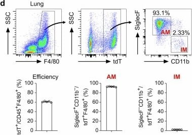
Generation and characterization of the CD68-rtTA mouse line.a Schematic diagram showing the knock-in strategy of the CD68-rtTA line by homologous recombination. b Genetic lineage tracing strategy by Cre-loxP recombination in CD68+ cells after doxycycline (Dox) treatment. c Schematic showing the experimental design. d Flow cytometric and quantification analyses of the percentage of tdT+ cells among CD45+F4/80+ macrophages (efficiency) and the percentage of SiglecF+ alveolar macrophages (AMs, left) and CD11b+ interstitial macrophages (IMs, right) among tdT+ macrophages from the lungs. Data are presented as the mean ± SEM; n = 5 mice per group. e Flow cytometric and quantification analyses of the percentage of tdT+ cells expressing F4/80 (specificity) in the lungs. Data are presented as the mean ± SEM; n = 5 mice per group. f Flow cytometric and quantification analyses of the percentage of CD45+ monocytes expressing tdT from blood. Data are presented as the mean ± SEM; n = 5 mice per group. g Immunostaining for F4/80, tdT, and SiglecF in lung tissue sections. The boxed region is magnified. Yellow arrowheads indicate labeled macrophages. h Quantitative analysis of the percentage of tdT+ cells among F4/80+ macrophages (efficiency), the percentage of F4/80+ cells among tdT+ cells (specificity), and the percentage of AMs and IMs among labeled tdT+ macrophages from images of immunostained lung sections. Data are presented as the mean ± SEM; n = 5 mice per group. i Cartoon image showing the effective labeling of tissue-resident macrophages (TRMs) in CD68-rtTA;TetO-Cre;R26-tdT mice. Scale bars, 100 µm. Each image is representative of five individual samples. Image collected and cropped by CiteAb from the following open publication (https://www.nature.com/articles/s41421-025-00859-0), licensed under a CC-BY license. Not internally tested by R&D Systems.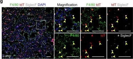
Detection of Siglec-F by Immunohistochemistry
Generation and characterization of the CD68-rtTA mouse line.a Schematic diagram showing the knock-in strategy of the CD68-rtTA line by homologous recombination. b Genetic lineage tracing strategy by Cre-loxP recombination in CD68+ cells after doxycycline (Dox) treatment. c Schematic showing the experimental design. d Flow cytometric and quantification analyses of the percentage of tdT+ cells among CD45+F4/80+ macrophages (efficiency) and the percentage of SiglecF+ alveolar macrophages (AMs, left) and CD11b+ interstitial macrophages (IMs, right) among tdT+ macrophages from the lungs. Data are presented as the mean ± SEM; n = 5 mice per group. e Flow cytometric and quantification analyses of the percentage of tdT+ cells expressing F4/80 (specificity) in the lungs. Data are presented as the mean ± SEM; n = 5 mice per group. f Flow cytometric and quantification analyses of the percentage of CD45+ monocytes expressing tdT from blood. Data are presented as the mean ± SEM; n = 5 mice per group. g Immunostaining for F4/80, tdT, and SiglecF in lung tissue sections. The boxed region is magnified. Yellow arrowheads indicate labeled macrophages. h Quantitative analysis of the percentage of tdT+ cells among F4/80+ macrophages (efficiency), the percentage of F4/80+ cells among tdT+ cells (specificity), and the percentage of AMs and IMs among labeled tdT+ macrophages from images of immunostained lung sections. Data are presented as the mean ± SEM; n = 5 mice per group. i Cartoon image showing the effective labeling of tissue-resident macrophages (TRMs) in CD68-rtTA;TetO-Cre;R26-tdT mice. Scale bars, 100 µm. Each image is representative of five individual samples. Image collected and cropped by CiteAb from the following open publication (https://www.nature.com/articles/s41421-025-00859-0), licensed under a CC-BY license. Not internally tested by R&D Systems.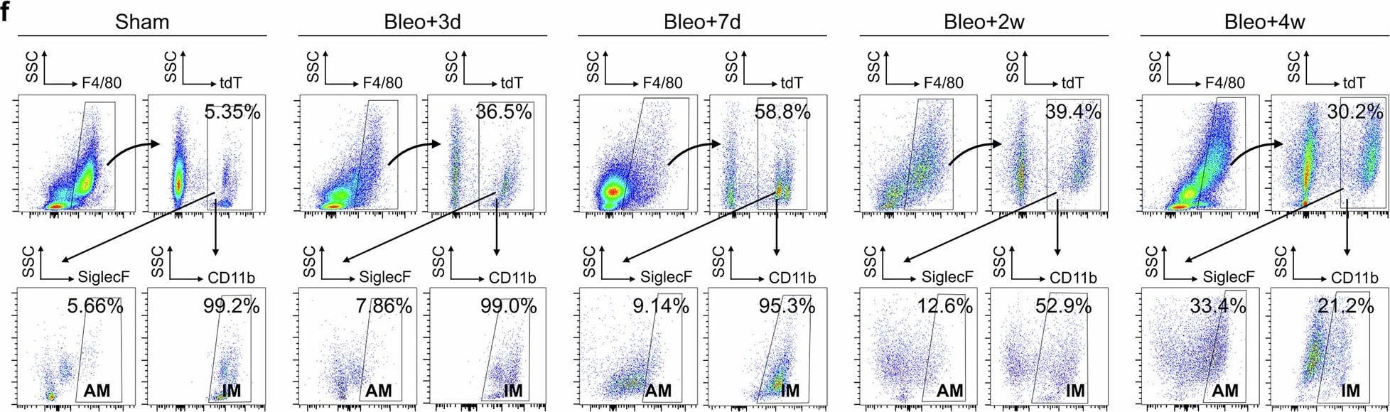
Detection of Siglec-F by Flow Cytometry
MDMs significantly increased following bleomycin-induced injury.a Schematic diagram showing the experimental design. Tam tamoxifen, Bleo bleomycin. b Whole-mount bright field, epifluorescence, and Sirius red images of lungs after bleomycin treatment. The boxed region is magnified. c Immunostaining for tdT, F4/80, and SiglecF in lung tissue sections. The boxed region is magnified. White arrowheads indicate MDMs. d Quantitative analysis of the percentage of F4/80+ macrophages expressing tdT (left) and the percentage of alveolar macrophages among labeled tdT+ macrophages (right) in immunostained lung sections. Data are presented as the mean ± SEM; n = 5 mice per group. e Quantification of the numbers of MDMs (tdT+F4/80+) and Mo-AMs (tdT+SiglecF+) in each 10× field from immunostained lung sections. Data are presented as the mean ± SEM; n = 5 mice per group. f, g Flow cytometric and quantification analyses of the percentage of tdT+ cells among CD45+F4/80+ macrophages (MDMs), the percentage of SiglecF+ alveolar macrophages among tdT+ macrophages (Mo-AMs), and the percentage of CD11b+ interstitial macrophages among tdT+ macrophages (Mo-IMs) from the lungs. Data are presented as the mean ± SEM; n = 5 mice per group. h Cartoon image showing the dynamic changes in the number of recruited macrophages in the lungs after bleomycin-induced injury. MDMs monocyte-derived macrophages, Mo-AMs monocyte-derived alveolar macrophages, Mo-IMs monocyte-derived interstitial macrophages. Scale bars, yellow, 1 mm; white and black, 100 µm. Each image is representative of five individual samples. Image collected and cropped by CiteAb from the following open publication (https://www.nature.com/articles/s41421-025-00859-0), licensed under a CC-BY license. Not internally tested by R&D Systems.Applications for Mouse Siglec-F Antibody
Western Blot
Sample: Recombinant Mouse Siglec-F Fc Chimera (Catalog # 1706-SF)
Formulation, Preparation, and Storage
Purification
Reconstitution
Reconstitute at 0.2 mg/mL in sterile PBS. For liquid material, refer to CoA for concentration.
Formulation
Shipping
Stability & Storage
- 12 months from date of receipt, -20 to -70 °C as supplied.
- 1 month, 2 to 8 °C under sterile conditions after reconstitution.
- 6 months, -20 to -70 °C under sterile conditions after reconstitution.
Calculators
Background: Siglec-F
References
- Crocker, P.R. et al. (1998) Glycobiology 8:v.
- Powell, L.D. et al. (1995) J. Biol. Chem. 270:14243.
- May, A.R. et al. (1998) Mol. Cell 1998. 1:719.
- Crocker, P.R. and A. Varki (2001) Trends Immunol. 22:337.
- Crocker, P.R. et al. (2001) Immunology 103:137.
- Angata, T. et al. (2002) J. Biol Chem. 277:24466.
- Angata, T. et al. (2001) J. Biol Chem. 276:45128.
Long Name
Alternate Names
Entrez Gene IDs
Gene Symbol
UniProt
Additional Siglec-F Products
Product Documents for Mouse Siglec-F Antibody
Certificate of Analysis
To download a Certificate of Analysis, please enter a lot or batch number in the search box below.
Note: Certificate of Analysis not available for kit components.
Product Specific Notices for Mouse Siglec-F Antibody
For research use only
Citations for Mouse Siglec-F Antibody
 Powered by Bioz
Customer Reviews for Mouse Siglec-F Antibody
There are currently no reviews for this product. Be the first to review Mouse Siglec-F Antibody and earn rewards!
Have you used Mouse Siglec-F Antibody?
Submit a review and receive an Amazon gift card!
$25/€18/£15/$25CAN/¥2500 for a review with an image
$10/€7/£6/$10CAN/¥1110 for a review without an image
Submit a review
Protocols
Find general support by application which include: protocols, troubleshooting, illustrated assays, videos and webinars.
- Cellular Response to Hypoxia Protocols
- R&D Systems Quality Control Western Blot Protocol
- Troubleshooting Guide: Western Blot Figures
- Western Blot Conditions
- Western Blot Protocol
- Western Blot Protocol for Cell Lysates
- Western Blot Troubleshooting
- Western Blot Troubleshooting Guide
- View all Protocols, Troubleshooting, Illustrated assays and Webinars