The type I transmembrane glycoprotein Signaling Lymphocytic Activation Molecule (SLAM), also known as CD150, is the prototypic member of the SLAM subgroup of the CD2 protein family. CD2 family proteins function as adhesion molecules and modulators of the immune response (1). Mouse SLAM consists of a 218 amino acid (aa) extracellular domain (ECD) with two Ig-like domains, a 23 aa transmembrane segment, and a 78 aa cytoplasmic domain with three immunoreceptor tyrosine switch motifs (ITSM) (2). Alternate splicing generates an isoform with a substituted cytoplasmic domain (2). Within the ECD, mouse SLAM shares 58% and 83% aa sequence identity with human and rat SLAM, respectively. It is expressed as a 75 kDa molecule of which approximately 30 kDa is N-linked carbohydrate (2). SLAM is expressed on T cells, B cells, thymocytes, macrophages, dendritic cells, platelets, and hematopoietic stem cells (2‑7). It is upregulated on activated B cells and CD4+ and CD8+ T cells, although it is downregulated on Th2 polarized cells (2, 3, 8). SLAM interacts homophilically with low affinity, and this interaction induces a Th0/Th1 response characterized by clonal expansion, production of IFN-gamma, and increased cytolytic activity of CD8+ T cells (2, 3, 9‑11). SLAM ligation also promotes B cell activation, allergen-induced eosinophil and mast cell activation, and macrophage responsiveness to LPS (4, 8, 12). In humans, SLAM functions as a cellular entry receptor for measles virus (13, 14).

Key Product Details
Validated by
Biological Validation
Species Reactivity
Mouse
Applications
Western Blot, Flow Cytometry, CyTOF-ready
Label
Unconjugated
Antibody Source
Polyclonal Sheep IgG
Loading...
Product Specifications
Immunogen
Mouse myeloma cell line NS0-derived recombinant mouse SLAM/CD150
Thr25-Pro242
Accession # Q9QUM4
Thr25-Pro242
Accession # Q9QUM4
Specificity
Detects mouse SLAM/CD150 in direct ELISAs and Western blots. In direct ELISAs, less than 1% cross-reactivity with recombinant human SLAM is observed.
Clonality
Polyclonal
Host
Sheep
Isotype
IgG
Scientific Data Images for Mouse SLAM/CD150 Antibody
Detection of SLAM/CD150 in Mouse Splenocytes by Flow Cytometry.
Mouse splenocytes were treated for 2 days with 50 ng/mL PMA and 500 ng/mL Ca2+ionomycin then stained with Sheep Anti-Mouse SLAM/CD150 Antigen Affinity-purified Poly-clonal Antibody (Catalog # AF4330, filled histogram) or isotype control antibody (Catalog # 5-001-A, open histogram), followed by NorthernLights™ 637-conjugated Anti-Sheep IgG Secondary Antibody (Catalog # NL011).Applications for Mouse SLAM/CD150 Antibody
Application
Recommended Usage
CyTOF-ready
Ready to be labeled using established conjugation methods. No BSA or other carrier proteins that could interfere with conjugation.
Flow Cytometry
0.25 µg/106 cells
Sample: Mouse splenocytes treated with PMA and Ca2+ ionomycin
Sample: Mouse splenocytes treated with PMA and Ca2+ ionomycin
Western Blot
0.1 µg/mL
Sample: Recombinant Mouse SLAM/CD150 (Catalog # 4330-SL)
Sample: Recombinant Mouse SLAM/CD150 (Catalog # 4330-SL)
Reviewed Applications
Read 1 review rated 4 using AF4330 in the following applications:
Flow Cytometry Panel Builder
Bio-Techne Knows Flow Cytometry
Save time and reduce costly mistakes by quickly finding compatible reagents using the Panel Builder Tool.
Advanced Features
- Spectra Viewer - Custom analysis of spectra from multiple fluorochromes
- Spillover Popups - Visualize the spectra of individual fluorochromes
- Antigen Density Selector - Match fluorochrome brightness with antigen density

Formulation, Preparation, and Storage
Purification
Antigen Affinity-purified
Reconstitution
Reconstitute at 0.2 mg/mL in sterile PBS. For liquid material, refer to CoA for concentration.
Loading...
Formulation
Lyophilized from a 0.2 μm filtered solution in PBS with Trehalose. *Small pack size (SP) is supplied either lyophilized or as a 0.2 µm filtered solution in PBS.
Shipping
Lyophilized product is shipped at ambient temperature. Liquid small pack size (-SP) is shipped with polar packs. Upon receipt, store immediately at the temperature recommended below.
Stability & Storage
Use a manual defrost freezer and avoid repeated freeze-thaw cycles.
- 12 months from date of receipt, -20 to -70 °C as supplied.
- 1 month, 2 to 8 °C under sterile conditions after reconstitution.
- 6 months, -20 to -70 °C under sterile conditions after reconstitution.
Calculators
Background: SLAM/CD150
References
- Ma, C.S. et al. (2007) Annu. Rev. Immunol. 25:337.
- Castro, A.G. et al. (1999) J. Immunol. 163:5860.
- Cocks, B.G. et al. (1995) Nature 376:260.
- Wang, N. et al. (2004) J. Exp. Med. 199:1255.
- Hahm, B. et al. (2004) Virology 323:292.
- Nanda, N. et al. (2005) Blood 106:3028.
- Kiel, M.J. et al. (2005) Cell 121:1109.
- Punnonen, J. et al. (1997) J. Exp. Med. 185:993.
- Mavaddat, N. et al. (2000) J. Biol. Chem. 275:28100.
- Aversa, G. et al. (1997) J. Immunol. 158:4036.
- Mehrle, S. et al. (2008) Mol. Immunol. 45:796.
- Wang, N. et al. (2006) Am. J. Respir. Cell Mol. Biol. 35:206.
- Tatsuo, H. et al. (2000) Nature 406:893.
- Hsu, E.C. et al. (2001) Virology 279:9.
Long Name
Signaling Lymphocytic Activation Molecule
Alternate Names
CD150, IPO-3, SLAMF1
Gene Symbol
SLAMF1
UniProt
Additional SLAM/CD150 Products
Product Documents for Mouse SLAM/CD150 Antibody
Certificate of Analysis
To download a Certificate of Analysis, please enter a lot or batch number in the search box below.
Note: Certificate of Analysis not available for kit components.
Product Specific Notices for Mouse SLAM/CD150 Antibody
For research use only
Related Research Areas
Citations for Mouse SLAM/CD150 Antibody
 Powered by Bioz
Customer Reviews for Mouse SLAM/CD150 Antibody (1)
4 out of 5
1 Customer Rating
Have you used Mouse SLAM/CD150 Antibody?
Submit a review and receive an Amazon gift card!
$25/€18/£15/$25CAN/¥2500 Yen for a review with an image
$10/€7/£6/$10CAN/¥1110 Yen for a review without an image
Submit a review
Customer Images
Showing
1
-
1 of
1 review
Showing All
Filter By:
-
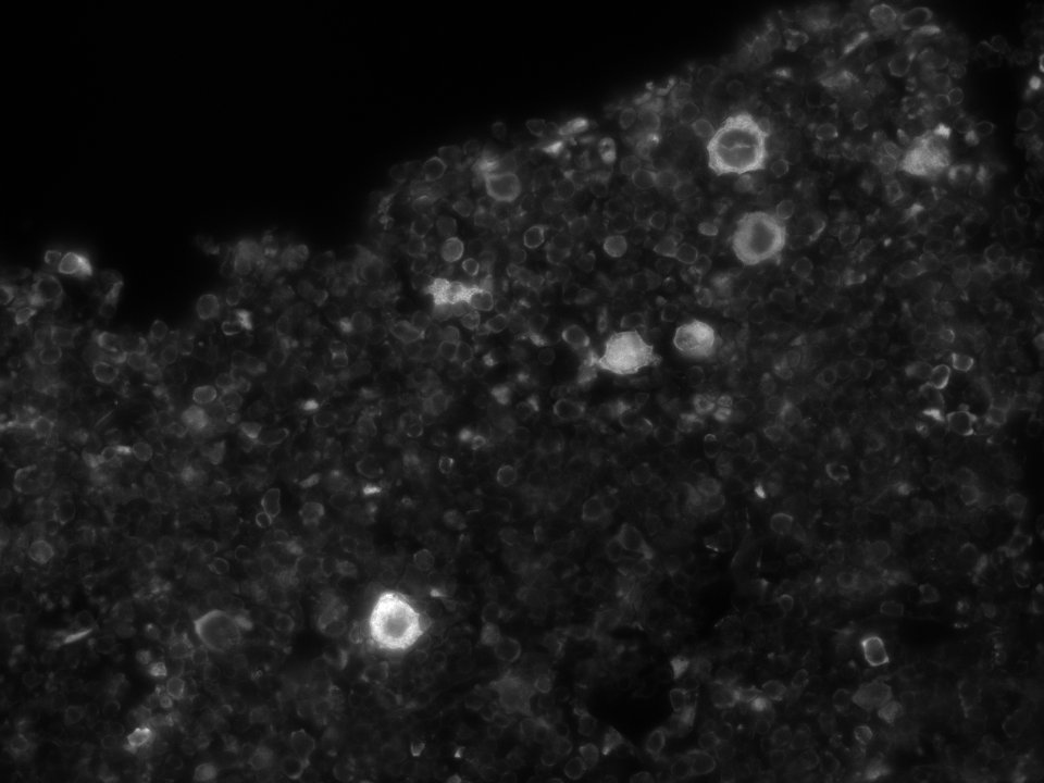
Application: Immunofluorescence - fixed-frozenSample Tested: bone marrowSpecies: MouseVerified Customer | Posted 10/21/2018Positivity in Megakaryocytes and few progenitors. acceptable background.Whole Femur Section, Fixed-frozen and decalcified

There are no reviews that match your criteria.
Protocols
Find general support by application which include: protocols, troubleshooting, illustrated assays, videos and webinars.
- 7-Amino Actinomycin D (7-AAD) Cell Viability Flow Cytometry Protocol
- Cellular Response to Hypoxia Protocols
- Extracellular Membrane Flow Cytometry Protocol
- Flow Cytometry Protocol for Cell Surface Markers
- Flow Cytometry Protocol for Staining Membrane Associated Proteins
- Flow Cytometry Staining Protocols
- Flow Cytometry Troubleshooting Guide
- Intracellular Flow Cytometry Protocol Using Alcohol (Methanol)
- Intracellular Flow Cytometry Protocol Using Detergents
- Intracellular Nuclear Staining Flow Cytometry Protocol Using Detergents
- Intracellular Staining Flow Cytometry Protocol Using Alcohol Permeabilization
- Intracellular Staining Flow Cytometry Protocol Using Detergents to Permeabilize Cells
- Propidium Iodide Cell Viability Flow Cytometry Protocol
- Protocol for Liperfluo
- Protocol for the Characterization of Human Th22 Cells
- Protocol for the Characterization of Human Th9 Cells
- Protocol: Annexin V and PI Staining by Flow Cytometry
- Protocol: Annexin V and PI Staining for Apoptosis by Flow Cytometry
- R&D Systems Quality Control Western Blot Protocol
- Troubleshooting Guide: Fluorokine Flow Cytometry Kits
- Troubleshooting Guide: Western Blot Figures
- Western Blot Conditions
- Western Blot Protocol
- Western Blot Protocol for Cell Lysates
- Western Blot Troubleshooting
- Western Blot Troubleshooting Guide
- View all Protocols, Troubleshooting, Illustrated assays and Webinars
Loading...
Associated Pathways

