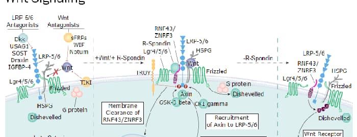
Wnt proteins are secreted glycoproteins with a conserved pattern of 23-24 cysteine residues that play critical roles in both carcinogenesis and embryonic development. Wnts bind to receptors of the Frizzled family, sometimes in conjunction with other membrane-associated proteins such as LRPs or proteoglycans. Downstream effects of Wnt signaling occur through different intracellular components, depending on which pathway is activated. Three pathways have been characterized: the canonical Wnt/ beta -catenin pathway, the Wnt/Ca2+ pathway, and the planar cell polarity (1-3). Wnt-5b is a 49 kDa glycoprotein of the subgroup of Wnts that is implicated in the Wnt/Ca2+ pathway (3-6). It is not axis-inducing in Xenopus embryos and only weakly transforms C57MG mammary epithelial cells. The non-canonical Wnt pathway can inhibit canonical Wnt/ beta -catenin signaling (3). Mouse Wnt-5b is synthesized as a 359 amino acid (aa) precursor that contains a 17 aa signal sequence and a 342 aa mature region. It is ubiquitously expressed at low but increasing levels throughout embryonic development (4, 5, 7). In adult mice, Wnt-5b is expressed in heart, liver, brain, lung, testes, kidney, and pancreas (4, 8). Wnt-5b appears to promote adipogenesis. It is upregulated in early adipogenesis. Also, Wnt-5b overexpression in 3T3-L1 cells partially inhibits canonical Wnt suppression of adipogenesis (9, 10). Human Wnt-5b polymorphisms have been associated with Type II diabetes (9). Although Wnt-5a and Wnt-5b share 83% aa identity, they show differential expression and regulation of cyclin D1 and p130 during endochondral bone development. Together, they appear to coordinate chondrocyte proliferation and differentiation (11). Mature mouse Wnt-5b shows 94%, 98%, 90%, and 88% aa sequence identity with mature human, rat, chick, and Xenopus Wnt-5b, respectively.

Key Product Details
Species Reactivity
Mouse
Applications
Immunocytochemistry
Label
Unconjugated
Antibody Source
Monoclonal Rat IgG2A Clone # 325004
Loading...
Product Specifications
Immunogen
E. coli-derived recombinant mouse Wnt‑5b
Ser55-Ser95 and Lys246-Glu315 connected by a Gly-Ser linker
Accession # NP_033551
Ser55-Ser95 and Lys246-Glu315 connected by a Gly-Ser linker
Accession # NP_033551
Specificity
Detects mouse Wnt-5b in direct ELISAs. In direct ELISAs, no cross-reactivity with recombinant mouse Wnt-5a, -8a, -8b, -9b, -10a, -10b, or recombinant human Wnt-7b is observed.
Clonality
Monoclonal
Host
Rat
Isotype
IgG2A
Scientific Data Images for Mouse Wnt-5b Antibody (325004)
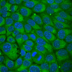
Wnt‑5b in NMuMG Mouse Cell Line.
Wnt-5b was detected in immersion fixed NMuMG mouse mammary gland epithelial cell line using Rat Anti-Mouse Wnt-5b Monoclonal Antibody (Catalog # MAB3006) at 10 µg/mL for 3 hours at room temperature. Cells were stained using the NorthernLights™ 493-conjugated Anti-Rat IgG Secondary Antibody (green; Catalog # NL015) and counterstained with DAPI (blue). Specific staining was localized to cytoplasm. View our protocol for Fluorescent ICC Staining of Cells on Coverslips.Applications for Mouse Wnt-5b Antibody (325004)
Application
Recommended Usage
Immunocytochemistry
8-25 µg/mL
Sample: Immersion fixed NMuMG mouse mammary gland epithelial cell line
Sample: Immersion fixed NMuMG mouse mammary gland epithelial cell line
Formulation, Preparation, and Storage
Purification
Protein A or G purified from hybridoma culture supernatant
Reconstitution
Reconstitute at 0.5 mg/mL in sterile PBS. For liquid material, refer to CoA for concentration.
Loading...
Formulation
Lyophilized from a 0.2 μm filtered solution in PBS with Trehalose. *Small pack size (SP) is supplied either lyophilized or as a 0.2 µm filtered solution in PBS.
Shipping
Lyophilized product is shipped at ambient temperature. Liquid small pack size (-SP) is shipped with polar packs. Upon receipt, store immediately at the temperature recommended below.
Stability & Storage
Use a manual defrost freezer and avoid repeated freeze-thaw cycles.
- 12 months from date of receipt, -20 to -70 °C as supplied.
- 1 month, 2 to 8 °C under sterile conditions after reconstitution.
- 6 months, -20 to -70 °C under sterile conditions after reconstitution.
Calculators
Background: Wnt-5b
References
- Miller, J.R. (2002) Genome Biol. 3:3001.
- Roelink, H. and R. Nusse (1991) Genes Dev. 5:381.
- Veeman, M.T. et al. (2003) Developmental Cell 5:367.
- Gavin, B. J. et al. (1990) Genes Dev. 4:2319.
- Nusse, R. and H.E. Varmus (1992) Cell 69:1073.
- Wong, G.T. et al. (1994) Mol. Cell. Biol. 14:6278.
- Kemp, C. et al. (2005) Dev. Dyn. 233:1064.
- Heller, R.S. et al. (2002) Dev. Dyn. 225:260.
- Kanazawa, A. et al. (2004) Am. J. Hum. Genet. 75:832.
- Kanazawa, A. et al. (2005) Biochem. Biophys. Res. Com. 330:505.
- Yang, Y. et al. (2003) Development 130:1003.
Long Name
Wingless-type MMTV Integration Site Family, Member 5b
Alternate Names
Wnt5b
Gene Symbol
WNT5B
UniProt
Additional Wnt-5b Products
Product Documents for Mouse Wnt-5b Antibody (325004)
Certificate of Analysis
To download a Certificate of Analysis, please enter a lot or batch number in the search box below.
Note: Certificate of Analysis not available for kit components.
Product Specific Notices for Mouse Wnt-5b Antibody (325004)
For research use only
Related Research Areas
Customer Reviews for Mouse Wnt-5b Antibody (325004)
There are currently no reviews for this product. Be the first to review Mouse Wnt-5b Antibody (325004) and earn rewards!
Have you used Mouse Wnt-5b Antibody (325004)?
Submit a review and receive an Amazon gift card!
$25/€18/£15/$25CAN/¥2500 for a review with an image
$10/€7/£6/$10CAN/¥1110 for a review without an image
Submit a review
Protocols
Find general support by application which include: protocols, troubleshooting, illustrated assays, videos and webinars.
- Appropriate Fixation of IHC/ICC Samples
- Cellular Response to Hypoxia Protocols
- ClariTSA™ Fluorophore Kits
- Detection & Visualization of Antibody Binding
- ICC Cell Smear Protocol for Suspension Cells
- ICC Immunocytochemistry Protocol Videos
- ICC for Adherent Cells
- Immunocytochemistry (ICC) Protocol
- Immunocytochemistry Troubleshooting
- Immunofluorescence of Organoids Embedded in Cultrex Basement Membrane Extract
- Immunohistochemistry (IHC) and Immunocytochemistry (ICC) Protocols
- Preparing Samples for IHC/ICC Experiments
- Preventing Non-Specific Staining (Non-Specific Binding)
- Primary Antibody Selection & Optimization
- Protocol for VisUCyte™ HRP Polymer Detection Reagent
- Protocol for the Fluorescent ICC Staining of Cell Smears - Graphic
- Protocol for the Fluorescent ICC Staining of Cultured Cells on Coverslips - Graphic
- Protocol for the Preparation and Fluorescent ICC Staining of Cells on Coverslips
- Protocol for the Preparation and Fluorescent ICC Staining of Non-adherent Cells
- Protocol for the Preparation and Fluorescent ICC Staining of Stem Cells on Coverslips
- Protocol for the Preparation of a Cell Smear for Non-adherent Cell ICC - Graphic
- TUNEL and Active Caspase-3 Detection by IHC/ICC Protocol
- The Importance of IHC/ICC Controls
- View all Protocols, Troubleshooting, Illustrated assays and Webinars