NCAM-1/CD56 Antibody (123C3.D5)
Novus Biologicals | Catalog # NBP2-15184
Key Product Details
Species Reactivity
Validated:
Human, Rat
Cited:
Rat
Applications
Validated:
Immunohistochemistry, Immunohistochemistry-Paraffin, Flow Cytometry, Flow (Cell Surface), Immunocytochemistry/ Immunofluorescence, Simple Western
Cited:
Immunohistochemistry-Paraffin
Label
Unconjugated
Antibody Source
Monoclonal Mouse IgG1 kappa Clone # 123C3.D5
Loading...
Product Specifications
Immunogen
Membrane preparation of a small cell lung carcinoma
Localization
Cell surface
Marker
Neuronal Cell Marker
Specificity
This monoclonal antibody reacts with an extracellular domain (close to transmembrane) of CD56/NCAM. Three isoforms of neural cell adhesion molecule (NCAM) are produced by differential splicing of the RNA transcript from a single gene. The 135kDa isoform is the basic molecule, which is glycosylated or sialylated to produce the mature species. Anti-CD56 recognizes two proteins of the neural cell adhesion molecule, the basic molecule expressed on most neuroectodermally derived tissues and neoplasms (e.g. retinoblastoma, medulloblastomas, astrocytomas, neuroblastomas, and small cell carcinomas). It is also expressed on some mesodermally derived tumors (rhabdomyosarcoma). Anti-CD56 plays an important role in the diagnosis of nodal and nasal NK/T-cell lymphomas.
Clonality
Monoclonal
Host
Mouse
Isotype
IgG1 kappa
Description
200ug/ml of antibody purified from Bioreactor Concentrate by Protein A or G. Prepared in 10 mM PBS with 0.05% BSA & 0.05% azide. Also available WITHOUT BSA & azide at 1.0 mg/ml. (NBP2-33132)
Antibody with azide - store at 2 to 8C. Antibody without azide - store at -20 to -80C.
Antibody with azide - store at 2 to 8C. Antibody without azide - store at -20 to -80C.
Scientific Data Images for NCAM-1/CD56 Antibody (123C3.D5)
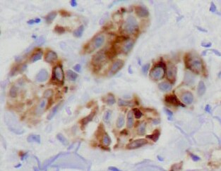
![Immunohistochemistry-Paraffin: NCAM-1/CD56 Antibody (123C3.D5) [NBP2-15184]](https://resources.rndsystems.com/images/products/NCAM-1-CD56-Antibody-123C3-D5-Immunohistochemistry-Paraffin-NBP2-15184-img0019.jpg "Immunohistochemistry-Paraffin: NCAM-1/CD56 Antibody (123C3.D5) [NBP2-15184]")
Immunohistochemistry-Paraffin: NCAM-1/CD56 Antibody (123C3.D5) [NBP2-15184]
Immunohistochemistry-Paraffin: NCAM-1/CD56 Antibody (123C3.D5) [NBP2-15184] - Analysis of NCAM-1/CD56 antibody (123C3.D5) on Human colon tissue. Incubation: 1 ug/ml for 30 minutes at room temperature. Image from verified customer review.![Flow Cytometry: NCAM-1/CD56 Antibody (123C3.D5) [NBP2-15184]](https://resources.rndsystems.com/images/products/NCAM-1-CD56-Antibody-123C3-D5-Flow-Cytometry-NBP2-15184-img0020.jpg "Flow Cytometry: NCAM-1/CD56 Antibody (123C3.D5) [NBP2-15184]")
Flow Cytometry: NCAM-1/CD56 Antibody (123C3.D5) [NBP2-15184]
Flow Cytometry: NCAM-1/CD56 Antibody (123C3.D5) [NBP2-15184] - Flow cytometry of lymphocyte gated PBMCs unstained (gray) orstained with CF568-labeled NCAM-1/CD56 antibody (123C3.D5) (orange).![Immunohistochemistry-Paraffin: NCAM-1/CD56 Antibody (123C3.D5) [NBP2-15184]](https://resources.rndsystems.com/images/products/NCAM-1-CD56-Antibody-123C3-D5-Immunohistochemistry-Paraffin-NBP2-15184-img0016.jpg "Immunohistochemistry-Paraffin: NCAM-1/CD56 Antibody (123C3.D5) [NBP2-15184]")
Immunohistochemistry-Paraffin: NCAM-1/CD56 Antibody (123C3.D5) [NBP2-15184]
Immunohistochemistry-Paraffin: NCAM-1/CD56 Antibody (123C3.D5) [NBP2-15184] - Formalin-fixed, paraffin-embedded human colon Ganglion stained with CD56 Monoclonal Antibody (123C3.D5)![Flow Cytometry: NCAM-1/CD56 Antibody (123C3.D5) [NBP2-15184]](https://resources.rndsystems.com/images/products/NCAM-1-CD56-Antibody-123C3-D5-Flow-Cytometry-NBP2-15184-img0014.jpg "Flow Cytometry: NCAM-1/CD56 Antibody (123C3.D5) [NBP2-15184]")
Flow Cytometry: NCAM-1/CD56 Antibody (123C3.D5) [NBP2-15184]
Flow Cytometry: NCAM-1/CD56 Antibody (123C3.D5) [NBP2-15184] - Analysis using the Azide and BSA Free version of NBP2-15184. Staining of AF700 conjugated CD56 in human PBMC using anti-CD56 antibody. The primary antibody was used at a dilution of 1:100, incubated for 25 min at 4C in 2% human serum, 0.5 mM EDTA in DPBS. Image from verified customer review.![Simple Western: NCAM-1/CD56 Antibody (123C3.D5) [NBP2-15184]](https://resources.rndsystems.com/images/products/NCAM-1-CD56-Antibody-123C3-D5-Simple-Western-NBP2-15184-img0007.jpg "Simple Western: NCAM-1/CD56 Antibody (123C3.D5) [NBP2-15184]")
Simple Western: NCAM-1/CD56 Antibody (123C3.D5) [NBP2-15184]
Simple Western: NCAM-1/CD56 Antibody (123C3.D5) [NBP2-15184] - Simple Western lane view shows a specific band for NCAM-1/CD56 in 0.2 mg/ml of IMR-32 lysate(s). This experiment was performed under reducing conditions using the 12-230 kDa separation system.![Simple Western: NCAM-1/CD56 Antibody (123C3.D5) [NBP2-15184]](https://resources.rndsystems.com/images/products/NCAM-1-CD56-Antibody-123C3-D5-Simple-Western-NBP2-15184-img0012.jpg "Simple Western: NCAM-1/CD56 Antibody (123C3.D5) [NBP2-15184]")
Simple Western: NCAM-1/CD56 Antibody (123C3.D5) [NBP2-15184]
Simple Western: NCAM-1/CD56 Antibody (123C3.D5) [NBP2-15184] - Electropherogram image of the corresponding Simple Western lane. NCAM-1/CD56 antibody was used at 10 ug/ml dilution of IMR-32 lysates(s) respectively.![NCAM-1/CD56 Antibody (123C3.D5) Western Blot: NCAM-1/CD56 Antibody (123C3.D5) [NBP2-15184] -](https://resources.rndsystems.com/images/products/nbp2-15184_mouse-monoclonal-ncam-1-cd56-antibody-123c3-d5-295202516384910.jpeg "Western Blot: NCAM-1/CD56 Antibody (123C3.D5) [NBP2-15184] -")
Western Blot: NCAM-1/CD56 Antibody (123C3.D5) [NBP2-15184] -
Western blot analysis of Human Brain tissue lysates using NCAM-1/CD56 Antibody (123C3.D5).Applications for NCAM-1/CD56 Antibody (123C3.D5)
Application
Recommended Usage
Flow Cytometry
1-2 ug/million cells
Immunocytochemistry/ Immunofluorescence
1-2 ug/ml
Immunohistochemistry-Paraffin
1-2 ug/ml
Simple Western
10 ug/ml
Application Notes
Immunohistochemistry (Formalin-fixed): 1-2ug/ml for 30 minutes at RT. Staining of formalin-fixed tissues requires heating tissue sections in 10mM Tris with 1mM EDTA, pH 9.0, for 45 min at 95C followed by cooling at RT for 20 minutes.
Optimal dilution for a specific application should be determined.
In Simple Western only 10 - 15 ul of the recommended dilution is used per data point.
See Simple Western Antibody Database for Simple Western validation: Tested in IMR-32 lysate(s), separated by Size, antibody dilution of 10 ug/mL, apparent MW was 183 kDa.
Reviewed Applications
Read 2 reviews rated 4 using NBP2-15184 in the following applications:
Flow Cytometry Panel Builder
Bio-Techne Knows Flow Cytometry
Save time and reduce costly mistakes by quickly finding compatible reagents using the Panel Builder Tool.
Advanced Features
- Spectra Viewer - Custom analysis of spectra from multiple fluorochromes
- Spillover Popups - Visualize the spectra of individual fluorochromes
- Antigen Density Selector - Match fluorochrome brightness with antigen density

Formulation, Preparation, and Storage
Purification
Protein A or G purified
Formulation
10 mM PBS with 0.05% BSA
Preservative
0.05% Sodium Azide
Concentration
0.2 mg/ml
Shipping
The product is shipped with polar packs. Upon receipt, store it immediately at the temperature recommended below.
Stability & Storage
Store at 4C.
Background: NCAM-1/CD56
Long Name
Neural Cell Adhesion Molecule
Alternate Names
CD56, NCAM1
Gene Symbol
NCAM1
UniProt
Additional NCAM-1/CD56 Products
Product Documents for NCAM-1/CD56 Antibody (123C3.D5)
Certificate of Analysis
To download a Certificate of Analysis, please enter a lot or batch number in the search box below.
Product Specific Notices for NCAM-1/CD56 Antibody (123C3.D5)
This product is for research use only and is not approved for use in humans or in clinical diagnosis. Primary Antibodies are guaranteed for 1 year from date of receipt.
Related Research Areas
Citations for NCAM-1/CD56 Antibody (123C3.D5)
 Powered by Bioz
Customer Reviews for NCAM-1/CD56 Antibody (123C3.D5) (2)
4 out of 5
2 Customer Ratings
Have you used NCAM-1/CD56 Antibody (123C3.D5)?
Submit a review and receive an Amazon gift card!
$25/€18/£15/$25CAN/¥2500 Yen for a review with an image
$10/€7/£6/$10CAN/¥1110 Yen for a review without an image
Submit a review
Customer Images
Showing
1
-
2 of
2 reviews
Showing All
Filter By:
-
Application: Immunohistochemistry-ParaffinSample Tested: Colon tissueSpecies: HumanVerified Customer | Posted 02/12/2022Human colon tissueIncubation: 1 ug/ml for 30 minutes at room temperature.

-
Application: ImmunofluorescenceSample Tested: Cortical neuronsSpecies: MouseVerified Customer | Posted 09/21/2017
There are no reviews that match your criteria.
Protocols
Find general support by application which include: protocols, troubleshooting, illustrated assays, videos and webinars.
- 7-Amino Actinomycin D (7-AAD) Cell Viability Flow Cytometry Protocol
- Antigen Retrieval Protocol (PIER)
- Antigen Retrieval for Frozen Sections Protocol
- Appropriate Fixation of IHC/ICC Samples
- Cellular Response to Hypoxia Protocols
- Chromogenic IHC Staining of Formalin-Fixed Paraffin-Embedded (FFPE) Tissue Protocol
- Chromogenic Immunohistochemistry Staining of Frozen Tissue
- ClariTSA™ Fluorophore Kits
- Detection & Visualization of Antibody Binding
- Extracellular Membrane Flow Cytometry Protocol
- Flow Cytometry Protocol for Cell Surface Markers
- Flow Cytometry Protocol for Staining Membrane Associated Proteins
- Flow Cytometry Staining Protocols
- Flow Cytometry Troubleshooting Guide
- Fluorescent IHC Staining of Frozen Tissue Protocol
- Graphic Protocol for Heat-induced Epitope Retrieval
- Graphic Protocol for the Preparation and Fluorescent IHC Staining of Frozen Tissue Sections
- Graphic Protocol for the Preparation and Fluorescent IHC Staining of Paraffin-embedded Tissue Sections
- Graphic Protocol for the Preparation of Gelatin-coated Slides for Histological Tissue Sections
- ICC Cell Smear Protocol for Suspension Cells
- ICC Immunocytochemistry Protocol Videos
- ICC for Adherent Cells
- IHC Sample Preparation (Frozen sections vs Paraffin)
- Immunocytochemistry (ICC) Protocol
- Immunocytochemistry Troubleshooting
- Immunofluorescence of Organoids Embedded in Cultrex Basement Membrane Extract
- Immunofluorescent IHC Staining of Formalin-Fixed Paraffin-Embedded (FFPE) Tissue Protocol
- Immunohistochemistry (IHC) and Immunocytochemistry (ICC) Protocols
- Immunohistochemistry Frozen Troubleshooting
- Immunohistochemistry Paraffin Troubleshooting
- Intracellular Flow Cytometry Protocol Using Alcohol (Methanol)
- Intracellular Flow Cytometry Protocol Using Detergents
- Intracellular Nuclear Staining Flow Cytometry Protocol Using Detergents
- Intracellular Staining Flow Cytometry Protocol Using Alcohol Permeabilization
- Intracellular Staining Flow Cytometry Protocol Using Detergents to Permeabilize Cells
- Preparing Samples for IHC/ICC Experiments
- Preventing Non-Specific Staining (Non-Specific Binding)
- Primary Antibody Selection & Optimization
- Propidium Iodide Cell Viability Flow Cytometry Protocol
- Protocol for Heat-Induced Epitope Retrieval (HIER)
- Protocol for Liperfluo
- Protocol for Making a 4% Formaldehyde Solution in PBS
- Protocol for VisUCyte™ HRP Polymer Detection Reagent
- Protocol for the Characterization of Human Th22 Cells
- Protocol for the Characterization of Human Th9 Cells
- Protocol for the Fluorescent ICC Staining of Cell Smears - Graphic
- Protocol for the Fluorescent ICC Staining of Cultured Cells on Coverslips - Graphic
- Protocol for the Preparation & Fixation of Cells on Coverslips
- Protocol for the Preparation and Chromogenic IHC Staining of Frozen Tissue Sections
- Protocol for the Preparation and Chromogenic IHC Staining of Frozen Tissue Sections - Graphic
- Protocol for the Preparation and Chromogenic IHC Staining of Paraffin-embedded Tissue Sections
- Protocol for the Preparation and Chromogenic IHC Staining of Paraffin-embedded Tissue Sections - Graphic
- Protocol for the Preparation and Fluorescent ICC Staining of Cells on Coverslips
- Protocol for the Preparation and Fluorescent ICC Staining of Non-adherent Cells
- Protocol for the Preparation and Fluorescent ICC Staining of Stem Cells on Coverslips
- Protocol for the Preparation and Fluorescent IHC Staining of Frozen Tissue Sections
- Protocol for the Preparation and Fluorescent IHC Staining of Paraffin-embedded Tissue Sections
- Protocol for the Preparation of Gelatin-coated Slides for Histological Tissue Sections
- Protocol for the Preparation of a Cell Smear for Non-adherent Cell ICC - Graphic
- Protocol: Annexin V and PI Staining by Flow Cytometry
- Protocol: Annexin V and PI Staining for Apoptosis by Flow Cytometry
- TUNEL and Active Caspase-3 Detection by IHC/ICC Protocol
- The Importance of IHC/ICC Controls
- Troubleshooting Guide: Fluorokine Flow Cytometry Kits
- Troubleshooting Guide: Immunohistochemistry
- View all Protocols, Troubleshooting, Illustrated assays and Webinars
Loading...
Associated Pathways


