Receptor Tyrosine Kinases (RTKs) are widely expressed transmembrane proteins that act as receptors for growth factors, neurotrophic factors, and other extracellular signaling molecules. Upon ligand binding, they undergo tyrosine phosphorylation at specific residues in the cytoplasmic tail. This leads to the binding of protein substrates and/or the establishment docking sites for adaptor proteins involved in RTK-mediated signal transduction. RTKs have critical functions in several developmental processes including regulating cell survival, proliferation, and motility. When unregulated, they play prominent roles in cancer formation. R&D Systems offers several different multiplex assay formats that allow the user to assess the activity of many RTKs in a single sample. Available RTK multiplex kits include bead-based multiplex kits for the Luminex platform, membrane-based antibody arrays, and microplate-based antibody arrays.
Proteome Profiler Mouse Phospho-RTK Array Kit
R&D Systems | Catalog # ARY014

Key Product Details
Species
Product Summary for Proteome Profiler Mouse Phospho-RTK Array Kit
Kit Summary
A membrane-based antibody array for the parallel determination of the relative levels of mouse receptor tyrosine kinase phosphorylation. Validated for analyte detection in cell lysates.
Key Benefits
- Detects phosphorylation of 39 mouse receptors simultaneously
- Requires no specialized equipment
Principle of the Assay
The Proteome Profiler Mouse Phospho-RTK Array Kit is a membrane-based sandwich immunoassay. Capture antibodies spotted in duplicate on nitrocellulose membranes bind to specific target proteins present in the sample (Step 1). Tyrosine phosphorylation of the captured proteins is detected with an HRP-conjugated pan phospho-tyrosine antibody (Step 2) and then visualized using chemiluminescent detection reagents (Step 3). The signal produced is proportional to the amount phosphorylation in the bound analyte.
Why Use an Antibody Array to Detect Receptor Phosphorylation?
Determining the phosphorylation of multiple receptors in a single sample can be expensive, time consuming and can require specialized equipment. Performing multiple immunoprecipitations and Western blots requires time, labor, and reagents. The use of a multiplex antibody array to detect multiple phosphorylations in a single sample can be cost-effective and also save time and sample.
- 4 Array Membranes
- 4-Well Multi-dish
- Array Buffers
- Lysis Buffer
- Wash Buffer
- Anti-Phospho-Tyrosine-HRP Detection Antibody
- Chemiluminescent Detection Reagents
- Transparency Overlay Template
- Detailed Protocol
For a complete list of the kit contents and necessary materials, please see the Materials Provided/Other Supplies Required sections of the product datasheet.
Stability and Storage
Store the unopened kit at 2 °C to 8 °C. Do not use past kit expiration date.
| Simultaneously detect the relative phosphorylation of these RTKs in a single sample | ||
|---|---|---|
| EGF R | PDGF R alpha | VEGF R3 |
| ErbB2 | PDGF R beta | MuSK |
| ErbB3 | SCF R | EphA1 |
| ErbB4 | Flt-3 | EphA2 |
| FGF R2 (IIIc) | M-CSF R | EphA3 |
| FGF R3 | c-Ret | EphA6 |
| FGF R4 | Tie-1 | EphA7 |
| Insulin R | Tie-2 | EphA8 |
| IGF-I R | TrkA | EphB1 |
| Axl | TrkB | EphB2 |
| DtK | TrkC | EphB4 |
| Mer | VEGF R1 | EphB6 |
| HGF R | VEGF R2 | |
| MSP R | ||
Assays for analytes represented in the Mouse Phospho-Receptor Tyrosine Kinase Array Kit
| DuoSet® ELISA Development Reagents | DuoSet® IC ELISA Development Reagents (Total) | DuoSet® IC ELISA Development Reagents (Phospho) | Quantikine® ELISA Kits | Cell-based ELISA Kits | |
|---|---|---|---|---|---|
| EGF R | |||||
| ErbB2 | |||||
| ErbB3 | |||||
| ErbB4 | |||||
| FGF R2 (IIIc) | |||||
| FGF R3 | |||||
| FGF R4 | |||||
| Insulin R | |||||
| IGF-I R | |||||
| Axl | DY854 | ||||
| DtK | DY759 | ||||
| Mer | DY591 | ||||
| HGF R | DY527 | ||||
| MSP R | |||||
| PDGF R alpha | |||||
| PDGF R beta | |||||
| SCF R | |||||
| Flt-3 | |||||
| M-CSF R | |||||
| c-Ret | |||||
| Tie-1 | |||||
| Tie-2 | DYC2816 | ||||
| TrkA | |||||
| TrkB | |||||
| TrkC | |||||
| VEGF R1 | DY471 | MVR100 | |||
| VEGF R2 | DY1558B | MVR200B | |||
| VEGF R3 | DY743 | ||||
| MuSK | |||||
| EphA1 | |||||
| EphA2 | |||||
| EphA3 | |||||
| EphA6 | |||||
| EphA7 | |||||
| EphA8 | |||||
| EphB1 | |||||
| EphB2 | |||||
| EphB4 | |||||
| EphB6 |
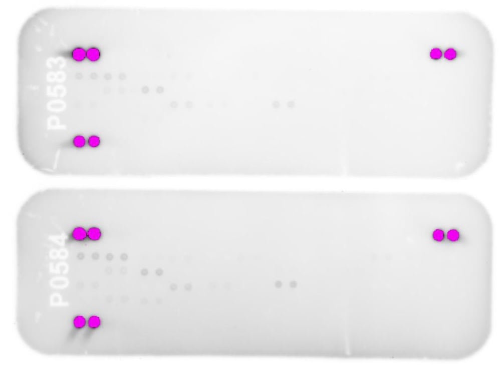
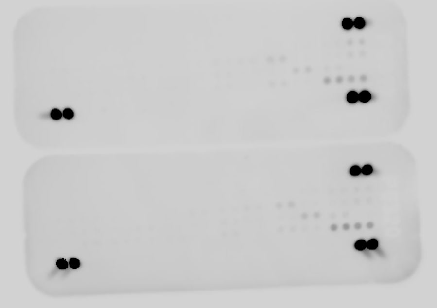
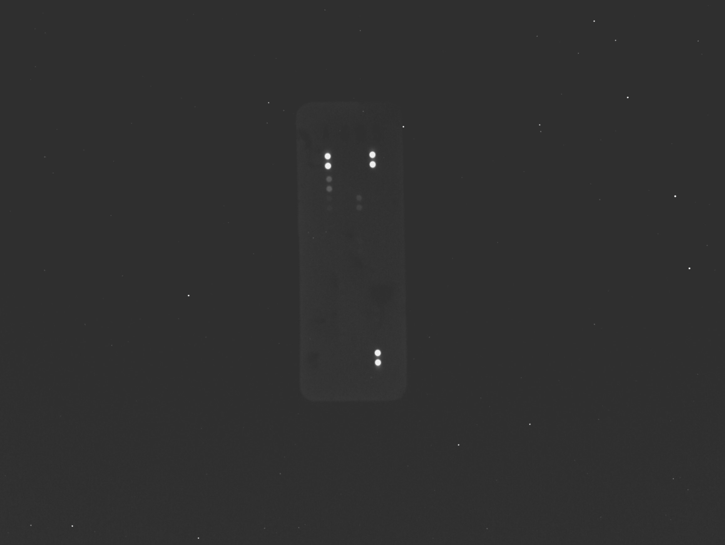
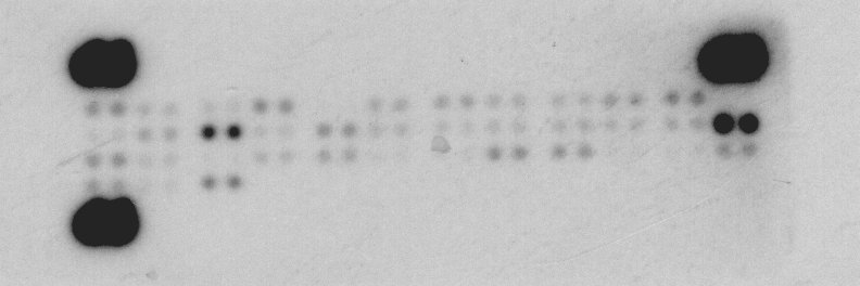
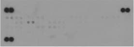
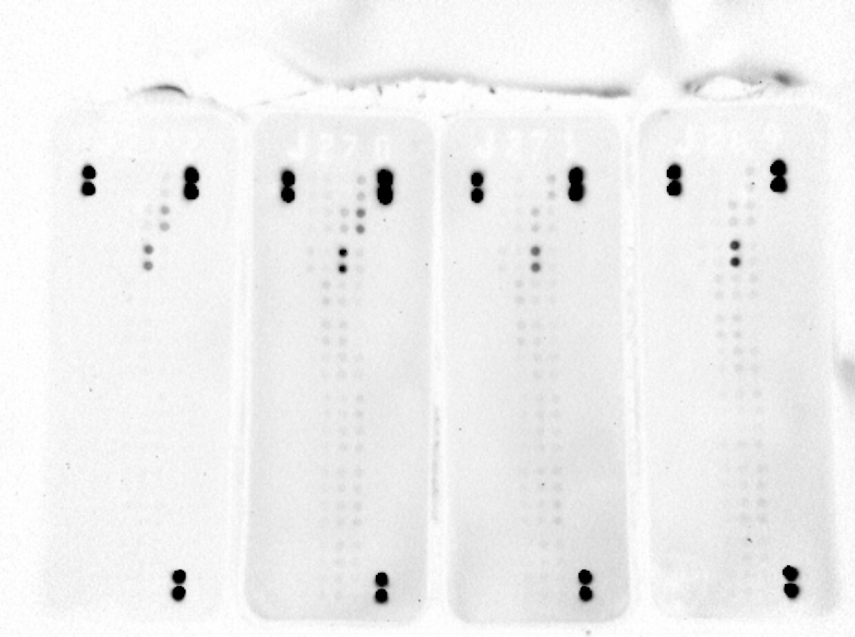
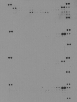
Scientific Data Images for Proteome Profiler Mouse Phospho-RTK Array Kit
Detection of Multiple Tyrosine Phosphorylated Receptors in Cell Lysates by the Mouse Phospho-RTK Array.
The amount of lysate incubated with each array is indicated in the figure. Data shown are from 2 minute (Panels A, B, and C) or 5 minute (Panel D) exposure to X-ray film.A. M1 mouse myeloid leukemia cells were either untreated or treated with 500 ng/mL recombinant mouse Flt-3 Ligand (Catalog # 427-FL) for 5 minutes.B. NMuMG mouse mammary gland epithelial cells were either untreated or treated with 200 ng/mL recombinant mouse EGF (Catalog # 2028-EG) for 5 minutes.C. Hepa 1-6 mouse hepatoma cells were either untreated or treated with 1 µg/mL recombinant human insulin (Sigma, Catalog # I9278).D. HEK293 human embryonic kidney cells transfected with mouse EphA1 were either untreated or treated with 3 µg/mL mouse Ephrin-A1 (Catalog # 602-A1) and 0.3 µg/mL goat anti-human IgG Fc (Catalog # G-102-C) for 20 minutes.Formulation, Preparation, and Storage
Shipping
Storage
Background: RTK Assay Kits
Additional RTK Assay Kits Products
Product Documents for Proteome Profiler Mouse Phospho-RTK Array Kit
Certificate of Analysis
To download a Certificate of Analysis, please enter a lot or batch number in the search box below.
Note: Certificate of Analysis not available for kit components.
Product Specific Notices for Proteome Profiler Mouse Phospho-RTK Array Kit
For research use only
Related Research Areas
Citations for Proteome Profiler Mouse Phospho-RTK Array Kit
 Powered by Bioz
Customer Reviews for Proteome Profiler Mouse Phospho-RTK Array Kit (8)
Have you used Proteome Profiler Mouse Phospho-RTK Array Kit?
Submit a review and receive an Amazon gift card!
$25/€18/£15/$25CAN/¥2500 Yen for a review with an image
$10/€7/£6/$10CAN/¥1110 Yen for a review without an image
Submit a review
Customer Images
-
Verified Customer | Posted 06/09/2023Use of RTK array to assay FACS sorted cells from murine lungs.

-
Verified Customer | Posted 10/16/2022Sample from FACS sorted cells, so limited amount of protein but still able to see some phospho RTK. Would definitely use again.
-
Verified Customer | Posted 04/20/2019Array works and produces publishable results. Not happy with the pho-PDGFR antibody, sinse it appear to cross react with something in mouse tumor lysate
-
Verified Customer | Posted 03/27/2019
-
Verified Customer | Posted 04/16/2018
-
Verified Customer | Posted 07/05/2017Lysates from mouse embryonic stem cells applied to membranes using instructions provided. Western blots were clean and worked great!
-
Verified Customer | Posted 05/16/2017Worked really well on mouse tumour lysates.
-
Verified Customer | Posted 06/29/2016Mammary tumors extracts from MMTV-Neu mice with different p53 genotypes (p53wt vs mutp53)
There are no reviews that match your criteria.
FAQs for Proteome Profiler Mouse Phospho-RTK Array Kit
-
Q: Can the Mouse Phospho-RTK Array be used for measuring the relative levels of total RTKs present in a sample?
A: No. Although the array kit uses capture antibodies that recognize both phosphorylated and unphosphorylated RTKs, the detection antibody is a pan anti-phospho-tyrosine antibody which only detects phosphorylated tyrosines on activated RTKs.
-
Q: To confirm positive signal for an RTK by IP-Western blot, are the capture antibodies from the Mouse Phospho-RTK Array Kit offered separately?
A: The identities of the capture antibodies on the Human Phospho-RTK Array are considered proprietary. Antibodies for each RTK analyte in the kit are available as well as Phospho-Tyrosine HRP-conjugated Antibody (Catalog #HAM1676). It can be challenging to directly compare Western Blot results of the linearized and reduced protein to Array detection of the native form. In ELISA, the protein will also be in the native form. ELISA kits are offered for many of the analytes targeted by the Array. These ELISA options are listed in a table on the ARY014 product webpage.
-
Q: What are the phosphorylation site(s) of the 49 different tyrosine residues on the RTKs?
A: This kit uses a pan anti-phospho-tyrosine antibody as the detection antibody, which means it is capable of detecting phosphorylation at any available tyrosine. It is not designed to be specific for one phosphorylation site on each molecule. For instance, PDGF R alpha has multiple sites of tyrosine phosphorylation (see the “PTM/Processing” section of http://www.uniprot.org/uniprot/P16234 for a list of the currently identified sites).
-
Q: Can the Mouse Phospho-RTK Array be used for measuring the relative levels of total RTKs present in a sample?
A: No. Although the array kit uses capture antibodies that recognize both phosphorylated and unphosphorylated RTKs, the detection antibody is a pan anti-phospho-tyrosine antibody which only detects phosphorylated tyrosines on activated RTKs.
-
Q: To confirm positive signal for an RTK by IP-Western blot, are the capture antibodies from the Mouse Phospho-RTK Array Kit offered separately?
A: The identities of the capture antibodies on the Human Phospho-RTK Array are considered proprietary. Antibodies for each RTK analyte in the kit are available as well as Phospho-Tyrosine HRP-conjugated Antibody (Catalog #HAM1676). It can be challenging to directly compare Western Blot results of the linearized and reduced protein to Array detection of the native form. In ELISA, the protein will also be in the native form. ELISA kits are offered for many of the analytes targeted by the Array. These ELISA options are listed in a table on the ARY014 product webpage.
-
Q: What are the phosphorylation site(s) of the 49 different tyrosine residues on the RTKs?
A: This kit uses a pan anti-phospho-tyrosine antibody as the detection antibody, which means it is capable of detecting phosphorylation at any available tyrosine. It is not designed to be specific for one phosphorylation site on each molecule. For instance, PDGF R alpha has multiple sites of tyrosine phosphorylation (see the “PTM/Processing” section of http://www.uniprot.org/uniprot/P16234 for a list of the currently identified sites).