STAT5b Antibody (2D1) - Azide and BSA Free
Novus Biologicals | Catalog # H00006777-M03
![Western Blot: STAT5b Antibody (2D1) [H00006777-M03]](https://resources.rndsystems.com/images/products/STAT5b-Antibody-2D1-Western-Blot-H00006777-M03-img0011.jpg "Western Blot: STAT5b Antibody (2D1) [H00006777-M03]")
Key Product Details
Species Reactivity
Validated:
Cited:
Applications
Validated:
Cited:
Label
Antibody Source
Format
Product Specifications
Immunogen
Specificity
Clonality
Host
Isotype
Description
Scientific Data Images for STAT5b Antibody (2D1) - Azide and BSA Free
Western Blot: STAT5b Antibody (2D1) [H00006777-M03]
Western Blot: STAT5b Antibody (2D1) [H00006777-M03] - Analysis of STAT5B expression in HeLa (Cat # L013V1).![Immunocytochemistry/ Immunofluorescence: STAT5b Antibody (2D1) [H00006777-M03]](https://resources.rndsystems.com/images/products/STAT5b-Antibody-2D1-Immunocytochemistry-Immunofluorescence-H00006777-M03-img0009.jpg "Immunocytochemistry/ Immunofluorescence: STAT5b Antibody (2D1) [H00006777-M03]")
Immunocytochemistry/ Immunofluorescence: STAT5b Antibody (2D1) [H00006777-M03]
Immunocytochemistry/Immunofluorescence: STAT5b Antibody (2D1) [H00006777-M03] - Analysis of monoclonal antibody to STAT5B on HeLa cell. Antibody concentration 10 ug/ml![Immunohistochemistry-Paraffin: STAT5b Antibody (2D1) [H00006777-M03]](https://resources.rndsystems.com/images/products/STAT5b-Antibody-2D1-Immunohistochemistry-Paraffin-H00006777-M03-img0010.jpg "Immunohistochemistry-Paraffin: STAT5b Antibody (2D1) [H00006777-M03]")
Immunohistochemistry-Paraffin: STAT5b Antibody (2D1) [H00006777-M03]
Immunohistochemistry-Paraffin: STAT5b Antibody (2D1) [H00006777-M03] - Analysis of monoclonal antibody to STAT5B on formalin-fixed paraffin-embedded human lymph node. Antibody concentration 1.5 ug/ml![ELISA: STAT5b Antibody (2D1) [H00006777-M03]](https://resources.rndsystems.com/images/products/STAT5b-Antibody-2D1-ELISA-H00006777-M03-img0005.jpg "ELISA: STAT5b Antibody (2D1) [H00006777-M03]")
ELISA: STAT5b Antibody (2D1) [H00006777-M03]
ELISA: STAT5b Antibody (2D1) [H00006777-M03] - Detection limit for recombinant GST tagged STAT5B is approximately 0.3ng/ml as a capture antibody.Applications for STAT5b Antibody (2D1) - Azide and BSA Free
ELISA
Immunocytochemistry/ Immunofluorescence
Immunohistochemistry
Immunohistochemistry-Paraffin
Western Blot
Reviewed Applications
Read 1 review rated 1 using H00006777-M03 in the following applications:
Formulation, Preparation, and Storage
Purification
Formulation
Format
Preservative
Concentration
Shipping
Stability & Storage
Background: STAT5b
Long Name
Alternate Names
Entrez Gene IDs
Gene Symbol
OMIM
UniProt
Additional STAT5b Products
Product Documents for STAT5b Antibody (2D1) - Azide and BSA Free
Certificate of Analysis
To download a Certificate of Analysis, please enter a lot or batch number in the search box below.
Product Specific Notices for STAT5b Antibody (2D1) - Azide and BSA Free
This product is produced by and distributed for Abnova, a company based in Taiwan.
This product is for research use only and is not approved for use in humans or in clinical diagnosis. Primary Antibodies are guaranteed for 1 year from date of receipt.
Citations for STAT5b Antibody (2D1) - Azide and BSA Free
 Powered by Bioz
Customer Reviews for STAT5b Antibody (2D1) - Azide and BSA Free (1)
Have you used STAT5b Antibody (2D1) - Azide and BSA Free?
Submit a review and receive an Amazon gift card!
$25/€18/£15/$25CAN/¥2500 Yen for a review with an image
$10/€7/£6/$10CAN/¥1110 Yen for a review without an image
Submit a review
Customer Images
-
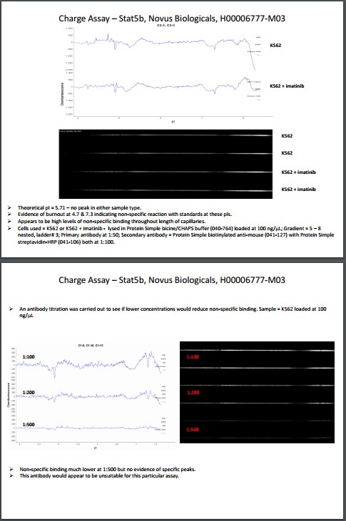
Application: Simple WesternSample Tested: K562 human chronic myelogenous leukemia cell lineSpecies: HumanVerified Customer | Posted 02/06/2017Stat5bCells used = K562 or K562 + imatinib - lysed in Protein Simple bicine/CHAPS buffer (040-764) loaded at 100 ng/µL; Gradient = 5 – 8 nested, ladder# 3; Primary antibody at 1:50; Secondary antibody = Protein Simple biotinylated anti-mouse (041-127) with Protein Simple streptavidin-HRP (041-106) both at 1:100.

There are no reviews that match your criteria.
Protocols
Find general support by application which include: protocols, troubleshooting, illustrated assays, videos and webinars.
- Antigen Retrieval Protocol (PIER)
- Antigen Retrieval for Frozen Sections Protocol
- Appropriate Fixation of IHC/ICC Samples
- Cellular Response to Hypoxia Protocols
- Chromogenic IHC Staining of Formalin-Fixed Paraffin-Embedded (FFPE) Tissue Protocol
- Chromogenic Immunohistochemistry Staining of Frozen Tissue
- ClariTSA™ Fluorophore Kits
- Detection & Visualization of Antibody Binding
- ELISA Sample Preparation & Collection Guide
- ELISA Troubleshooting Guide
- Fluorescent IHC Staining of Frozen Tissue Protocol
- Graphic Protocol for Heat-induced Epitope Retrieval
- Graphic Protocol for the Preparation and Fluorescent IHC Staining of Frozen Tissue Sections
- Graphic Protocol for the Preparation and Fluorescent IHC Staining of Paraffin-embedded Tissue Sections
- Graphic Protocol for the Preparation of Gelatin-coated Slides for Histological Tissue Sections
- How to Run an R&D Systems DuoSet ELISA
- How to Run an R&D Systems Quantikine ELISA
- How to Run an R&D Systems Quantikine™ QuicKit™ ELISA
- ICC Cell Smear Protocol for Suspension Cells
- ICC Immunocytochemistry Protocol Videos
- ICC for Adherent Cells
- IHC Sample Preparation (Frozen sections vs Paraffin)
- Immunocytochemistry (ICC) Protocol
- Immunocytochemistry Troubleshooting
- Immunofluorescence of Organoids Embedded in Cultrex Basement Membrane Extract
- Immunofluorescent IHC Staining of Formalin-Fixed Paraffin-Embedded (FFPE) Tissue Protocol
- Immunohistochemistry (IHC) and Immunocytochemistry (ICC) Protocols
- Immunohistochemistry Frozen Troubleshooting
- Immunohistochemistry Paraffin Troubleshooting
- Preparing Samples for IHC/ICC Experiments
- Preventing Non-Specific Staining (Non-Specific Binding)
- Primary Antibody Selection & Optimization
- Protocol for Heat-Induced Epitope Retrieval (HIER)
- Protocol for Making a 4% Formaldehyde Solution in PBS
- Protocol for VisUCyte™ HRP Polymer Detection Reagent
- Protocol for the Fluorescent ICC Staining of Cell Smears - Graphic
- Protocol for the Fluorescent ICC Staining of Cultured Cells on Coverslips - Graphic
- Protocol for the Preparation & Fixation of Cells on Coverslips
- Protocol for the Preparation and Chromogenic IHC Staining of Frozen Tissue Sections
- Protocol for the Preparation and Chromogenic IHC Staining of Frozen Tissue Sections - Graphic
- Protocol for the Preparation and Chromogenic IHC Staining of Paraffin-embedded Tissue Sections
- Protocol for the Preparation and Chromogenic IHC Staining of Paraffin-embedded Tissue Sections - Graphic
- Protocol for the Preparation and Fluorescent ICC Staining of Cells on Coverslips
- Protocol for the Preparation and Fluorescent ICC Staining of Non-adherent Cells
- Protocol for the Preparation and Fluorescent ICC Staining of Stem Cells on Coverslips
- Protocol for the Preparation and Fluorescent IHC Staining of Frozen Tissue Sections
- Protocol for the Preparation and Fluorescent IHC Staining of Paraffin-embedded Tissue Sections
- Protocol for the Preparation of Gelatin-coated Slides for Histological Tissue Sections
- Protocol for the Preparation of a Cell Smear for Non-adherent Cell ICC - Graphic
- Quantikine HS ELISA Kit Assay Principle, Alkaline Phosphatase
- Quantikine HS ELISA Kit Principle, Streptavidin-HRP Polymer
- R&D Systems Quality Control Western Blot Protocol
- Sandwich ELISA (Colorimetric) – Biotin/Streptavidin Detection Protocol
- Sandwich ELISA (Colorimetric) – Direct Detection Protocol
- TUNEL and Active Caspase-3 Detection by IHC/ICC Protocol
- The Importance of IHC/ICC Controls
- Troubleshooting Guide: ELISA
- Troubleshooting Guide: Immunohistochemistry
- Troubleshooting Guide: Western Blot Figures
- Western Blot Conditions
- Western Blot Protocol
- Western Blot Protocol for Cell Lysates
- Western Blot Troubleshooting
- Western Blot Troubleshooting Guide
- View all Protocols, Troubleshooting, Illustrated assays and Webinars
FAQs for STAT5b Antibody (2D1) - Azide and BSA Free
-
Q: For antibodies : STAT5b H00006777-M03; STAT 5a NBR2-00622; STAT3 NBP2-22471; STAT 6 H00006778-M01 - do these detect phosphorylated, unphosphorylated or both forms of STATs?
A: Antibodies H00006777-M03, NBR2-00622, NBP2-22471, and H00006778-M01 were raised to recognize total STAT proteins, they do not differentiate between any posttranslational modifications and will recognize both phosphorylated and non-phosphorylated protein.
Associated Pathways


