
Organelles: Mitochondria
Overview
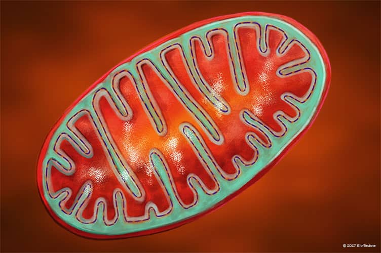
Mitochondria are double membrane organelles that provide energy for the cell. The electron transport chain generates ATP through the oxidative phosphorylation of amino acids, carbohydrates, and fatty acids.
Data Examples

AIF in HeLa Human Cell Line. AIF was detected in formaldehyde fixed HeLa human cervical epithelial carcinoma cell line using Rabbit Anti-Human/Mouse/Rat AIF Alexa Fluor® 488-conjugated Antigen Affinity-purified Polyclonal Antibody (green; Catalog # IC1457G) at 1:10 dilution overnight at 4°C and counterstained with DAPI (blue). Specific staining was localized to mitochondria. View our protocol for Fluorescent ICC Staining of Cells on Coverslips.

COX4‑I1 in HeLa Human Cell Line. COX4-I1 was detected in immersion fixed HeLa human cervical epithelial carcinoma cell line using Goat Anti-Human/Mouse COX4-I1 Antigen Affinity-purified Polyclonal Antibody (Catalog # AF5814) at 5 µg/mL for 3 hours at room temperature. Cells were stained using the NorthernLights™ 557-conjugated Anti-Goat IgG Secondary Antibody (yellow; Catalog # NL001) and counterstained with DAPI (blue). Specific staining was localized to mitochondria. View our protocol for Fluorescent ICC Staining of Cells on Coverslips.

HSP60 in HeLa Human Cell Line. HSP60 was detected in immersion fixed HeLa human cervical epithelial carcinoma cell line using Mouse Anti-Human/Mouse/Rat HSP60 Monoclonal Antibody (Catalog # MAB1800) at 25 µg/mL for 3 hours at room temperature. Cells were stained using the NorthernLights™ 557-conjugated Anti-Mouse IgG Secondary Antibody (red; Catalog # NL007) and counterstained with DAPI (blue). Specific staining was localized to cytoplasm. View our protocol for Fluorescent ICC Staining of Cells on Coverslips.

Visualization of the Mitochondria in Live HeLa Cells with MitoMark Green I. MitoMark Green I (200 nM; Tocris, Catalog # 6444) was used to visualize the mitochondria (green) in live HeLa human cervical epithelial carcinoma cells.
