CD40 is a type I transmembrane glycoprotein belonging to the TNF receptor superfamily. The mature hCD40 consists of a 172 amino acid (aa) extracellular domain, a 22 aa transmembrane region and a 62 aa cytoplasmic domain (1). Human and mouse CD40 share 62% aa identity. CD40 is expressed in B cells, follicular dendritic cells, dendritic cells, activated monocytes, macrophages, endothelial cells, vascular smooth muscle cells, and several tumor cell lines (2). The extracellular domain has the cysteine-rich repeat regions, which are characteristic for many of the receptors of the TNF superfamily. Interaction of CD40 with its ligand, CD40L, leads to aggregation of CD40 molecules, which in turn interact with cytoplasmic components to initiate signaling pathways. Early studies on the CD40-CD40L system revealed its role in humoral immunity. Interaction between CD40L on T cells and CD40 on B cells stimulated B cell proliferation and provided the signal for immunoglobulin isotype switching (3). Mutations in the CD40L gene, which resulted in a CD40L molecule unable to interact with CD40, are responsible for the hyper-IgM syndrome (4). Cross-linking of CD40 with antibodies or by CD40 binding to CD40L produces cell type-specific responses which include costimulation and induction of proliferation, induction of cytokine production, rescue from apoptosis, and upregulation of adhesion molecules (5). Some of the early events of intracellular signaling by the CD40-CD40L system include the association of the CD40 with TRAFs and the activation of various kinases (6 - 8).

Key Product Details
Species Reactivity
Validated:
Human
Cited:
Human, Mouse
Applications
Validated:
Western Blot, Simple Western, Agonist Activity
Cited:
Immunohistochemistry, Western Blot, Neutralization, Bioassay
Label
Unconjugated
Antibody Source
Polyclonal Goat IgG
Loading...
Product Specifications
Immunogen
Mouse myeloma cell line NS0-derived recombinant human CD40
Glu21-Arg193
Accession # P25942
Glu21-Arg193
Accession # P25942
Specificity
Detects human CD40 in direct ELISAs and Western blots.
Clonality
Polyclonal
Host
Goat
Isotype
IgG
Endotoxin Level
<0.10 EU per 1 μg of the antibody by the LAL method.
Scientific Data Images for Human CD40/TNFRSF5 Antibody
Detection of Human CD40/TNFRSF5 by Western Blot.
Western blot shows lysates of Raji human Burkitt's lymphoma cell line and Daudi human Burkitt's lymphoma cell line. PVDF membrane was probed with 2 µg/mL of Goat Anti-Human CD40/TNFRSF5 Antigen Affinity-purified Polyclonal Antibody (Catalog # AF632) followed by HRP-conjugated Anti-Goat IgG Secondary Antibody (HAF017). A specific band was detected for CD40/TNFRSF5 at approximately 40-45 kDa (as indicated). This experiment was conducted under reducing conditions and using Immunoblot Buffer Group 1.
Human CD40/TNFRSF5 Antibody Stimulates Cell Proliferation in Human B Cells.
Goat Anti-Human CD40/TNFRSF5 Antigen Affinity-purified Polyclonal Antibody (Catalog # AF632) stimulates human B cell proliferation in the presence of Recombinant Human IL-4 (204-IL) in a dose-dependent manner, as measured by Resazurin (AR002). The ED50 for this effect is typically 3-12 ng/mL
Detection of Human CD40/TNFRSF5 by Simple WesternTM.
Simple Western lane view shows lysates of Raji human Burkitt's lymphoma cell line and Daudi human Burkitt's lymphoma cell line, loaded at 0.2 mg/mL. A specific band was detected for CD40/TNFRSF5 at approximately 57 kDa (as indicated) using 50 µg/mL of Goat Anti-Human CD40/TNFRSF5 Antigen Affinity-purified Polyclonal Antibody (Catalog # AF632). This experiment was conducted under reducing conditions and using the 12-230 kDa separation system.
Detection of Fungus CD40/TNFRSF5 by Western Blot
SDS–PAGE and Western blot analysis of recombinant CD40-N expressed in P. pastoris. a A 20-μL sample of supernatant was loaded onto a 12 % SDS–PAGE gel and stained with Coomassie brilliant blue G-250. All of the SDS-PAGE experiments were performed under the same conditions. Samples were collected at 12-h intervals for 84 h. The sizes of molecular weight markers (kDa) are shown (M), and recombinant CD40-N protein is indicated with an arrow. b Western blot analysis of human Flag-tagged CD40-N (not undergoing codon optimization) and CD40-N-S (with signal sequence and not undergoing codon optimization) expressed in HEK293T cells. pCDNA3.3-CD40-N and pCDNA3.3-CD40-N-S were introduced into HEK293T cells, and the cell lysates were harvested after 48 h. A 10-μl sample was loaded onto a 12 % SDS–PAGE gel. Anti-Flag, anti-CD40-N and anti-GAPDH antibodies were used to detect the proteins. c Western blot analysis of proteins at different times in P. pastoris (0, 12, 24, 36, 48, 60, 72, and 84 h). P indicates the positive control (CD40-N expressed in HEK293T cells). Image collected and cropped by CiteAb from the following open publication (https://pubmed.ncbi.nlm.nih.gov/26809818), licensed under a CC-BY license. Not internally tested by R&D Systems.
Detection of Yeast CD40/TNFRSF5 by Western Blot
SDS–PAGE and Western blot analysis of recombinant CD40-N expressed in P. pastoris. a A 20-μL sample of supernatant was loaded onto a 12 % SDS–PAGE gel and stained with Coomassie brilliant blue G-250. All of the SDS-PAGE experiments were performed under the same conditions. Samples were collected at 12-h intervals for 84 h. The sizes of molecular weight markers (kDa) are shown (M), and recombinant CD40-N protein is indicated with an arrow. b Western blot analysis of human Flag-tagged CD40-N (not undergoing codon optimization) and CD40-N-S (with signal sequence and not undergoing codon optimization) expressed in HEK293T cells. pCDNA3.3-CD40-N and pCDNA3.3-CD40-N-S were introduced into HEK293T cells, and the cell lysates were harvested after 48 h. A 10-μl sample was loaded onto a 12 % SDS–PAGE gel. Anti-Flag, anti-CD40-N and anti-GAPDH antibodies were used to detect the proteins. c Western blot analysis of proteins at different times in P. pastoris (0, 12, 24, 36, 48, 60, 72, and 84 h). P indicates the positive control (CD40-N expressed in HEK293T cells) Image collected and cropped by CiteAb from the following open publication (https://pubmed.ncbi.nlm.nih.gov/26809818), licensed under a CC-BY license. Not internally tested by R&D Systems.Applications for Human CD40/TNFRSF5 Antibody
Application
Recommended Usage
Agonist Activity
Measured in a cell proliferation assay using B cell enriched human peripheral blood lymphocytes in the presence of IL-4. Banchereau, J. et al. (1991) Science 251:70. The ED50 for this effect is typically 3-12 ng/mL.
Simple Western
50 µg/mL
Sample: Raji human Burkitt's lymphoma cell line and Daudi human Burkitt's lymphoma cell line
Sample: Raji human Burkitt's lymphoma cell line and Daudi human Burkitt's lymphoma cell line
Western Blot
2 µg/mL
Sample: Raji human Burkitt's lymphoma cell line and Daudi human Burkitt's lymphoma cell line
Sample: Raji human Burkitt's lymphoma cell line and Daudi human Burkitt's lymphoma cell line
Reviewed Applications
Read 1 review rated 5 using AF632 in the following applications:
Formulation, Preparation, and Storage
Purification
Antigen Affinity-purified
Reconstitution
Reconstitute at 0.2 mg/mL in sterile PBS. For liquid material, refer to CoA for concentration.
Loading...
Formulation
Lyophilized from a 0.2 μm filtered solution in PBS with Trehalose. *Small pack size (SP) is supplied either lyophilized or as a 0.2 µm filtered solution in PBS.
Shipping
Lyophilized product is shipped at ambient temperature. Liquid small pack size (-SP) is shipped with polar packs. Upon receipt, store immediately at the temperature recommended below.
Stability & Storage
Use a manual defrost freezer and avoid repeated freeze-thaw cycles.
- 12 months from date of receipt, -20 to -70 °C as supplied.
- 1 month, 2 to 8 °C under sterile conditions after reconstitution.
- 6 months, -20 to -70 °C under sterile conditions after reconstitution.
Calculators
Background: CD40/TNFRSF5
References
- Torres, R.M. and E.A. Clark (1992) J. Immunol. 148:620.
- Schonbeck, U. et al. (1997) J. Biol. Chem. 272:19569.
- Armitage, R.J. et al. (1993) J. Immunol. 150:3671.
- Callard, R.E. et al. (1993) Immunol. Today 14:559.
- Stout, R.D. and J. Suttles (1996) Immunol. Today 17:487.
- Pullen, S.S. et al. (1999) Biochemistry 38:10168.
- Faris, M. et al. (1994) J. Exp. Med. 179:1923.
- Hanissian, S.H. and R.S Geha (1997) Immunity 6:379.
Alternate Names
CD40, TNFRSF5
Gene Symbol
CD40
UniProt
Additional CD40/TNFRSF5 Products
Product Documents for Human CD40/TNFRSF5 Antibody
Certificate of Analysis
To download a Certificate of Analysis, please enter a lot or batch number in the search box below.
Note: Certificate of Analysis not available for kit components.
Product Specific Notices for Human CD40/TNFRSF5 Antibody
For research use only
Related Research Areas
Citations for Human CD40/TNFRSF5 Antibody
 Powered by Bioz
Customer Reviews for Human CD40/TNFRSF5 Antibody (1)
5 out of 5
1 Customer Rating
Have you used Human CD40/TNFRSF5 Antibody?
Submit a review and receive an Amazon gift card!
$25/€18/£15/$25CAN/¥2500 Yen for a review with an image
$10/€7/£6/$10CAN/¥1110 Yen for a review without an image
Submit a review
Customer Images
Showing
1
-
1 of
1 review
Showing All
Filter By:
-
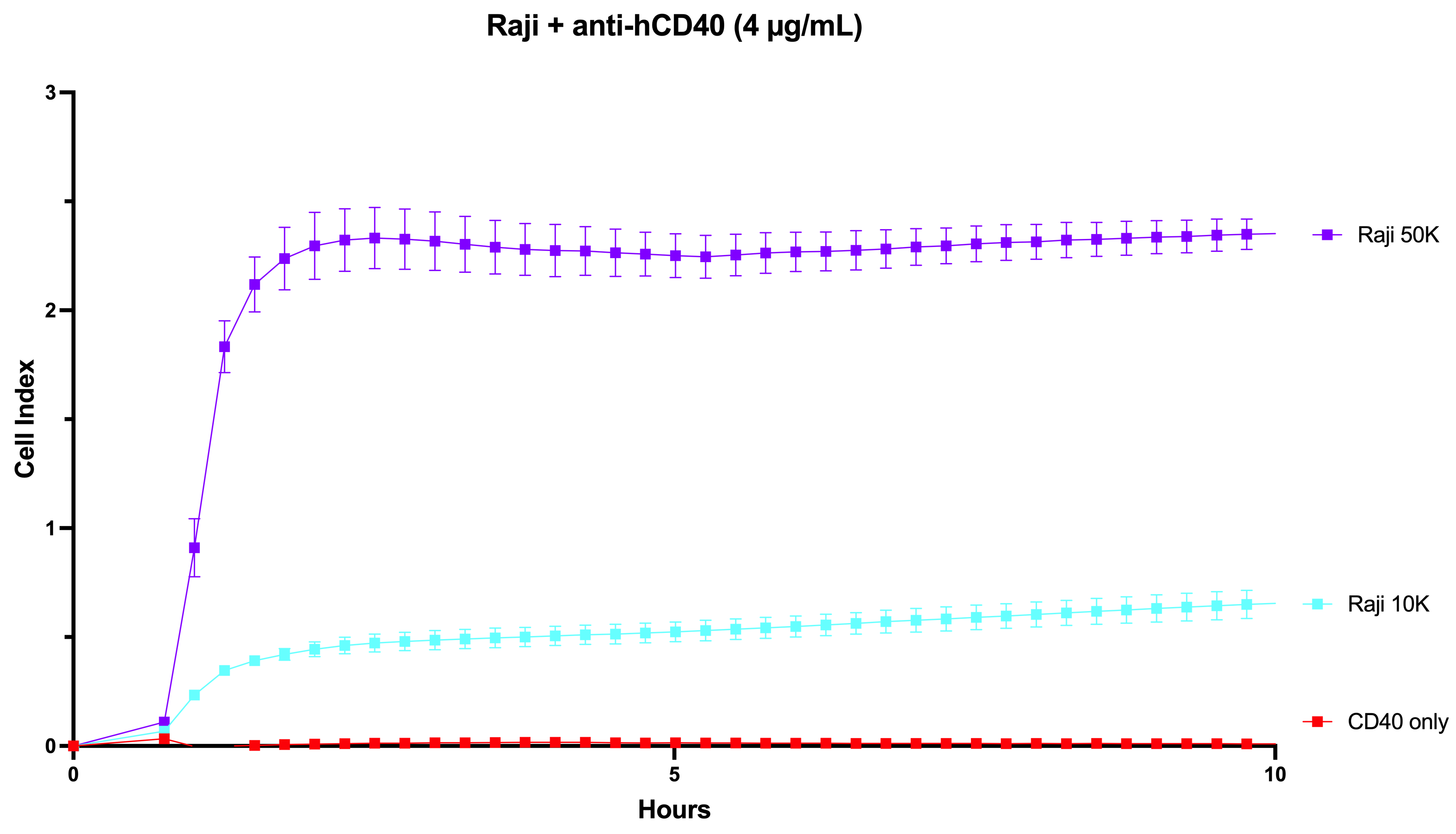
Application: Tethering reagent to attach non-adherent cells (Raji cells)Sample Tested: Raji human Burkitt's lymphoma cell lineSpecies: HumanVerified Customer | Posted 07/19/2023Cell Proliferation measured as cell impedance by Xcelligence assay.

There are no reviews that match your criteria.
Protocols
Find general support by application which include: protocols, troubleshooting, illustrated assays, videos and webinars.
- Cellular Response to Hypoxia Protocols
- R&D Systems Quality Control Western Blot Protocol
- Troubleshooting Guide: Western Blot Figures
- Western Blot Conditions
- Western Blot Protocol
- Western Blot Protocol for Cell Lysates
- Western Blot Troubleshooting
- Western Blot Troubleshooting Guide
- View all Protocols, Troubleshooting, Illustrated assays and Webinars
Loading...
Associated Pathways

