COL4A1 (collagen IV alpha 1) is a 185 kDa member of the type IV collagen family. It is a secreted glycoprotein that is expressed by multiple cell types, including fibroblasts, keratinocytes and endothelial cells. Two COL4A1 molecules interact with a 170 kDa alpha 2 chain to form a collagen IV triple helix. This helix further interacts with other helices to generate covalent oligomers that form a scaffold in the basement membrane. Mature human COL4A1 is 1642 amino acids (aa) in length. It has an N-terminal “7S” proregion (aa 28-172), a central collagenous domain that contains multiple Gly-based repeats (aa 173-1440), and a C-terminal domain that is proteolytically cleaved to generate a 25-28 kDa NC1 globular segment that has potent antiangiogenic activity (aa 1441-1669). Multiple splice forms exist. One shows a deletion of aa 499-849, a second shows a seven aa substitution for aa 513-1669, and a third shows a seven aa substitution for aa 958-1669. Over aa 1441-1669, human COL4A1 is 97% aa identical to mouse COL4A1.
Human Collagen IV alpha 1 Antibody (577238)
R&D Systems | Catalog # MAB6308

Key Product Details
Species Reactivity
Validated:
Human
Cited:
Human
Applications
Validated:
Immunocytochemistry
Cited:
Immunohistochemistry, ELISA Capture
Label
Unconjugated
Antibody Source
Monoclonal Mouse IgG1 Clone # 577238
Loading...
Product Specifications
Immunogen
Mouse myeloma cell line NS0-derived recombinant human Collagen IV alpha 1
Ser1441-Thr1669
Accession # P02462
Ser1441-Thr1669
Accession # P02462
Specificity
Detects human Collagen IV alpha 1 in direct ELISAs. In direct ELISAs, no cross-reactivity with recombinant human Collagen I alpha 1, III alpha 1, XIII alpha 1v4, XXIII alpha 1, or recombinant mouse Collagen XXV alpha 1 is observed.
Clonality
Monoclonal
Host
Mouse
Isotype
IgG1
Scientific Data Images for Human Collagen IV alpha 1 Antibody (577238)
Collagen IV alpha 1 in T98G Human Cell Line.
Collagen IV a1 was detected in immersion fixed T98G human glioblastoma cell line using Mouse Anti-Human Collagen IV alpha 1 Monoclonal Antibody (Catalog # MAB6308) at 10 µg/mL for 3 hours at room temperature. Cells were stained using the NorthernLights™ 557-conjugated Anti-Mouse IgG Secondary Antibody (red; Catalog # NL007) and counterstained with DAPI (blue). Specific staining was localized to perinuclear areas and cytoplasm. View our protocol for Fluorescent ICC Staining of Cells on Coverslips.
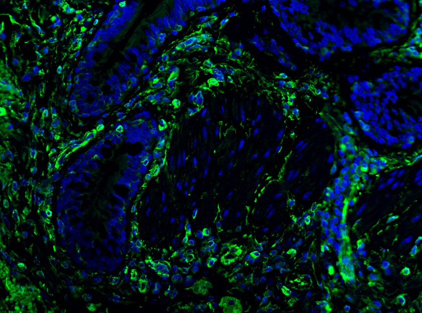
Collagen IV alpha 1 in THP‑1 Human Cell Line.
Collagen IV a1 was detected in immersion fixed THP-1 human acute monocytic leukemia cell line using Mouse Anti-Human Collagen IV a1 Monoclonal Antibody (Catalog # MAB6308) at 8 µg/mL for 3 hours at room temperature. Cells were stained using the NorthernLights™ 557-conjugated Anti-Mouse IgG Secondary Antibody (red; Catalog # NL007) and counterstained with DAPI (blue). Specific staining was localized to cytoplasm. View our protocol for Fluorescent ICC Staining of Non-adherent Cells.Applications for Human Collagen IV alpha 1 Antibody (577238)
Application
Recommended Usage
Immunocytochemistry
8-25 µg/mL
Sample: Immersion fixed T98G human glioblastoma cell line and THP-1 human acute monocytic leukemia cell line
Sample: Immersion fixed T98G human glioblastoma cell line and THP-1 human acute monocytic leukemia cell line
Reviewed Applications
Read 1 review rated 5 using MAB6308 in the following applications:
Formulation, Preparation, and Storage
Purification
Protein A or G purified from hybridoma culture supernatant
Reconstitution
Sterile PBS to a final concentration of 0.5 mg/mL. For liquid material, refer to CoA for concentration.
Loading...
Formulation
Lyophilized from a 0.2 μm filtered solution in PBS with Trehalose. *Small pack size (SP) is supplied either lyophilized or as a 0.2 µm filtered solution in PBS.
Shipping
Lyophilized product is shipped at ambient temperature. Liquid small pack size (-SP) is shipped with polar packs. Upon receipt, store immediately at the temperature recommended below.
Stability & Storage
Use a manual defrost freezer and avoid repeated freeze-thaw cycles.
- 12 months from date of receipt, -20 to -70 °C as supplied.
- 1 month, 2 to 8 °C under sterile conditions after reconstitution.
- 6 months, -20 to -70 °C under sterile conditions after reconstitution.
Calculators
Background: Collagen IV alpha 1
Alternate Names
Arresten, COL4A1
Gene Symbol
COL4A1
UniProt
Additional Collagen IV alpha 1 Products
Product Documents for Human Collagen IV alpha 1 Antibody (577238)
Certificate of Analysis
To download a Certificate of Analysis, please enter a lot or batch number in the search box below.
Note: Certificate of Analysis not available for kit components.
Product Specific Notices for Human Collagen IV alpha 1 Antibody (577238)
For research use only
Related Research Areas
Citations for Human Collagen IV alpha 1 Antibody (577238)
 Powered by Bioz
Customer Reviews for Human Collagen IV alpha 1 Antibody (577238) (1)
5 out of 5
1 Customer Rating
Have you used Human Collagen IV alpha 1 Antibody (577238)?
Submit a review and receive an Amazon gift card!
$25/€18/£15/$25CAN/¥2500 Yen for a review with an image
$10/€7/£6/$10CAN/¥1110 Yen for a review without an image
Submit a review
Customer Images
Showing
1
-
1 of
1 review
Showing All
Filter By:
-
Application: ImmunohistochemistrySample Tested: Lung tissueSpecies: HumanVerified Customer | Posted 12/07/2016

There are no reviews that match your criteria.
Protocols
Find general support by application which include: protocols, troubleshooting, illustrated assays, videos and webinars.
- Appropriate Fixation of IHC/ICC Samples
- Cellular Response to Hypoxia Protocols
- ClariTSA™ Fluorophore Kits
- Detection & Visualization of Antibody Binding
- ICC Cell Smear Protocol for Suspension Cells
- ICC Immunocytochemistry Protocol Videos
- ICC for Adherent Cells
- Immunocytochemistry (ICC) Protocol
- Immunocytochemistry Troubleshooting
- Immunofluorescence of Organoids Embedded in Cultrex Basement Membrane Extract
- Immunohistochemistry (IHC) and Immunocytochemistry (ICC) Protocols
- Preparing Samples for IHC/ICC Experiments
- Preventing Non-Specific Staining (Non-Specific Binding)
- Primary Antibody Selection & Optimization
- Protocol for VisUCyte™ HRP Polymer Detection Reagent
- Protocol for the Fluorescent ICC Staining of Cell Smears - Graphic
- Protocol for the Fluorescent ICC Staining of Cultured Cells on Coverslips - Graphic
- Protocol for the Preparation and Fluorescent ICC Staining of Cells on Coverslips
- Protocol for the Preparation and Fluorescent ICC Staining of Non-adherent Cells
- Protocol for the Preparation and Fluorescent ICC Staining of Stem Cells on Coverslips
- Protocol for the Preparation of a Cell Smear for Non-adherent Cell ICC - Graphic
- TUNEL and Active Caspase-3 Detection by IHC/ICC Protocol
- The Importance of IHC/ICC Controls
- View all Protocols, Troubleshooting, Illustrated assays and Webinars


