CTLA-4 (cytotoxic T-lymphocyte associated protein‑4, designated CD152), is a type I transmembrane T cell inhibitory molecule that is a member of the Ig superfamily (1, 2). Human or mouse CTLA-4 cDNA encodes 223 amino acids (aa) including a 35 aa signal sequence, a 126 aa extracellular domain (ECD) with one Ig-like V-type domain, a 21 aa transmembrane (TM) sequence, and a 41 aa cytoplasmic sequence. It is found as a covalent homodimer of 41-43 kDa (2) Within the ECD, human CTLA-4 shares 68%, 71% and 83‑86% aa sequence identity with mouse, rat and porcine/bovine/rabbit/feline/canine CTLA-4, respectively. A 174 aa form that lacks TM and cytoplasmic sequences (sCTLA-4) is possibly secreted (3-5). Isoforms of 56-79 aa that mainly contain parts of the cytoplasmic domain are reported. In mouse, an isoform lacking the Ig-like domain has ligand-independent inhibitory activity and is termed liCTLA-4 (6). CD28, which is structurally related to CTLA-4, is constitutively expressed on naïve T cells and promotes T cell activation when engaged by B7-2 on antigen-presenting cells (APC) within the immunological synapse (IS) (1, 7, 8). In contrast, CTLA-4 is recruited from intracellular vesicles to the IS beginning 1-2 days after T cell activation (2, 7, 8). It forms a linear lattice with B7-1 on APC, inducing negative regulatory signals and ending T cell activation (9). Abatacept, a therapeutic human CTLA-4-Ig fusion protein (trade name Orencia), competes with CD28 for B7-1 and B7-2 binding and has been used to antagonize T cell activation in autoimmune conditions and to enhance transplant survival (10). Mice deleted for CTLA-4 show no abnormalities until after birth, but then develop lethal autoimmune reactions due to continued T cell activation and poor control by regulatory T cells, which constitutively express CTLA-4 in wild-type mice and humans (11-13).
Human CTLA-4 Antibody (2188A)
R&D Systems | Catalog # MAB3252
Recombinant Monoclonal Antibody.

Key Product Details
Species Reactivity
Human
Applications
Flow Cytometry, Immunocytochemistry
Label
Unconjugated
Antibody Source
Recombinant Monoclonal Rabbit IgG Clone # 2188A
Loading...
Product Specifications
Immunogen
Chinese hamster ovary cell line CHO-derived recombinant human CTLA-4
Ala37-Phe162
Accession # P16410
Ala37-Phe162
Accession # P16410
Specificity
Detects human CTLA-4 in direct ELISAs.
Clonality
Monoclonal
Host
Rabbit
Isotype
IgG
Scientific Data Images for Human CTLA-4 Antibody (2188A)
Detection of CTLA‑4 in NS0 Mouse Cell Line Transfected with Human CTLA-4 and eGFP by Flow Cytometry.
NS0 mouse myeloma cell line transfected with (A) human CTLA-4 or (B) irrelevant transfectants and eGFP was stained with Rabbit Anti-Human CTLA-4 Monoclonal Antibody (Catalog # MAB3252) followed by APC-conjugated Goat anti-Rabbit IgG Secondary Antibody (Catalog # F0111). Quadrants were set based on Normal Rabbit IgG Control Antibody (Catalog # MAB1050, data not shown). View our protocol for Staining Membrane-associated Proteins.
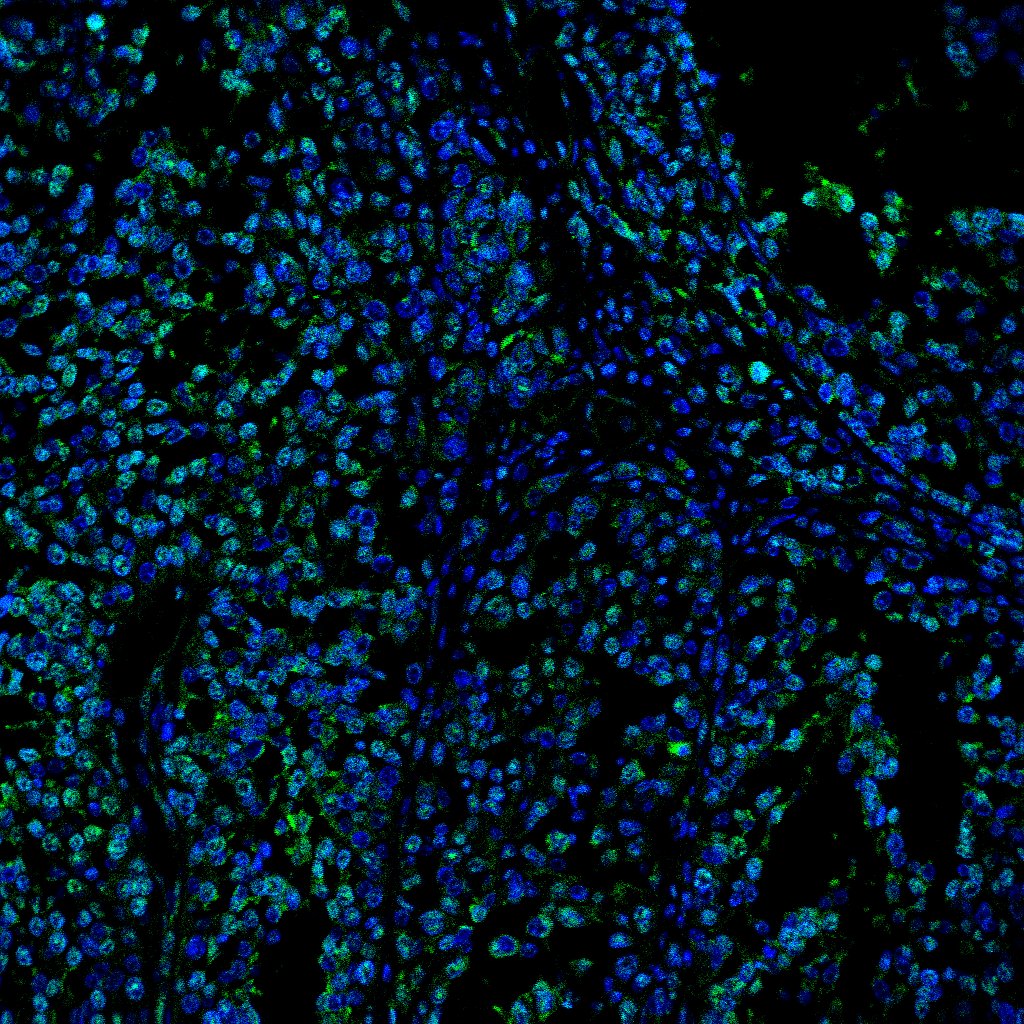
CTLA‑4 in Human PBMCs.
CTLA-4 was detected in immersion fixed human peripheral blood mononuclear cells (PBMCs) using Rabbit Anti-Human CTLA-4 Monoclonal Antibody (Catalog # MAB3252) at 3 µg/mL for 3 hours at room temperature. Cells were stained using the NorthernLights™ 557-conjugated Anti-Rabbit IgG Secondary Antibody (red; Catalog # NL004) and counterstained with DAPI (blue). Specific staining was localized to cell surfaces. View our protocol for Fluorescent ICC Staining of Non-adherent Cells.Applications for Human CTLA-4 Antibody (2188A)
Application
Recommended Usage
Flow Cytometry
0.25 µg/106 cells
Sample: NS0 mouse myeloma cell line transfected with human CTLA-4 and eGFP
Sample: NS0 mouse myeloma cell line transfected with human CTLA-4 and eGFP
Immunocytochemistry
3-25 µg/mL
Sample: Immersion fixed human peripheral blood mononuclear cells (PBMCs)
Sample: Immersion fixed human peripheral blood mononuclear cells (PBMCs)
Reviewed Applications
Read 1 review rated 3 using MAB3252 in the following applications:
Flow Cytometry Panel Builder
Bio-Techne Knows Flow Cytometry
Save time and reduce costly mistakes by quickly finding compatible reagents using the Panel Builder Tool.
Advanced Features
- Spectra Viewer - Custom analysis of spectra from multiple fluorochromes
- Spillover Popups - Visualize the spectra of individual fluorochromes
- Antigen Density Selector - Match fluorochrome brightness with antigen density

Formulation, Preparation, and Storage
Purification
Protein A or G purified from cell culture supernatant
Reconstitution
Reconstitute at 0.5 mg/mL in sterile PBS. For liquid material, refer to CoA for concentration.
Loading...
Formulation
Lyophilized from a 0.2 μm filtered solution in PBS with Trehalose. *Small pack size (SP) is supplied either lyophilized or as a 0.2 µm filtered solution in PBS.
Shipping
Lyophilized product is shipped at ambient temperature. Liquid small pack size (-SP) is shipped with polar packs. Upon receipt, store immediately at the temperature recommended below.
Stability & Storage
Use a manual defrost freezer and avoid repeated freeze-thaw cycles.
- 12 months from date of receipt, -20 to -70 °C as supplied.
- 1 month, 2 to 8 °C under sterile conditions after reconstitution.
- 6 months, -20 to -70 °C under sterile conditions after reconstitution.
Calculators
Background: CTLA-4
References
- Harper, K. et al. (1991) J. Immunol. 147:1037.
- Teft, W.A. et al. (2006) Annu. Rev. Immunol. 24:65.
- Magistrelli, G. et al. (1999) Eur. J. Immunol. 29:3596.
- Tector, M. et al. (2009) BMC Immunol. 10:51.
- Oaks, M.K. and K.M. Hallett (2000) J. Immunol. 164:5015.
- Vijayakrishnan, L. et al. (2004) Immunity 20:563.
- Pentcheva-Hoang, T. et al. (2004) Immunity 21:401.
- Jansson, A. et al. (2005) J. Immunol 175:1575.
- Darlington, P.J. et al. (2005) J. Immunol. 175:996.
- Platt, A.M. et al. (2010) J. Immunol. 185:1558.
- Wing, K. et al. (2008) Science 322:271.
- Friedline, R.H. et al. (2009) J. Exp. Med. 206:421.
- Jain, N. et al. (2010) Proc. Natl. Acad. Sci. USA 107:1524.
Long Name
Cytotoxic T-lymphocyte-associated Molecule 4
Alternate Names
CD152, CTLA4
Gene Symbol
CTLA4
UniProt
Additional CTLA-4 Products
Product Documents for Human CTLA-4 Antibody (2188A)
Certificate of Analysis
To download a Certificate of Analysis, please enter a lot or batch number in the search box below.
Note: Certificate of Analysis not available for kit components.
Product Specific Notices for Human CTLA-4 Antibody (2188A)
For research use only
Customer Reviews for Human CTLA-4 Antibody (2188A) (1)
3 out of 5
1 Customer Rating
Have you used Human CTLA-4 Antibody (2188A)?
Submit a review and receive an Amazon gift card!
$25/€18/£15/$25CAN/¥2500 Yen for a review with an image
$10/€7/£6/$10CAN/¥1110 Yen for a review without an image
Submit a review
Customer Images
Showing
1
-
1 of
1 review
Showing All
Filter By:
-
Application: Immunocytochemistry/ImmunofluorescenceSample Tested: Melanoma tissueSpecies: HumanVerified Customer | Posted 04/11/2022

There are no reviews that match your criteria.
Protocols
Find general support by application which include: protocols, troubleshooting, illustrated assays, videos and webinars.
- 7-Amino Actinomycin D (7-AAD) Cell Viability Flow Cytometry Protocol
- Appropriate Fixation of IHC/ICC Samples
- Cellular Response to Hypoxia Protocols
- ClariTSA™ Fluorophore Kits
- Detection & Visualization of Antibody Binding
- Extracellular Membrane Flow Cytometry Protocol
- Flow Cytometry Protocol for Cell Surface Markers
- Flow Cytometry Protocol for Staining Membrane Associated Proteins
- Flow Cytometry Staining Protocols
- Flow Cytometry Troubleshooting Guide
- ICC Cell Smear Protocol for Suspension Cells
- ICC Immunocytochemistry Protocol Videos
- ICC for Adherent Cells
- Immunocytochemistry (ICC) Protocol
- Immunocytochemistry Troubleshooting
- Immunofluorescence of Organoids Embedded in Cultrex Basement Membrane Extract
- Immunohistochemistry (IHC) and Immunocytochemistry (ICC) Protocols
- Intracellular Flow Cytometry Protocol Using Alcohol (Methanol)
- Intracellular Flow Cytometry Protocol Using Detergents
- Intracellular Nuclear Staining Flow Cytometry Protocol Using Detergents
- Intracellular Staining Flow Cytometry Protocol Using Alcohol Permeabilization
- Intracellular Staining Flow Cytometry Protocol Using Detergents to Permeabilize Cells
- Preparing Samples for IHC/ICC Experiments
- Preventing Non-Specific Staining (Non-Specific Binding)
- Primary Antibody Selection & Optimization
- Propidium Iodide Cell Viability Flow Cytometry Protocol
- Protocol for Liperfluo
- Protocol for VisUCyte™ HRP Polymer Detection Reagent
- Protocol for the Characterization of Human Th22 Cells
- Protocol for the Characterization of Human Th9 Cells
- Protocol for the Fluorescent ICC Staining of Cell Smears - Graphic
- Protocol for the Fluorescent ICC Staining of Cultured Cells on Coverslips - Graphic
- Protocol for the Preparation and Fluorescent ICC Staining of Cells on Coverslips
- Protocol for the Preparation and Fluorescent ICC Staining of Non-adherent Cells
- Protocol for the Preparation and Fluorescent ICC Staining of Stem Cells on Coverslips
- Protocol for the Preparation of a Cell Smear for Non-adherent Cell ICC - Graphic
- Protocol: Annexin V and PI Staining by Flow Cytometry
- Protocol: Annexin V and PI Staining for Apoptosis by Flow Cytometry
- TUNEL and Active Caspase-3 Detection by IHC/ICC Protocol
- The Importance of IHC/ICC Controls
- Troubleshooting Guide: Fluorokine Flow Cytometry Kits
- View all Protocols, Troubleshooting, Illustrated assays and Webinars
Loading...