CXCL10 was originally identified as an IFN-gamma -inducible gene in monocytes, fibroblasts and endothelial cells. It has since been shown that CXCL10 mRNA is also induced by LPS, IL-1 beta, TNF-alpha, IL-12, and viruses. Additional cell types that have been shown to express CXCL10 include activated T-lymphocytes, splenocytes, keratinocytes, osteoblasts, astrocytes, and smooth muscle cells. CXCL10 is also expressed in psoriatic and lepromatous lesions of skin. The mouse homologue of human CXCL10, CRG-2, has been cloned and shown to share approximately 67% amino acid sequence identity with human CXCL10. Human CXCL10 cDNA encodes a 98 amino acid (aa) residue precursor protein with a 21 aa residue signal peptide that is cleaved to form the 77 aa residue secreted protein. The amino acid sequence of CXCL10 identified the protein as a member of the chemokine alpha subfamily that lacks the ELR domain. CXCL10 has been shown to be a chemoattractant for activated T-lymphocytes. CXCL10 has been reported to be a potent inhibitor of angiogenesis and to display a potent thymus-dependent antitumor effect. A chemokine receptor specific for CXCL10 and Mig has been cloned and shown to be highly expressed in IL-2-activated T-lymphocytes.
Human CXCL10/IP-10/CRG-2 Antibody (33036)
R&D Systems | Catalog # MAB266

Key Product Details
Species Reactivity
Validated:
Human
Cited:
Human, Mouse, Primate - Macaca mulatta (Rhesus Macaque), Primate - Pan troglodytes (Chimpanzee)
Applications
Validated:
ELISA Capture (Matched Antibody Pair), Neutralization, Intracellular Staining by Flow Cytometry, CyTOF-ready
Cited:
Immunohistochemistry, Immunohistochemistry-Paraffin, Neutralization, Flow Cytometry, Immunocytochemistry, Bioassay, Cell Selection, ELISA Capture, ELISA Development, ELISA Development (Capture), Functional Assay, Immunoassay Development, Immunodepletion, Luminex Development
Label
Unconjugated
Antibody Source
Monoclonal Mouse IgG1 Clone # 33036
Loading...
Product Specifications
Immunogen
E. coli-derived recombinant human CXCL10/IP-10/CRG-2
Val22-Pro98
Accession # P02778.2
Val22-Pro98
Accession # P02778.2
Specificity
Detects human CXCL10/IP-10/CRG-2 in ELISAs.
Clonality
Monoclonal
Host
Mouse
Isotype
IgG1
Endotoxin Level
<0.10 EU per 1 μg of the antibody by the LAL method.
Scientific Data Images for Human CXCL10/IP-10/CRG-2 Antibody (33036)
Chemotaxis Induced by CXCL10/IP‑10 and Neutralization by Human CXCL10/IP‑10 Antibody.
Recombinant Human CXCL10/ IP-10 (Catalog # 266-IP) chemo-attracts the BaF3 mouse pro-B cell line transfected with human CXCR3 in a dose-dependent manner (orange line). The amount of cells that migrated through to the lower chemotaxis chamber was measured by Resazurin (Catalog # AR002). Chemotaxis elicited by Recombinant Human CXCL10/ IP-10 (0.2 µg/mL) is neutralized (green line) by increasing concentrations of Mouse Anti-Human CXCL10/IP-10 Mono-clonal Antibody (Catalog # MAB266). The ND50 is typically 0.5-2.0 µg/mL.
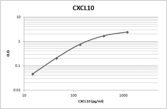
Human CXCL10 / IP-10 / CRG-2 ELISA Standard Curve
Recombinant Human CXCL10/IP‑10/CRG‑2 (Catalog # 266-IP) was serially diluted and captured by Mouse Anti-Human CXCL10/IP‑10/CRG‑2 Monoclonal Antibody (Catalog # MAB266) coated on a Clear Polystyrene Microplate (Catalog # DY990). Goat Anti-Human CXCL10/IP‑10/CRG‑2 Antigen Affinity-purified Polyclonal Antibody (Catalog # AF-266-NA) was biotinylated and incubated with the protein captured on the plate. Detection of the standard curve was achieved by incubating Streptavidin-HRP (Catalog # DY998)Applications for Human CXCL10/IP-10/CRG-2 Antibody (33036)
Application
Recommended Usage
CyTOF-ready
Ready to be labeled using established conjugation methods. No BSA or other carrier proteins that could interfere with conjugation.
Intracellular Staining by Flow Cytometry
2.5 µg/106 cells
Sample: Human peripheral blood monocytes treated with Recombinant Human IFN‑ gamma (Catalog # 285‑IF), fixed with paraformaldehyde, and permeabilized with saponin
Sample: Human peripheral blood monocytes treated with Recombinant Human IFN‑ gamma (Catalog # 285‑IF), fixed with paraformaldehyde, and permeabilized with saponin
Neutralization
Measured by its ability to neutralize CXCL10/IP‑10/CRG‑2-induced chemotaxis in the BaF3 mouse pro‑B cell line transfected with human CXCR3. The Neutralization Dose (ND50) is typically 0.5-2.0 µg/mL in the presence of 0.2 µg/mL Recombinant Human CXCL10/IP‑10/CRG‑2.
Human CXCL10/IP-10 Sandwich Immunoassay
Please Note: Optimal dilutions of this antibody should be experimentally determined.
Reviewed Applications
Read 5 reviews rated 4.6 using MAB266 in the following applications:
Flow Cytometry Panel Builder
Bio-Techne Knows Flow Cytometry
Save time and reduce costly mistakes by quickly finding compatible reagents using the Panel Builder Tool.
Advanced Features
- Spectra Viewer - Custom analysis of spectra from multiple fluorochromes
- Spillover Popups - Visualize the spectra of individual fluorochromes
- Antigen Density Selector - Match fluorochrome brightness with antigen density

Formulation, Preparation, and Storage
Purification
Protein A or G purified from ascites
Reconstitution
Reconstitute at 0.5 mg/mL in sterile PBS. For liquid material, refer to CoA for concentration.
Loading...
Formulation
Lyophilized from a 0.2 μm filtered solution in PBS with Trehalose. *Small pack size (SP) is supplied either lyophilized or as a 0.2 µm filtered solution in PBS.
Shipping
Lyophilized product is shipped at ambient temperature. Liquid small pack size (-SP) is shipped with polar packs. Upon receipt, store immediately at the temperature recommended below.
Stability & Storage
Use a manual defrost freezer and avoid repeated freeze-thaw cycles.
- 12 months from date of receipt, -20 to -70 °C as supplied.
- 1 month, 2 to 8 °C under sterile conditions after reconstitution.
- 6 months, -20 to -70 °C under sterile conditions after reconstitution.
Calculators
Background: CXCL10/IP-10/CRG-2
References
- Loetscher, M. et al. (1996) J. Exp. Med. 184:963.
- Wang, X. et al. (1996) J. Biol. Chem. 271:24286.
Alternate Names
CRG-2, CRG2, IP-10
Gene Symbol
CXCL10
UniProt
Additional CXCL10/IP-10/CRG-2 Products
Product Documents for Human CXCL10/IP-10/CRG-2 Antibody (33036)
Certificate of Analysis
To download a Certificate of Analysis, please enter a lot or batch number in the search box below.
Note: Certificate of Analysis not available for kit components.
Product Specific Notices for Human CXCL10/IP-10/CRG-2 Antibody (33036)
For research use only
Citations for Human CXCL10/IP-10/CRG-2 Antibody (33036)
 Powered by Bioz
Customer Reviews for Human CXCL10/IP-10/CRG-2 Antibody (33036) (5)
4.6 out of 5
5 Customer Ratings
Have you used Human CXCL10/IP-10/CRG-2 Antibody (33036)?
Submit a review and receive an Amazon gift card!
$25/€18/£15/$25CAN/¥2500 Yen for a review with an image
$10/€7/£6/$10CAN/¥1110 Yen for a review without an image
Submit a review
Customer Images

Showing
1
-
5 of
5 reviews
Showing All
Filter By:
-
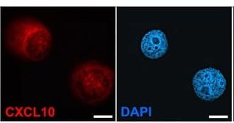
Application: Immunocytochemistry/ImmunofluorescenceSample Tested: Breast cancer cellsSpecies: HumanVerified Customer | Posted 11/12/2025
 Bio-Techne ResponseThis review reflects a new species or application tested on a primary antibody.
 -
Application: ELISASample Tested: SerumSpecies: PrimateVerified Customer | Posted 11/28/2022Worked as capture antibody
-
Application: ELISASample Tested: EDTA PlasmaSpecies: HumanVerified Customer | Posted 08/17/2018
-
Application: ELISASample Tested: PlasmaSpecies: HumanVerified Customer | Posted 07/13/2018
-
Application: ELISASample Tested: Serum and PlasmaSpecies: HumanVerified Customer | Posted 11/17/2017Sandwich ELISA using MAB266 on solid phase and BAF266 as detection.
There are no reviews that match your criteria.
Protocols
Find general support by application which include: protocols, troubleshooting, illustrated assays, videos and webinars.
- 7-Amino Actinomycin D (7-AAD) Cell Viability Flow Cytometry Protocol
- Extracellular Membrane Flow Cytometry Protocol
- Flow Cytometry Protocol for Cell Surface Markers
- Flow Cytometry Protocol for Staining Membrane Associated Proteins
- Flow Cytometry Staining Protocols
- Flow Cytometry Troubleshooting Guide
- Intracellular Flow Cytometry Protocol Using Alcohol (Methanol)
- Intracellular Flow Cytometry Protocol Using Detergents
- Intracellular Nuclear Staining Flow Cytometry Protocol Using Detergents
- Intracellular Staining Flow Cytometry Protocol Using Alcohol Permeabilization
- Intracellular Staining Flow Cytometry Protocol Using Detergents to Permeabilize Cells
- Propidium Iodide Cell Viability Flow Cytometry Protocol
- Protocol for Liperfluo
- Protocol for the Characterization of Human Th22 Cells
- Protocol for the Characterization of Human Th9 Cells
- Protocol: Annexin V and PI Staining by Flow Cytometry
- Protocol: Annexin V and PI Staining for Apoptosis by Flow Cytometry
- Troubleshooting Guide: Fluorokine Flow Cytometry Kits
- View all Protocols, Troubleshooting, Illustrated assays and Webinars
Loading...
Associated Pathways
