Cyclin D1 is a 44 kDa cell cycle regulatory protein whose expression level and nuclear/cytoplasmic distribution are tightly regulated in synchrony with the cell cycle. Cyclin D1 complexes containing Cdk4 or Cdk6 induce phosphorylation of Rb, a requirement for progression through the G1/S cell cycle transition. Human Cyclin D1 shares 93% amino acid sequence identity with mouse and rat Cyclin D1.
Human Cyclin D1 Antibody (328303)
R&D Systems | Catalog # MAB4314

Key Product Details
Species Reactivity
Validated:
Human
Cited:
Human, Mouse
Applications
Validated:
Immunocytochemistry
Cited:
Western Blot
Label
Unconjugated
Antibody Source
Monoclonal Mouse IgG1 Clone # 328303
Loading...
Product Specifications
Immunogen
E. coli-derived recombinant human Cyclin D1
Met1-Ile295
Accession # P24385
Met1-Ile295
Accession # P24385
Specificity
Detects human Cyclin D1 in direct ELISAs.
Clonality
Monoclonal
Host
Mouse
Isotype
IgG1
Scientific Data Images for Human Cyclin D1 Antibody (328303)
Cyclin D1 in COLO 205 Human Cell Line.
Cyclin D1 was detected in immersion fixed COLO 205 human colorectal adenocarcinoma cell line using Mouse Anti-Human Cyclin D1 Monoclonal Antibody (Catalog # MAB4314) at 10 µg/mL for 3 hours at room temperature. Cells were stained using the NorthernLights™ 557-conjugated Anti-Mouse IgG Secondary Antibody (yellow, upper panel; Catalog # NL007) and counterstained with DAPI (blue, lower panel). View our protocol for Fluorescent ICC Staining of Cells on Coverslips.
Detection of Cyclin D1 by Western Blot
LPS suppressed EPCs cell viability via inducing cell pyroptosis. (a, b) NLRP3 was silenced in EPCs cells. (c) MTT assay was used to examine cell viability. (d) The expression status of Cyclin D1 and CDK2 were determined by Western blot analysis. Individual experiment repeated 3 times, and *P < 0.05 Image collected and cropped by CiteAb from the following open publication (https://pubmed.ncbi.nlm.nih.gov/34738867), licensed under a CC-BY license. Not internally tested by R&D Systems.Applications for Human Cyclin D1 Antibody (328303)
Application
Recommended Usage
Immunocytochemistry
8-25 µg/mL
Sample: Immersion fixed COLO 205 human colorectal adenocarcinoma cell line
Sample: Immersion fixed COLO 205 human colorectal adenocarcinoma cell line
Reviewed Applications
Read 2 reviews rated 4 using MAB4314 in the following applications:
Formulation, Preparation, and Storage
Purification
Protein A or G purified from hybridoma culture supernatant
Reconstitution
Reconstitute at 0.5 mg/mL in sterile PBS. For liquid material, refer to CoA for concentration.
Loading...
Formulation
Lyophilized from a 0.2 μm filtered solution in PBS with Trehalose. See Certificate of Analysis for details.
*Small pack size (-SP) is supplied either lyophilized or as a 0.2 µm filtered solution in PBS.
*Small pack size (-SP) is supplied either lyophilized or as a 0.2 µm filtered solution in PBS.
Shipping
Lyophilized product is shipped at ambient temperature. Liquid small pack size (-SP) is shipped with polar packs. Upon receipt, store immediately at the temperature recommended below.
Stability & Storage
Use a manual defrost freezer and avoid repeated freeze-thaw cycles.
- 12 months from date of receipt, -20 to -70 °C as supplied.
- 1 month, 2 to 8 °C under sterile conditions after reconstitution.
- 6 months, -20 to -70 °C under sterile conditions after reconstitution.
Calculators
Background: Cyclin D1
Alternate Names
Bcl-1, CCND1, Cyl-1, PRAD1
Gene Symbol
CCND1
UniProt
Additional Cyclin D1 Products
Product Documents for Human Cyclin D1 Antibody (328303)
Certificate of Analysis
To download a Certificate of Analysis, please enter a lot or batch number in the search box below.
Note: Certificate of Analysis not available for kit components.
Product Specific Notices for Human Cyclin D1 Antibody (328303)
For research use only
Related Research Areas
Citations for Human Cyclin D1 Antibody (328303)
 Powered by Bioz
Customer Reviews for Human Cyclin D1 Antibody (328303) (2)
4 out of 5
2 Customer Ratings
Have you used Human Cyclin D1 Antibody (328303)?
Submit a review and receive an Amazon gift card!
$25/€18/£15/$25CAN/¥2500 Yen for a review with an image
$10/€7/£6/$10CAN/¥1110 Yen for a review without an image
Submit a review
Customer Images
Showing
1
-
2 of
2 reviews
Showing All
Filter By:
-
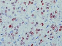
Application: ImmunohistochemistrySample Tested: Prostate carcinomaSpecies: HumanVerified Customer | Posted 11/19/2021

-
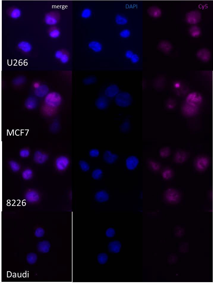
Application: ImmunocytochemistrySample Tested: U266Species: HumanVerified Customer | Posted 11/29/2016Immunocytochemistry/immunofluorescent staining of U266, MCF7, RPMI-8226 and Daudi with MAB4314.Cells immersion-fixed in 4% paraformaldehyde 10 minutes room temperature; permeabilized 0.5% Triton X-100 5 minutes; blocked 10% donkey serum/PBS 1 hour at room temperature; MAB4314 1/30 overnight at 4oC; Alexa 647 donkey F(ab')2 anti-mouse IgG 1/1000 1 hour at room termperature; images acquired on Zeiss AxioImager Cy5 filter 500ms exposure.
There are no reviews that match your criteria.
Protocols
Find general support by application which include: protocols, troubleshooting, illustrated assays, videos and webinars.
- Appropriate Fixation of IHC/ICC Samples
- Cellular Response to Hypoxia Protocols
- ClariTSA™ Fluorophore Kits
- Detection & Visualization of Antibody Binding
- ICC Cell Smear Protocol for Suspension Cells
- ICC Immunocytochemistry Protocol Videos
- ICC for Adherent Cells
- Immunocytochemistry (ICC) Protocol
- Immunocytochemistry Troubleshooting
- Immunofluorescence of Organoids Embedded in Cultrex Basement Membrane Extract
- Immunohistochemistry (IHC) and Immunocytochemistry (ICC) Protocols
- Preparing Samples for IHC/ICC Experiments
- Preventing Non-Specific Staining (Non-Specific Binding)
- Primary Antibody Selection & Optimization
- Protocol for VisUCyte™ HRP Polymer Detection Reagent
- Protocol for the Fluorescent ICC Staining of Cell Smears - Graphic
- Protocol for the Fluorescent ICC Staining of Cultured Cells on Coverslips - Graphic
- Protocol for the Preparation and Fluorescent ICC Staining of Cells on Coverslips
- Protocol for the Preparation and Fluorescent ICC Staining of Non-adherent Cells
- Protocol for the Preparation and Fluorescent ICC Staining of Stem Cells on Coverslips
- Protocol for the Preparation of a Cell Smear for Non-adherent Cell ICC - Graphic
- TUNEL and Active Caspase-3 Detection by IHC/ICC Protocol
- The Importance of IHC/ICC Controls
- View all Protocols, Troubleshooting, Illustrated assays and Webinars
Loading...
Associated Pathways

