Human FFAR3/GPR41 Antibody (2590G)
R&D Systems | Catalog # MAB10562
Recombinant Monoclonal Antibody.

Key Product Details
Species Reactivity
Human
Applications
Western Blot, Flow Cytometry, CyTOF-ready
Label
Unconjugated
Antibody Source
Recombinant Monoclonal Rabbit IgG Clone # 2590G
Loading...
Product Specifications
Immunogen
Synthetic peptide containing human FFAR3
Specificity
Detects human FFAR3 in direct ELISAs.
Clonality
Monoclonal
Host
Rabbit
Isotype
IgG
Scientific Data Images for Human FFAR3/GPR41 Antibody (2590G)
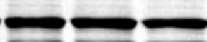
Detection of Human FFAR3/GPR41 by Western Blot.
Western blot shows lysates of A549 human lung carcinoma cell line and HeLa human cervical epithelial carcinoma cell line. PVDF membrane was probed with 1 µg/mL of Rabbit Anti-Human FFAR3/GPR41 Monoclonal Antibody (Catalog # MAB10562) followed by HRP-conjugated Anti-Rabbit IgG Secondary Antibody (HAF008). A specific band was detected for FFAR3/GPR41 at approximately 39 kDa (as indicated). This experiment was conducted under reducing conditions and using Western Blot Buffer Group 1.
Detection of FFAR3/GPR41 in U937 Human Cell Line by Flow Cytometry
U937 human histiocytic lymphoma cell line was stained with Rabbit anti-human FFAR3/GPR41 monoclonal antibody (Catalog # MAB10562, open histogram) or isotype control antibody (MAB1050, open histogram) followed by Allophycocyanin-conjugated anti-Rabbit IgG Secondary Antibody (F0111). Staining was performed using our Staining Membrane-Associated Proteins protocol.Applications for Human FFAR3/GPR41 Antibody (2590G)
Application
Recommended Usage
CyTOF-ready
Ready to be labeled using established conjugation methods. No BSA or other carrier proteins that could interfere with conjugation.
Flow Cytometry
0.25 µg/106 cells
Sample: U937 human histiocytic lymphoma cell line
Sample: U937 human histiocytic lymphoma cell line
Western Blot
1 µg/mL
Sample: A549 human lung carcinoma cell line and HeLa human cervical epithelial carcinoma cell line
Sample: A549 human lung carcinoma cell line and HeLa human cervical epithelial carcinoma cell line
Reviewed Applications
Read 1 review rated 5 using MAB10562 in the following applications:
Flow Cytometry Panel Builder
Bio-Techne Knows Flow Cytometry
Save time and reduce costly mistakes by quickly finding compatible reagents using the Panel Builder Tool.
Advanced Features
- Spectra Viewer - Custom analysis of spectra from multiple fluorochromes
- Spillover Popups - Visualize the spectra of individual fluorochromes
- Antigen Density Selector - Match fluorochrome brightness with antigen density

Formulation, Preparation, and Storage
Purification
Protein A or G purified from cell culture supernatant
Reconstitution
Reconstitute at 0.5 mg/mL in sterile PBS. For liquid material, refer to CoA for concentration.
Loading...
Formulation
Lyophilized from a 0.2 μm filtered solution in PBS with Trehalose. *Small pack size (SP) is supplied either lyophilized or as a 0.2 µm filtered solution in PBS.
Shipping
Lyophilized product is shipped at ambient temperature. Liquid small pack size (-SP) is shipped with polar packs. Upon receipt, store immediately at the temperature recommended below.
Stability & Storage
Use a manual defrost freezer and avoid repeated freeze-thaw cycles.
- 12 months from date of receipt, -20 to -70 °C as supplied.
- 1 month, 2 to 8 °C under sterile conditions after reconstitution.
- 6 months, -20 to -70 °C under sterile conditions after reconstitution.
Calculators
Background: FFAR3/GPR41
Long Name
Free Fatty Acid Receptor 3
Alternate Names
FFA3, FFA3R, GPR41
Gene Symbol
FFAR3
Additional FFAR3/GPR41 Products
Product Documents for Human FFAR3/GPR41 Antibody (2590G)
Certificate of Analysis
To download a Certificate of Analysis, please enter a lot or batch number in the search box below.
Note: Certificate of Analysis not available for kit components.
Product Specific Notices for Human FFAR3/GPR41 Antibody (2590G)
For research use only
Related Research Areas
Customer Reviews for Human FFAR3/GPR41 Antibody (2590G) (1)
5 out of 5
1 Customer Rating
Have you used Human FFAR3/GPR41 Antibody (2590G)?
Submit a review and receive an Amazon gift card!
$25/€18/£15/$25CAN/¥2500 Yen for a review with an image
$10/€7/£6/$10CAN/¥1110 Yen for a review without an image
Submit a review
Customer Images
Showing
1
-
1 of
1 review
Showing All
Filter By:
-
Application: Western BlotSample Tested: A549 human lung carcinoma cell lineSpecies: HumanVerified Customer | Posted 09/21/2021

There are no reviews that match your criteria.
Protocols
Find general support by application which include: protocols, troubleshooting, illustrated assays, videos and webinars.
- 7-Amino Actinomycin D (7-AAD) Cell Viability Flow Cytometry Protocol
- Cellular Response to Hypoxia Protocols
- Extracellular Membrane Flow Cytometry Protocol
- Flow Cytometry Protocol for Cell Surface Markers
- Flow Cytometry Protocol for Staining Membrane Associated Proteins
- Flow Cytometry Staining Protocols
- Flow Cytometry Troubleshooting Guide
- Intracellular Flow Cytometry Protocol Using Alcohol (Methanol)
- Intracellular Flow Cytometry Protocol Using Detergents
- Intracellular Nuclear Staining Flow Cytometry Protocol Using Detergents
- Intracellular Staining Flow Cytometry Protocol Using Alcohol Permeabilization
- Intracellular Staining Flow Cytometry Protocol Using Detergents to Permeabilize Cells
- Propidium Iodide Cell Viability Flow Cytometry Protocol
- Protocol for Liperfluo
- Protocol for the Characterization of Human Th22 Cells
- Protocol for the Characterization of Human Th9 Cells
- Protocol: Annexin V and PI Staining by Flow Cytometry
- Protocol: Annexin V and PI Staining for Apoptosis by Flow Cytometry
- R&D Systems Quality Control Western Blot Protocol
- Troubleshooting Guide: Fluorokine Flow Cytometry Kits
- Troubleshooting Guide: Western Blot Figures
- Western Blot Conditions
- Western Blot Protocol
- Western Blot Protocol for Cell Lysates
- Western Blot Troubleshooting
- Western Blot Troubleshooting Guide
- View all Protocols, Troubleshooting, Illustrated assays and Webinars
Loading...