GPR56 is a member of the LN-TM7 family of adhesion-type 7-transmembrane (TM) G-protein coupled receptors (GPCR) with long extracellular N-termini (1‑3). The 693 amino acid (aa) human GPR56 contains a 25 aa signal sequence, a 377 aa N-terminal extracellular domain (ECD) and seven TM regions separated by short intracellular and extracellular regions. Like other LN-TM7 members, the ECD contains a highly glycosylated mucin-like stalk followed by a GPCR proteolytic cleavage site (GPS) (1, 4). Cleavage of the 60 kDa N-terminus from the 80 kDa full length form is needed for efficient cell surface expression (5, 6). While the cleaved portion may remain non-covalently associated, it has also been found in conditioned medium of cultured cells (5). Human GPR56 shares 71%, 72%, 80%, 80% and 79% aa identity with mouse, rat, canine, equine, and bovine GPR56 within the cleaved ECD. A functional splice variant lacking the GPS site and a non-functional splice variant lacking portions of the TM domains have also been described (4). A human brain developmental disorder, bilateral frontoparietal polymicrogyria, is associated with GPR56 mutations that also show impaired GPS cleavage, intracellular trafficking, and expression at the cell surface (5). GPR56 is widely distributed, with highest mRNA or expressed sequence tag expression in brain, thyroid, skin and female reproductive system (3, 4). GPR56 expression is upregulated during cell transformation and is high in melanomas, glioblastomas and astrocytomas, but downregulated in melanomas with high metastatic potential (2, 6‑8). Although the function of GPR56 is not completely known, it is clearly an adhesion protein (6‑8). Tissue transglutaminase (TG2) is one reported ligand, binding of which inhibits melanoma growth and metastasis (6). Association of GPR56 with the tetraspanin CD81 stabilizes its complex with Gaq/11 for cell signaling (9).
Human GPR56 Antibody (2446B)
R&D Systems | Catalog # MAB46361
Recombinant Monoclonal Antibody.

Key Product Details
Species Reactivity
Human
Applications
Western Blot, Flow Cytometry, Immunocytochemistry, CyTOF-ready
Label
Unconjugated
Antibody Source
Recombinant Monoclonal Rabbit IgG Clone # 2446B
Loading...
Product Specifications
Immunogen
Chinese hamster ovary cell line CHO-derived recombinant human GPR56
Met1-Val345
Accession # Q9Y653
Met1-Val345
Accession # Q9Y653
Specificity
Detects human GPR56 in direct ELISAs.
Clonality
Monoclonal
Host
Rabbit
Isotype
IgG
Scientific Data Images for Human GPR56 Antibody (2446B)
Detection of Human GPR56 by Western Blot.
Western blot shows lysates of NS0 mouse myeloma cell line either mock transfected or transfected with human GPR56. PVDF membrane was probed with 2 µg/mL of Rabbit Anti-Human GPR56 Monoclonal Antibody (Catalog # MAB46361) followed by HRP-conjugated Anti-Rabbit IgG Secondary Antibody (Catalog # HAF008). A specific band was detected for GPR56 at approximately 75 kDa (as indicated). GAPDH (Catalog # AF5718) is shown as a loading control.This experiment was conducted under reducing conditions and using Immunoblot Buffer Group 1.
Detection of GPR56 in Human Peripheral Blood Cells by Flow Cytometry.
Human peripheral blood cells were stained with (A) Rabbit Anti-Human GPR56 Monoclonal Antibody (Catalog # MAB46361) or (B) Rabbit IgG isotype control antibody (Catalog # MAB1050) followed by APC-conjugated Anti-Rabbit IgG Secondary Antibody (Catalog # F0111) and Mouse anti-Human CD56 PE-conjugated monoclonal antibody (Catalog # FAB2408P). View our protocol for Staining Membrane-associated Proteins.
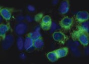
GPR56 in T47D human breast cancer cell line.
GPR56 was detected in immersion fixed T47D human breast cancer cell line (left panel; positive staining) and WM-115 human malignant melanoma cell line (right panel; negative staining) using Rabbit Anti-Human GPR56 Monoclonal Antibody (Catalog # MAB46361) at 3 µg/mL for 3 hours at room temperature. Cells were stained using the NorthernLights™ 557-conjugated Anti-Rabbit IgG Secondary Antibody (red; Catalog # NL004) and counterstained with DAPI (blue). Specific staining was localized to cytoplasm. View our protocol for Fluorescent ICC Staining of Cells on Coverslips.Applications for Human GPR56 Antibody (2446B)
Application
Recommended Usage
CyTOF-ready
Ready to be labeled using established conjugation methods. No BSA or other carrier proteins that could interfere with conjugation.
Flow Cytometry
0.25 µg/106 cells
Sample: Human Peripheral Blood Cells
Sample: Human Peripheral Blood Cells
Immunocytochemistry
3-25 µg/mL
Sample: Immersion fixed T47D human breast cancer cell line
Sample: Immersion fixed T47D human breast cancer cell line
Western Blot
2 µg/mL
Sample: NS0 mouse myeloma cell line transfected with human GPR56
Sample: NS0 mouse myeloma cell line transfected with human GPR56
Reviewed Applications
Read 1 review rated 5 using MAB46361 in the following applications:
Flow Cytometry Panel Builder
Bio-Techne Knows Flow Cytometry
Save time and reduce costly mistakes by quickly finding compatible reagents using the Panel Builder Tool.
Advanced Features
- Spectra Viewer - Custom analysis of spectra from multiple fluorochromes
- Spillover Popups - Visualize the spectra of individual fluorochromes
- Antigen Density Selector - Match fluorochrome brightness with antigen density

Formulation, Preparation, and Storage
Purification
Protein A or G purified from cell culture supernatant
Reconstitution
Reconstitute at 0.5 mg/mL in sterile PBS. For liquid material, refer to CoA for concentration.
Loading...
Formulation
Lyophilized from a 0.2 μm filtered solution in PBS with Trehalose. *Small pack size (SP) is supplied either lyophilized or as a 0.2 µm filtered solution in PBS.
Shipping
Lyophilized product is shipped at ambient temperature. Liquid small pack size (-SP) is shipped with polar packs. Upon receipt, store immediately at the temperature recommended below.
Stability & Storage
Use a manual defrost freezer and avoid repeated freeze-thaw cycles.
- 12 months from date of receipt, -20 to -70 °C as supplied.
- 1 month, 2 to 8 °C under sterile conditions after reconstitution.
- 6 months, -20 to -70 °C under sterile conditions after reconstitution.
Calculators
Background: GPR56
References
- Fredriksson, R. et al. (2002) FEBS Lett. 531:407.
- Zendman, A.J.W. et al. (1999) FEBS Lett. 446:292.
- Liu, M. et al. (1999) Genomics 55:296.
- Bjarnadottir, T.K. et al. (2007) Gene 387:38.
- Jin, Z. et al. (2007) Hum. Mol. Genet. 16:1972.
- Xu, L. et al. (2006) Proc. Natl. Acad. Sci. USA 103:9023.
- Shashidhar, S. et al. (2005) Oncogene 24:1673.
- Ke, N. et al. (2007) Mol. Cancer Ther. 6:1840.
- Little, K.D. et al. (2004) Mol. Biol. Cell 15:2375.
Long Name
G Protein-coupled Receptor 56
Alternate Names
BFPP, EGF-TM7-like, TM7LN4, TM7XN1
Gene Symbol
ADGRG1
UniProt
Additional GPR56 Products
Product Documents for Human GPR56 Antibody (2446B)
Certificate of Analysis
To download a Certificate of Analysis, please enter a lot or batch number in the search box below.
Note: Certificate of Analysis not available for kit components.
Product Specific Notices for Human GPR56 Antibody (2446B)
For research use only
Related Research Areas
Customer Reviews for Human GPR56 Antibody (2446B) (1)
5 out of 5
1 Customer Rating
Have you used Human GPR56 Antibody (2446B)?
Submit a review and receive an Amazon gift card!
$25/€18/£15/$25CAN/¥2500 Yen for a review with an image
$10/€7/£6/$10CAN/¥1110 Yen for a review without an image
Submit a review
Customer Images
Showing
1
-
1 of
1 review
Showing All
Filter By:
-
Application: Immunocytochemistry/ImmunofluorescenceSample Tested: neuroblastoma cellsSpecies: HumanVerified Customer | Posted 10/22/2022

There are no reviews that match your criteria.
Protocols
Find general support by application which include: protocols, troubleshooting, illustrated assays, videos and webinars.
- 7-Amino Actinomycin D (7-AAD) Cell Viability Flow Cytometry Protocol
- Appropriate Fixation of IHC/ICC Samples
- Cellular Response to Hypoxia Protocols
- ClariTSA™ Fluorophore Kits
- Detection & Visualization of Antibody Binding
- Extracellular Membrane Flow Cytometry Protocol
- Flow Cytometry Protocol for Cell Surface Markers
- Flow Cytometry Protocol for Staining Membrane Associated Proteins
- Flow Cytometry Staining Protocols
- Flow Cytometry Troubleshooting Guide
- ICC Cell Smear Protocol for Suspension Cells
- ICC Immunocytochemistry Protocol Videos
- ICC for Adherent Cells
- Immunocytochemistry (ICC) Protocol
- Immunocytochemistry Troubleshooting
- Immunofluorescence of Organoids Embedded in Cultrex Basement Membrane Extract
- Immunohistochemistry (IHC) and Immunocytochemistry (ICC) Protocols
- Intracellular Flow Cytometry Protocol Using Alcohol (Methanol)
- Intracellular Flow Cytometry Protocol Using Detergents
- Intracellular Nuclear Staining Flow Cytometry Protocol Using Detergents
- Intracellular Staining Flow Cytometry Protocol Using Alcohol Permeabilization
- Intracellular Staining Flow Cytometry Protocol Using Detergents to Permeabilize Cells
- Preparing Samples for IHC/ICC Experiments
- Preventing Non-Specific Staining (Non-Specific Binding)
- Primary Antibody Selection & Optimization
- Propidium Iodide Cell Viability Flow Cytometry Protocol
- Protocol for Liperfluo
- Protocol for VisUCyte™ HRP Polymer Detection Reagent
- Protocol for the Characterization of Human Th22 Cells
- Protocol for the Characterization of Human Th9 Cells
- Protocol for the Fluorescent ICC Staining of Cell Smears - Graphic
- Protocol for the Fluorescent ICC Staining of Cultured Cells on Coverslips - Graphic
- Protocol for the Preparation and Fluorescent ICC Staining of Cells on Coverslips
- Protocol for the Preparation and Fluorescent ICC Staining of Non-adherent Cells
- Protocol for the Preparation and Fluorescent ICC Staining of Stem Cells on Coverslips
- Protocol for the Preparation of a Cell Smear for Non-adherent Cell ICC - Graphic
- Protocol: Annexin V and PI Staining by Flow Cytometry
- Protocol: Annexin V and PI Staining for Apoptosis by Flow Cytometry
- R&D Systems Quality Control Western Blot Protocol
- TUNEL and Active Caspase-3 Detection by IHC/ICC Protocol
- The Importance of IHC/ICC Controls
- Troubleshooting Guide: Fluorokine Flow Cytometry Kits
- Troubleshooting Guide: Western Blot Figures
- Western Blot Conditions
- Western Blot Protocol
- Western Blot Protocol for Cell Lysates
- Western Blot Troubleshooting
- Western Blot Troubleshooting Guide
- View all Protocols, Troubleshooting, Illustrated assays and Webinars
Loading...