HAPLN1 (also known as link protein and CRTL1) is a member of the hyaladherin family of hyaluronic acid (HA) binding proteins. Hyaluronan binding proteins are of two types; those with link modules, and those without. Link modules are 100 amino acid (aa) HA and protein-binding sequences that contain two alpha -helices and two antiparallel beta -sheets (1, 3). There are three categories of link module-containing proteins. “A” domain-type proteins contain one link module; “B” domain-type proteins contain one link module with an N- and C-terminal flanking region; and “C” domain-type proteins have an extended structure with one N-terminal V-type Ig-like domain followed by two link modules (2). The HAPLN family is a group of four C domain-type proteins that share approximately 50% aa identity (4). HAPLN1 is synthesized as a 354 aa precursor that contains a 15 aa signal sequence and a 339 aa mature region (4 - 6). It contains one Ig-like domain and two 95 aa link modules (6). It is variably glycosylated with a native molecular weight between 41 - 48 kDa (7, 8). Mature human HAPLN1 is 97%, 96%, 96%, 96%, and 96% aa identical to equine, porcine, rat, mouse and bovine HAPLN1, respectively. HAPLN1 contributes to extracellular matrix stability and flexibility (9). In cartilage, HALPN1 forms a ternary complex with HA and aggrecan. This creates a gel-like substance with remarkable resistance to deformation (3). In this complex, HA forms a linear backbone with perpendicularly attached aggrecan and HAPLN1. Aggrecan and HAPLN1 lie parallel to each other, while HA runs between the two HAPLN1 link modules (2, 3, 10). The Ig domain of HAPLN1 binds to aggrecan, while the two link modules of HAPLN1 bind to HA. Although HA and aggrecan will associate, the tendency is towards dissociation (2, 3, 8). HAPLN1 provides a stabilizing influence on HA-aggrecan associations, thus creating a long-lived ternary functional complex.

Key Product Details
Species Reactivity
Validated:
Human
Cited:
Human, Mouse, Rat, Transgenic Mouse
Applications
Validated:
Western Blot
Cited:
Immunohistochemistry, Immunohistochemistry-Frozen, Western Blot, Immunocytochemistry, ELISA Capture
Label
Unconjugated
Antibody Source
Polyclonal Goat IgG
Loading...
Product Specifications
Immunogen
Mouse myeloma cell line NS0-derived recombinant human HAPLN1 (R&D Systems, Catalog # 2608-HP)
Asp16-Asn354
Accession # P10915
Asp16-Asn354
Accession # P10915
Specificity
Detects human HAPLN1 in direct ELISAs and Western blots. In these formats, this antibody shows approximately 5% cross‑reactivity with recombinant human HAPLN4.
Clonality
Polyclonal
Host
Goat
Isotype
IgG
Scientific Data Images for Human HAPLN1 Antibody
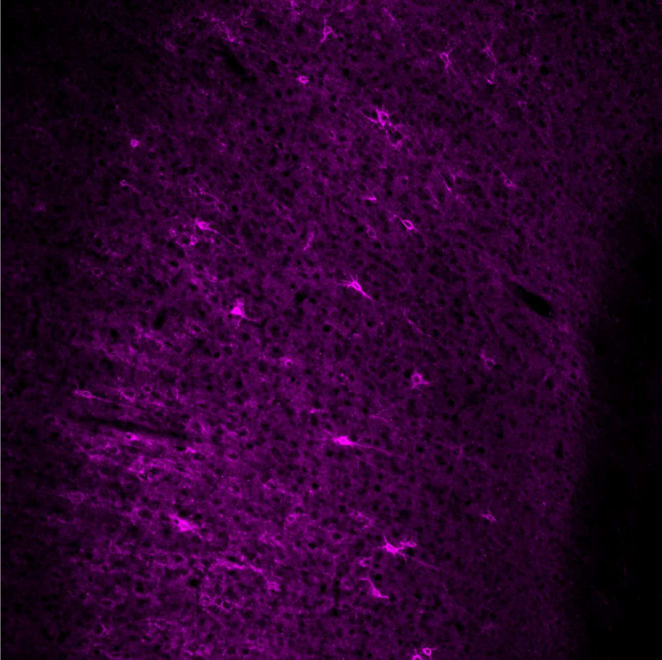
Detection of Human HAPLN1 by Immunocytochemistry/Immunofluorescence
Absent or fragmented omnipause neuron perineuronal net triple immunofluorescence staining.Triple immunofluorescence staining for different components of perineuronal nets, revealed by a confocal laser scanning microscope. In the control case, omnipause neurons (OPN) are ensheathed by prominent perineuronal nets showing the same appearance with antibodies against the link protein (HPLN1), chondroitin sulfate proteoglycan (CSPG) and aggrecan (ACAN) (A, D, G, arrow). In the saccadic palsy patient, the neurons of the superior olive (SO) from the same sections as OPN are ensheathed by prominent perineuronal nets revealed by immunostaining of HPLN1, CSPG, and ACAN (C, F, I, arrows). However, around OPN (asterisk) in the patient, only HPLN1-based perineuronal nets can be detected, which appear fragmented (B, arrow). CSPG- and ACAN-immunostaining does not reveal perineuronal nets, but only few fragments along a few dendrites (E, H, arrow). Scale bars A,D,G = 20μm; B,C,E,F,H,I = 200μm. Image collected and cropped by CiteAb from the following publication (https://pubmed.ncbi.nlm.nih.gov/26135580), licensed under a CC-BY license. Not internally tested by R&D Systems.
Detection of Human HAPLN1 by Immunocytochemistry/Immunofluorescence
Absent or fragmented omnipause neuron perineuronal net triple immunofluorescence staining.Triple immunofluorescence staining for different components of perineuronal nets, revealed by a confocal laser scanning microscope. In the control case, omnipause neurons (OPN) are ensheathed by prominent perineuronal nets showing the same appearance with antibodies against the link protein (HPLN1), chondroitin sulfate proteoglycan (CSPG) and aggrecan (ACAN) (A, D, G, arrow). In the saccadic palsy patient, the neurons of the superior olive (SO) from the same sections as OPN are ensheathed by prominent perineuronal nets revealed by immunostaining of HPLN1, CSPG, and ACAN (C, F, I, arrows). However, around OPN (asterisk) in the patient, only HPLN1-based perineuronal nets can be detected, which appear fragmented (B, arrow). CSPG- and ACAN-immunostaining does not reveal perineuronal nets, but only few fragments along a few dendrites (E, H, arrow). Scale bars A,D,G = 20μm; B,C,E,F,H,I = 200μm. Image collected and cropped by CiteAb from the following publication (https://pubmed.ncbi.nlm.nih.gov/26135580), licensed under a CC-BY license. Not internally tested by R&D Systems.Applications for Human HAPLN1 Antibody
Application
Recommended Usage
Western Blot
0.1 µg/mL
Sample: Recombinant Human HAPLN1 (Catalog # 2608-HP)
Sample: Recombinant Human HAPLN1 (Catalog # 2608-HP)
Reviewed Applications
Read 2 reviews rated 5 using AF2608 in the following applications:
Formulation, Preparation, and Storage
Purification
Antigen Affinity-purified
Reconstitution
Reconstitute at 0.2 mg/mL in sterile PBS. For liquid material, refer to CoA for concentration.
Loading...
Formulation
Lyophilized from a 0.2 μm filtered solution in PBS with Trehalose. *Small pack size (SP) is supplied either lyophilized or as a 0.2 µm filtered solution in PBS.
Shipping
Lyophilized product is shipped at ambient temperature. Liquid small pack size (-SP) is shipped with polar packs. Upon receipt, store immediately at the temperature recommended below.
Stability & Storage
Use a manual defrost freezer and avoid repeated freeze-thaw cycles.
- 12 months from date of receipt, -20 to -70 °C as supplied.
- 1 month, 2 to 8 °C under sterile conditions after reconstitution.
- 6 months, -20 to -70 °C under sterile conditions after reconstitution.
Calculators
Background: HAPLN1
References
- Day, A.J. and G.D. Prestwich (2002) J. Biol. Chem. 277:4585.
- Seyfried, N.T. et al. (2005) J. Biol. Chem. 280:5435.
- Matsumoto, K. et al. (2003) J. Biol. Chem. 278:41205.
- Spicer, A.P. et al. (2003) J. Biol. Chem. 278:21083.
- Dudhia, J. and T.E. Hardingham (1990) Nucleic Acids Res. 18:1292.
- Osborne-Lawrence, S.L. et al. (1990) Genomics 8:562.
- Roughley, P.J. et al. (1982) J. Biol. Chem. 257:11908.
- Shi, S. et al. (2004) J. Biol. Chem. 279:12060.
- Binette, F. et al. (1994) J. Biol. Chem. 269:19116.
- Perkins, S.J. et al. (1992) Biochem. J. 285:263.
Long Name
Hyaluronan and Proteoglycan Link Protein 1
Alternate Names
CRTL1
Entrez Gene IDs
1404 (Human)
Gene Symbol
HAPLN1
UniProt
Additional HAPLN1 Products
Product Documents for Human HAPLN1 Antibody
Certificate of Analysis
To download a Certificate of Analysis, please enter a lot or batch number in the search box below.
Note: Certificate of Analysis not available for kit components.
Product Specific Notices for Human HAPLN1 Antibody
For research use only
Related Research Areas
Citations for Human HAPLN1 Antibody
 Powered by Bioz
Customer Reviews for Human HAPLN1 Antibody (2)
5 out of 5
2 Customer Ratings
Have you used Human HAPLN1 Antibody?
Submit a review and receive an Amazon gift card!
$25/€18/£15/$25CAN/¥2500 Yen for a review with an image
$10/€7/£6/$10CAN/¥1110 Yen for a review without an image
Submit a review
Customer Images
Showing
1
-
2 of
2 reviews
Showing All
Filter By:
-
Application: Immunocytochemistry/ImmunofluorescenceSample Tested: Adult brainSpecies: Rat and MouseVerified Customer | Posted 04/10/2018Very strong and specific staining.

-
Application: Western BlotSample Tested: See PMID 23940048Species: RatVerified Customer | Posted 01/07/2015
There are no reviews that match your criteria.
Protocols
Find general support by application which include: protocols, troubleshooting, illustrated assays, videos and webinars.
- Cellular Response to Hypoxia Protocols
- R&D Systems Quality Control Western Blot Protocol
- Troubleshooting Guide: Western Blot Figures
- Western Blot Conditions
- Western Blot Protocol
- Western Blot Protocol for Cell Lysates
- Western Blot Troubleshooting
- Western Blot Troubleshooting Guide
- View all Protocols, Troubleshooting, Illustrated assays and Webinars
Loading...
Associated Pathways
