Interleukin 4 induced protein 1 (IL-4I1), also known as protein FIG-1 and L-amino acid oxidase, is encoded by a B-cell IL-4-inducible gene, FIG1, and is highly expressed in primary metastinal B-cell lymphomas (1-4). It belongs to the flavin monoamine oxidase family, FIG1 subfamily. Enzymological characterization reveals that IL-4I1 has L-amino acid oxidase activity with preference toward aromatic amino acids. Studies have shown that hIL-4I1 inhibited the proliferation of CD3‑stimulated T lymphocytes with a similar effect on CD4(+) and CD8(+) T cells (5). Its inhibitory effect was dependent on enzymatic activity and H2O2 production. Its restricted expression to lymphoid tissues indicates that it may play an important function in the immune system (1, 4).

Key Product Details
Validated by
Biological Validation
Species Reactivity
Human
Applications
Immunohistochemistry, Western Blot, Immunocytochemistry
Label
Unconjugated
Antibody Source
Monoclonal Rat IgG2B Clone # 1006202
Loading...
Product Specifications
Immunogen
Chinese Hamster Ovary cell line, CHO-derived human IL-4I1
Met1-His567
Accession # Q96RQ9
Met1-His567
Accession # Q96RQ9
Specificity
Detects human IL-4I1 in direct ELISAs.
Clonality
Monoclonal
Host
Rat
Isotype
IgG2B
Scientific Data Images for Human IL-4I1 Antibody (1006202)
Detection of Human IL‑4I1 by Western Blot.
Western blot shows lysates of THP-1 human acute monocytic leukemia cell line untreated (-) or treated (+) with 200 nM PMA for 24 hours and 10 µg/mL LPS for 3 hours. PVDF membrane was probed with 2 µg/mL of Rat Anti-Human IL-4I1 Monoclonal Antibody (Catalog # MAB5684) followed by HRP-conjugated Anti-Rat IgG Secondary Antibody (Catalog # HAF005). A specific band was detected for IL-4I1 at approximately 75 kDa (as indicated). This experiment was conducted under reducing conditions and using Immunoblot Buffer Group 1.
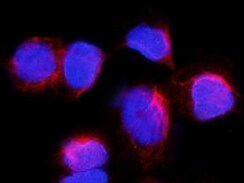
IL‑4I1 in HDLM‑2 Human Cell Line.
IL-4I1 was detected in immersion fixed HDLM-2 human Hodgkin's lymphoma cell line using Rat Anti-Human IL-4I1 Monoclonal Antibody (Catalog # MAB5684) at 8 µg/mL for 3 hours at room temperature. Cells were stained using the NorthernLights™ 557-conjugated Anti-Rat IgG Secondary Antibody (red; Catalog # NL013) and counterstained with DAPI (blue). Specific staining was localized to cytoplasm in lysosomes. View our protocol for Fluorescent ICC Staining of Non-adherent Cells.
IL‑4I1 in Human B Cell Lymphoma.
IL-4I1 was detected in immersion fixed paraffin-embedded sections of human B cell lymphoma using Rat Anti-Human IL-4I1 Monoclonal Antibody (Catalog # MAB5684) at 5 µg/mL for 1 hour at room temperature followed by incubation with the Anti-Rat IgG VisUCyte™ HRP Polymer Antibody (Catalog # VC005). Before incubation with the primary antibody, tissue was subjected to heat-induced epitope retrieval using Antigen Retrieval Reagent-Basic (Catalog # CTS013). Tissue was stained using DAB (brown) and counterstained with hematoxylin (blue). Specific staining was localized to cytoplasm in lymphocytes. View our protocol for IHC Staining with VisUCyte HRP Polymer Detection Reagents.
Detection of IL-4I1 by Western Blot
IFN-gamma and TNF-alpha increase the expression of IL4I1 in MuSCs through NF-kappa B and STAT6 pathway.A Volcano plot of differentially expressed genes in MuSCs after the stimulation of IFN-gamma and TNF-alpha (10 ng/ml each) for 24 h using RNA-seq. B Volcano plot of differentially expressed genes in MSCs after the stimulation of IFN-gamma and TNF-alpha (10 ng/ml each) for 24 h using RNA-seq. C The mRNA expression of IL4I1 in MSCs and MuSCs after the stimulation of IFN-gamma and TNF-alpha (10 ng/ml each) for 24 h was assayed by qRT-PCR. D The concentration of IL4I1 in the supernatants of MuSCs after the stimulation of IFN-gamma and TNF-alpha (10 ng/ml each) for 24 h was measured by ELISA. E The protein expression of IL4I1 and beta -ACTIN (loading control) in MuSCs after the stimulation of IFN-gamma and TNF-alpha (10 ng/ml each) for 24 h was determined by western blotting. F The mRNA and protein expression levels of IL4I1 in MuSCs stimulated with IFN-gamma and TNF-alpha (10 ng/ml each) for 24 h in the presence or absence of AS1517499 were respectively assayed by qRT-PCR and western blotting. G The protein expression levels of IL4I1 in MuSCs stimulated with IFN-gamma and TNF-alpha (10 ng/ml each) for 24 h in the presence or absence of BAY117082 were assayed by western blotting. Data were shown as means ± SEM. Data were representative of three experiments with similar results. For two-group comparison, statistical analysis was performed by Student’s t test. ***P < 0.001; ****P < 0.0001. Image collected and cropped by CiteAb from the following open publication (https://pubmed.ncbi.nlm.nih.gov/37507432), licensed under a CC-BY license. Not internally tested by R&D Systems.
Detection of IL-4I1 by Western Blot
IL4I1 mediates the therapeutic effect of MuSCs on ALI.A The therapeutic strategy of MuSCs in the LPS-induced ALI model. Mice were treated with 2 mg/kg LPS through endotracheal infusion. 1 h later, Ctrl-shRNA MuSCs or IL4I1-shRNA MuSCs (5 × 10⁵) pretreated with IFN-gamma and TNF-alpha (10 ng/ml each) for 24 h were intravenously injected into mice. Then, all experimental mice were euthanized after 23 h, and the lung samples were collected for further processing. B The efficiency of IL4I1 knockdown measured by western blotting analysis. C The total amount of IL4I1 protein in the lung tissue homogenates of ALI mice was determined by ELISA (PBS: n = 3, LPS: n = 3, LPS+Ctrl-shRNA MuSCs: n = 3, LPS + IL4I1-shRNA MuSCs: n = 3). D The expression levels of IL-6 mRNA (left) and protein (right) in the lung tissue homogenates of ALI mice were respectively determined by qRT-PCR and ELISA. E Lung tissues of mice with various treatments were fixed for H&E staining. Yellow arrowhead, area of widespread septal thickening with increased air-space cellularity and exudation and enhanced interstitial immune cell infiltration in the damaged lungs of ALI mice. Scale bars, 250 μm. F The expression levels of chemokines in the lung tissue homogenates of ALI mice were determined by qRT-PCR. G Lung tissues of mice with various treatments were stained with CXCL1 antibody. Scale bars, 250 μm (PBS: n = 3, LPS: n = 4, LPS+Ctrl-shRNA MuSCs: n = 5, LPS + IL4I1-shRNA MuSCs: n = 5). Data were shown as means ± SEM. Data were representative of three experiments with similar results. For multiple group comparison, statistical analysis was performed by one-way ANOVA test. *P < 0.05; **P < 0.01; ***P < 0.001. Image collected and cropped by CiteAb from the following open publication (https://pubmed.ncbi.nlm.nih.gov/37507432), licensed under a CC-BY license. Not internally tested by R&D Systems.
Detection of IL-4I1 by Western Blot
IFN-gamma and TNF-alpha increase the expression of IL4I1 in MuSCs through NF-kappa B and STAT6 pathway.A Volcano plot of differentially expressed genes in MuSCs after the stimulation of IFN-gamma and TNF-alpha (10 ng/ml each) for 24 h using RNA-seq. B Volcano plot of differentially expressed genes in MSCs after the stimulation of IFN-gamma and TNF-alpha (10 ng/ml each) for 24 h using RNA-seq. C The mRNA expression of IL4I1 in MSCs and MuSCs after the stimulation of IFN-gamma and TNF-alpha (10 ng/ml each) for 24 h was assayed by qRT-PCR. D The concentration of IL4I1 in the supernatants of MuSCs after the stimulation of IFN-gamma and TNF-alpha (10 ng/ml each) for 24 h was measured by ELISA. E The protein expression of IL4I1 and beta -ACTIN (loading control) in MuSCs after the stimulation of IFN-gamma and TNF-alpha (10 ng/ml each) for 24 h was determined by western blotting. F The mRNA and protein expression levels of IL4I1 in MuSCs stimulated with IFN-gamma and TNF-alpha (10 ng/ml each) for 24 h in the presence or absence of AS1517499 were respectively assayed by qRT-PCR and western blotting. G The protein expression levels of IL4I1 in MuSCs stimulated with IFN-gamma and TNF-alpha (10 ng/ml each) for 24 h in the presence or absence of BAY117082 were assayed by western blotting. Data were shown as means ± SEM. Data were representative of three experiments with similar results. For two-group comparison, statistical analysis was performed by Student’s t test. ***P < 0.001; ****P < 0.0001. Image collected and cropped by CiteAb from the following open publication (https://pubmed.ncbi.nlm.nih.gov/37507432), licensed under a CC-BY license. Not internally tested by R&D Systems.
Detection of IL-4I1 by Western Blot
IL4I1 mediates the therapeutic effect of MuSCs on ALI.A The therapeutic strategy of MuSCs in the LPS-induced ALI model. Mice were treated with 2 mg/kg LPS through endotracheal infusion. 1 h later, Ctrl-shRNA MuSCs or IL4I1-shRNA MuSCs (5 × 10⁵) pretreated with IFN-gamma and TNF-alpha (10 ng/ml each) for 24 h were intravenously injected into mice. Then, all experimental mice were euthanized after 23 h, and the lung samples were collected for further processing. B The efficiency of IL4I1 knockdown measured by western blotting analysis. C The total amount of IL4I1 protein in the lung tissue homogenates of ALI mice was determined by ELISA (PBS: n = 3, LPS: n = 3, LPS+Ctrl-shRNA MuSCs: n = 3, LPS + IL4I1-shRNA MuSCs: n = 3). D The expression levels of IL-6 mRNA (left) and protein (right) in the lung tissue homogenates of ALI mice were respectively determined by qRT-PCR and ELISA. E Lung tissues of mice with various treatments were fixed for H&E staining. Yellow arrowhead, area of widespread septal thickening with increased air-space cellularity and exudation and enhanced interstitial immune cell infiltration in the damaged lungs of ALI mice. Scale bars, 250 μm. F The expression levels of chemokines in the lung tissue homogenates of ALI mice were determined by qRT-PCR. G Lung tissues of mice with various treatments were stained with CXCL1 antibody. Scale bars, 250 μm (PBS: n = 3, LPS: n = 4, LPS+Ctrl-shRNA MuSCs: n = 5, LPS + IL4I1-shRNA MuSCs: n = 5). Data were shown as means ± SEM. Data were representative of three experiments with similar results. For multiple group comparison, statistical analysis was performed by one-way ANOVA test. *P < 0.05; **P < 0.01; ***P < 0.001. Image collected and cropped by CiteAb from the following open publication (https://pubmed.ncbi.nlm.nih.gov/37507432), licensed under a CC-BY license. Not internally tested by R&D Systems.
Detection of IL-4I1 by Western Blot
IFN-gamma and TNF-alpha increase the expression of IL4I1 in MuSCs through NF-kappa B and STAT6 pathway.A Volcano plot of differentially expressed genes in MuSCs after the stimulation of IFN-gamma and TNF-alpha (10 ng/ml each) for 24 h using RNA-seq. B Volcano plot of differentially expressed genes in MSCs after the stimulation of IFN-gamma and TNF-alpha (10 ng/ml each) for 24 h using RNA-seq. C The mRNA expression of IL4I1 in MSCs and MuSCs after the stimulation of IFN-gamma and TNF-alpha (10 ng/ml each) for 24 h was assayed by qRT-PCR. D The concentration of IL4I1 in the supernatants of MuSCs after the stimulation of IFN-gamma and TNF-alpha (10 ng/ml each) for 24 h was measured by ELISA. E The protein expression of IL4I1 and beta -ACTIN (loading control) in MuSCs after the stimulation of IFN-gamma and TNF-alpha (10 ng/ml each) for 24 h was determined by western blotting. F The mRNA and protein expression levels of IL4I1 in MuSCs stimulated with IFN-gamma and TNF-alpha (10 ng/ml each) for 24 h in the presence or absence of AS1517499 were respectively assayed by qRT-PCR and western blotting. G The protein expression levels of IL4I1 in MuSCs stimulated with IFN-gamma and TNF-alpha (10 ng/ml each) for 24 h in the presence or absence of BAY117082 were assayed by western blotting. Data were shown as means ± SEM. Data were representative of three experiments with similar results. For two-group comparison, statistical analysis was performed by Student’s t test. ***P < 0.001; ****P < 0.0001. Image collected and cropped by CiteAb from the following open publication (https://pubmed.ncbi.nlm.nih.gov/37507432), licensed under a CC-BY license. Not internally tested by R&D Systems.Applications for Human IL-4I1 Antibody (1006202)
Application
Recommended Usage
Immunocytochemistry
5-25 µg/mL
Sample: Immersion fixed HDLM-2 human Hodgkin's lymphoma cell line
Sample: Immersion fixed HDLM-2 human Hodgkin's lymphoma cell line
Immunohistochemistry
8-25 µg/mL
Sample: Immersion fixed paraffin-embedded sections of human B cell lymphoma
Sample: Immersion fixed paraffin-embedded sections of human B cell lymphoma
Western Blot
2 µg/mL
Sample: THP‑1 human acute monocytic leukemia cell line treated in PMA and LPS
Sample: THP‑1 human acute monocytic leukemia cell line treated in PMA and LPS
Reviewed Applications
Read 1 review rated 5 using MAB5684 in the following applications:
Formulation, Preparation, and Storage
Purification
Protein A or G purified from hybridoma culture supernatant
Reconstitution
Reconstitute at 0.5 mg/mL in sterile PBS. For liquid material, refer to CoA for concentration.
Loading...
Formulation
Lyophilized from a 0.2 μm filtered solution in PBS with Trehalose. *Small pack size (SP) is supplied either lyophilized or as a 0.2 µm filtered solution in PBS.
Shipping
Lyophilized product is shipped at ambient temperature. Liquid small pack size (-SP) is shipped with polar packs. Upon receipt, store immediately at the temperature recommended below.
Stability & Storage
Use a manual defrost freezer and avoid repeated freeze-thaw cycles.
- 12 months from date of receipt, -20 to -70 °C as supplied.
- 1 month, 2 to 8 °C under sterile conditions after reconstitution.
- 6 months, -20 to -70 °C under sterile conditions after reconstitution.
Calculators
Background: IL-4I1
References
- Chu, C.C. and W.E. Paul. (1997) Proc. Natl. Acad. Sci. USA 94:2507.
- Mason, J.M. et al. (2004) J. Immunol. 173:4561.
- Chavan, S.S. et al. (2002) Biochim. Biophys. Acta. 1576:70.
- Copie-Bergman, C. et al. (2003) Blood 101:2756.
- Boulland, M.L. et al. (2007) Blood 110:220.
Long Name
Interleukin 4-induced Protein-1
Alternate Names
FIG1, IL4I1
Gene Symbol
IL4I1
UniProt
Additional IL-4I1 Products
Product Documents for Human IL-4I1 Antibody (1006202)
Certificate of Analysis
To download a Certificate of Analysis, please enter a lot or batch number in the search box below.
Note: Certificate of Analysis not available for kit components.
Product Specific Notices for Human IL-4I1 Antibody (1006202)
For research use only
Related Research Areas
Citations for Human IL-4I1 Antibody (1006202)
 Powered by Bioz
Customer Reviews for Human IL-4I1 Antibody (1006202) (1)
5 out of 5
1 Customer Rating
Have you used Human IL-4I1 Antibody (1006202)?
Submit a review and receive an Amazon gift card!
$25/€18/£15/$25CAN/¥2500 Yen for a review with an image
$10/€7/£6/$10CAN/¥1110 Yen for a review without an image
Submit a review
Customer Images
Showing
1
-
1 of
1 review
Showing All
Filter By:
-
Application: Immunocytochemistry/ImmunofluorescenceSample Tested: HDLM-2 human Hodgkin's lymphoma cell lineSpecies: HumanVerified Customer | Posted 07/25/2022

There are no reviews that match your criteria.
Protocols
Find general support by application which include: protocols, troubleshooting, illustrated assays, videos and webinars.
- Antigen Retrieval Protocol (PIER)
- Antigen Retrieval for Frozen Sections Protocol
- Appropriate Fixation of IHC/ICC Samples
- Cellular Response to Hypoxia Protocols
- Chromogenic IHC Staining of Formalin-Fixed Paraffin-Embedded (FFPE) Tissue Protocol
- Chromogenic Immunohistochemistry Staining of Frozen Tissue
- ClariTSA™ Fluorophore Kits
- Detection & Visualization of Antibody Binding
- Fluorescent IHC Staining of Frozen Tissue Protocol
- Graphic Protocol for Heat-induced Epitope Retrieval
- Graphic Protocol for the Preparation and Fluorescent IHC Staining of Frozen Tissue Sections
- Graphic Protocol for the Preparation and Fluorescent IHC Staining of Paraffin-embedded Tissue Sections
- Graphic Protocol for the Preparation of Gelatin-coated Slides for Histological Tissue Sections
- ICC Cell Smear Protocol for Suspension Cells
- ICC Immunocytochemistry Protocol Videos
- ICC for Adherent Cells
- IHC Sample Preparation (Frozen sections vs Paraffin)
- Immunocytochemistry (ICC) Protocol
- Immunocytochemistry Troubleshooting
- Immunofluorescence of Organoids Embedded in Cultrex Basement Membrane Extract
- Immunofluorescent IHC Staining of Formalin-Fixed Paraffin-Embedded (FFPE) Tissue Protocol
- Immunohistochemistry (IHC) and Immunocytochemistry (ICC) Protocols
- Immunohistochemistry Frozen Troubleshooting
- Immunohistochemistry Paraffin Troubleshooting
- Preparing Samples for IHC/ICC Experiments
- Preventing Non-Specific Staining (Non-Specific Binding)
- Primary Antibody Selection & Optimization
- Protocol for Heat-Induced Epitope Retrieval (HIER)
- Protocol for Making a 4% Formaldehyde Solution in PBS
- Protocol for VisUCyte™ HRP Polymer Detection Reagent
- Protocol for the Fluorescent ICC Staining of Cell Smears - Graphic
- Protocol for the Fluorescent ICC Staining of Cultured Cells on Coverslips - Graphic
- Protocol for the Preparation & Fixation of Cells on Coverslips
- Protocol for the Preparation and Chromogenic IHC Staining of Frozen Tissue Sections
- Protocol for the Preparation and Chromogenic IHC Staining of Frozen Tissue Sections - Graphic
- Protocol for the Preparation and Chromogenic IHC Staining of Paraffin-embedded Tissue Sections
- Protocol for the Preparation and Chromogenic IHC Staining of Paraffin-embedded Tissue Sections - Graphic
- Protocol for the Preparation and Fluorescent ICC Staining of Cells on Coverslips
- Protocol for the Preparation and Fluorescent ICC Staining of Non-adherent Cells
- Protocol for the Preparation and Fluorescent ICC Staining of Stem Cells on Coverslips
- Protocol for the Preparation and Fluorescent IHC Staining of Frozen Tissue Sections
- Protocol for the Preparation and Fluorescent IHC Staining of Paraffin-embedded Tissue Sections
- Protocol for the Preparation of Gelatin-coated Slides for Histological Tissue Sections
- Protocol for the Preparation of a Cell Smear for Non-adherent Cell ICC - Graphic
- R&D Systems Quality Control Western Blot Protocol
- TUNEL and Active Caspase-3 Detection by IHC/ICC Protocol
- The Importance of IHC/ICC Controls
- Troubleshooting Guide: Immunohistochemistry
- Troubleshooting Guide: Western Blot Figures
- Western Blot Conditions
- Western Blot Protocol
- Western Blot Protocol for Cell Lysates
- Western Blot Troubleshooting
- Western Blot Troubleshooting Guide
- View all Protocols, Troubleshooting, Illustrated assays and Webinars
Loading...