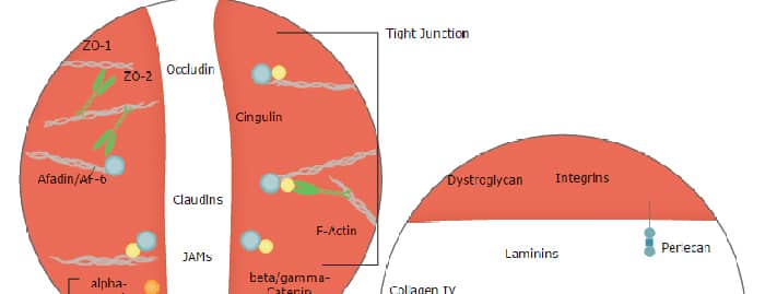
Integrin alpha V beta 3 together with alpha IIb beta 3, constitutes the only known beta 3 Integrins (1‑3). The non-covalent heterodimer of 170 kDa alpha V/CD51 and 93 kDa beta 3/CD61 subunits shows wide expression, notably by endothelial cells and osteoclasts (2‑4). Each subunit has a transmembrane sequence and a short cytoplasmic tail connected to the cytoskeleton. Active cell surface alpha V beta 3 adheres to matrix proteins including vitronectin, fibronectin, fibrinogen and thrombospondin (2, 3). The ligand binding site of alpha V beta 3 is in the N-terminal head region, formed by interaction of the beta 3 vWFA domain with the alpha V beta-propeller structure (4). The alpha V subunit contributes a thigh and a calf region, while the beta 3 subunit contains a PSI domain and four cysteine-rich I-EGF folds. The alpha V subunit domains termed thigh, calf-1 and calf-2 generate a “knee” region that is bent when the alpha V beta 3 is in its constitutively inactive state. Activation, either by “inside out” signaling or by Mg2+ or Mn2+ binding, extends the Integrin to expose its ligand binding site (1, 4). Two splice variants of beta 3 (b and c) diverge over the last 21 amino acids (aa) and lack cytoplasmic phosphorylation sites (5, 6). Another beta 3 splice variant diverges after the vWFA domain, producing a soluble 60 kDa form in platelets and endothelial cells (7). alpha V beta 3 is essential for the maturation of osteoclasts and their binding and resorption of bone; it also, however, promotes their apoptosis (8, 9). M-CSF R and alpha V beta 3 share signaling pathways during osteoclastogenesis, and deletion of either molecule causes osteopetrosis (8, 9). Also cell entry of several viruses is mediated by alpha V beta 3 (4, 10). The 962 aa human alpha V ECD (11) shares 92‑95% aa sequence identity with mouse, rat and cow alpha V while the 685 aa human beta 3 ECD (12) shares 95% aa identity with horse and dog, and 89‑92% aa identity with mouse, rat and pig beta 3.
Human Integrin alpha V beta 3 Antibody (23C6)
R&D Systems | Catalog # MAB3050

Scientific images may be resized, cropped, or adjusted for brightness or contrast for presentation purposes. These updates are limited to presentation and do not affect the underlying scientific interpretation of the data. Where available, additional source data may be provided upon request.
Key Product Details
Species Reactivity
Validated:
Cited:
Applications
Validated:
Cited:
Label
Antibody Source
Product Specifications
Immunogen
Specificity
Clonality
Host
Isotype
Endotoxin Level
Scientific Data Images for Human Integrin alpha V beta 3 Antibody (23C6)
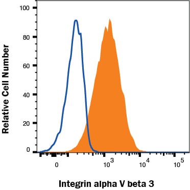
Detection of Integrin alpha V beta 3 in HT1080 cells by Flow Cytometry
HT1080 cells were stained with Mouse Anti-Human Integrin alpha V beta 3 Monoclonal Antibody (Catalog # MAB3050, filled histogram) or isotype control antibody (Catalog # MAB002, open histogram) followed by Allophycocyanin-conjugated Anti-Mouse IgG Secondary Antibody (Catalog # F0101B). View our protocol for Staining Membrane-associated Proteins.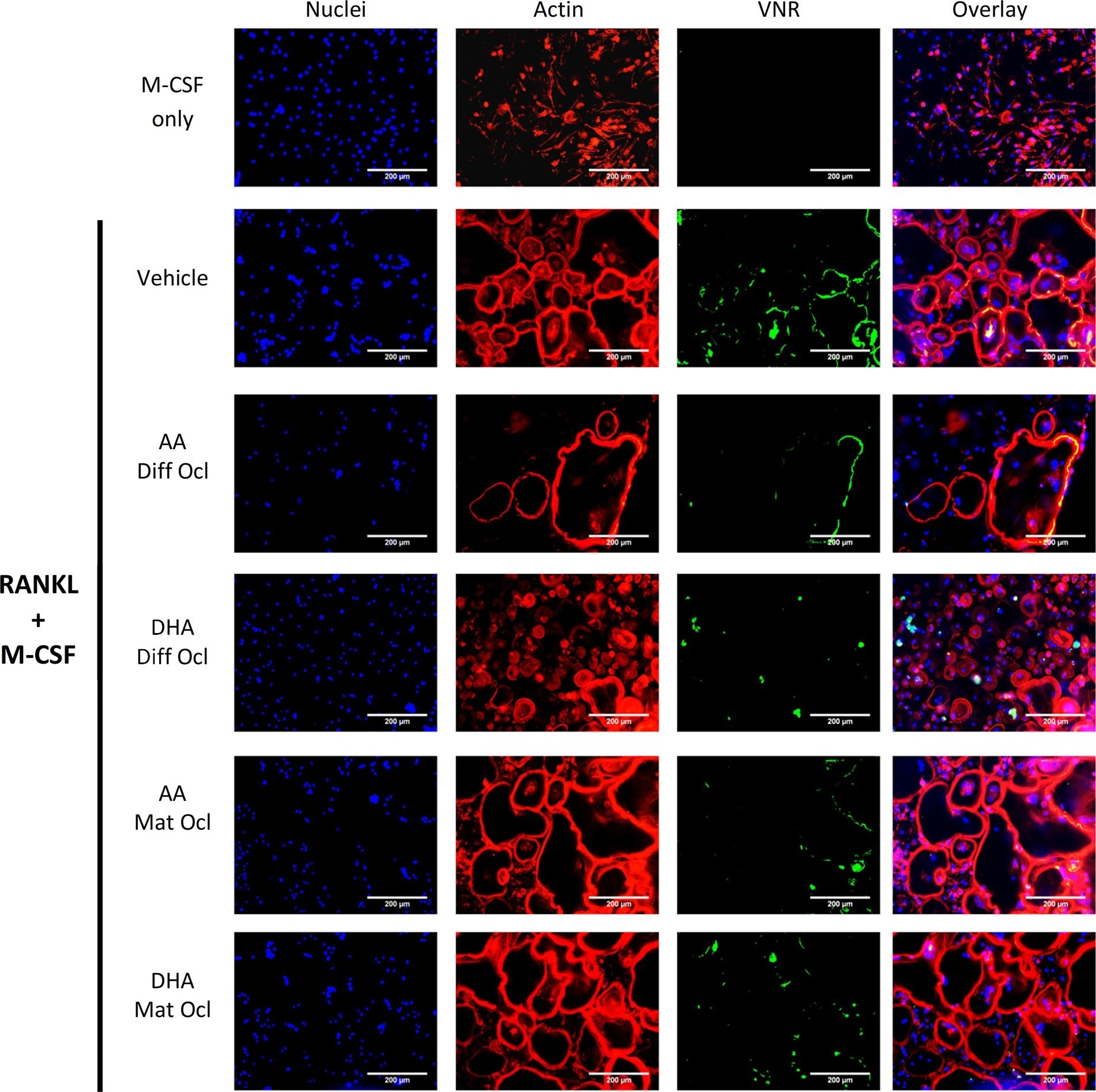
Detection of Human Integrin alpha V beta 3 by Immunocytochemistry/Immunofluorescence
Effects of AA and DHA on actin ring formation and VNR expression.Osteoclast differentiation was stimulated in CD14+ monocytes by the addition of 25 ng ml-1 M-CSF and 30 ng ml-1 RANKL as described in Materials and Methods. Vehicle (0.08% ethanol) or LCPUFAs (40 μorption (day 12–14) in mature osteoclasts. After 3 weeks of culture, cells were fixed and stained with Hoechst (blue) for nuclei, phalloidin (red) for actin rings, and anti-VNR antibody (green). The results are representative of three independent experiments conducted in triplicate. Scale bar = 200 μm. Diff Ocl—differentiating osteoclasts. Mat Ocl—mature osteoclasts. Image collected and cropped by CiteAb from the following publication (https://pubmed.ncbi.nlm.nih.gov/25867515), licensed under a CC-BY license. Not internally tested by R&D Systems.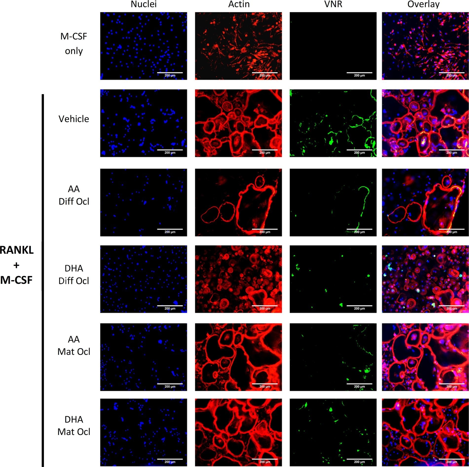
Detection of Human Human Integrin alpha V beta 3 Antibody by Immunocytochemistry/ Immunofluorescence
Effects of AA and DHA on actin ring formation and VNR expression.Osteoclast differentiation was stimulated in CD14+ monocytes by the addition of 25 ng ml-1 M-CSF and 30 ng ml-1 RANKL as described in Materials and Methods. Vehicle (0.08% ethanol) or LCPUFAs (40 μorption (day 12–14) in mature osteoclasts. After 3 weeks of culture, cells were fixed and stained with Hoechst (blue) for nuclei, phalloidin (red) for actin rings, and anti-VNR antibody (green). The results are representative of three independent experiments conducted in triplicate. Scale bar = 200 μm. Diff Ocl—differentiating osteoclasts. Mat Ocl—mature osteoclasts. Image collected and cropped by CiteAb from the following publication (https://pubmed.ncbi.nlm.nih.gov/25867515), licensed under a CC-BY license. Not internally tested by R&D Systems.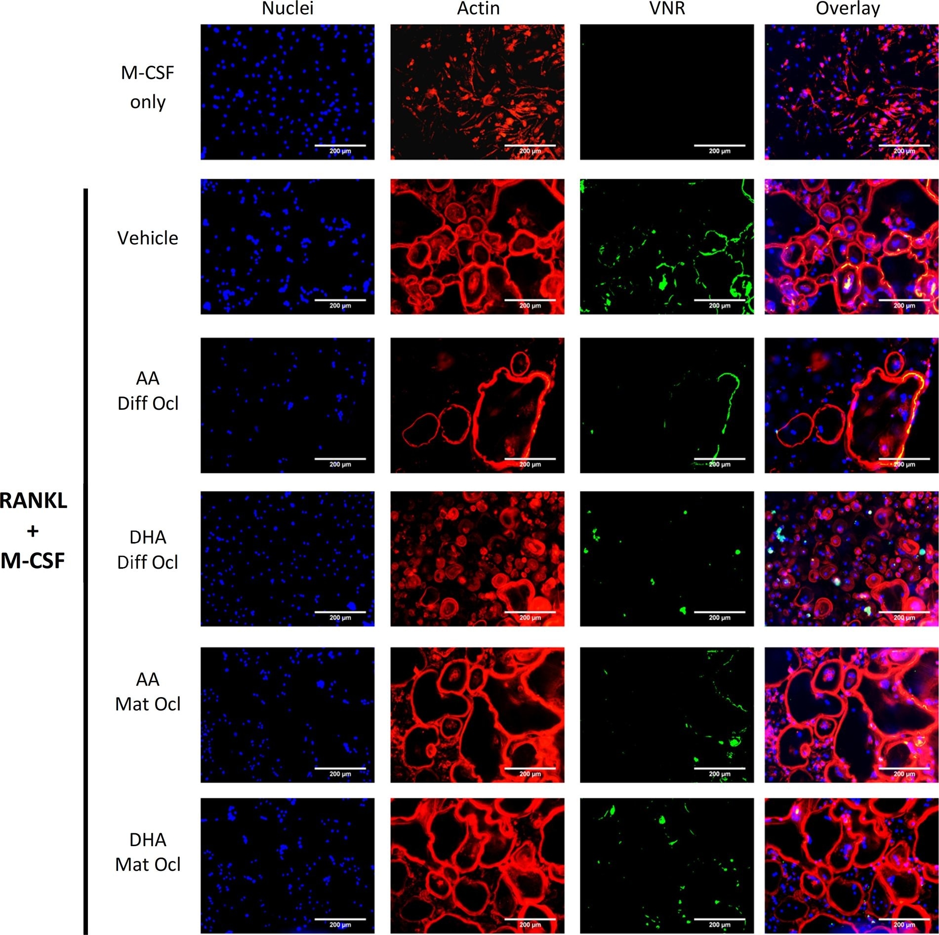
Detection of Human Integrin alpha V beta 3 by Immunocytochemistry/ Immunofluorescence
Effects of AA and DHA on actin ring formation and VNR expression.Osteoclast differentiation was stimulated in CD14+ monocytes by the addition of 25 ng ml-1 M-CSF and 30 ng ml-1 RANKL as described in Materials and Methods. Vehicle (0.08% ethanol) or LCPUFAs (40 μorption (day 12–14) in mature osteoclasts. After 3 weeks of culture, cells were fixed and stained with Hoechst (blue) for nuclei, phalloidin (red) for actin rings, and anti-VNR antibody (green). The results are representative of three independent experiments conducted in triplicate. Scale bar = 200 μm. Diff Ocl—differentiating osteoclasts. Mat Ocl—mature osteoclasts. Image collected and cropped by CiteAb from the following open publication (https://pubmed.ncbi.nlm.nih.gov/25867515), licensed under a CC-BY license. Not internally tested by R&D Systems.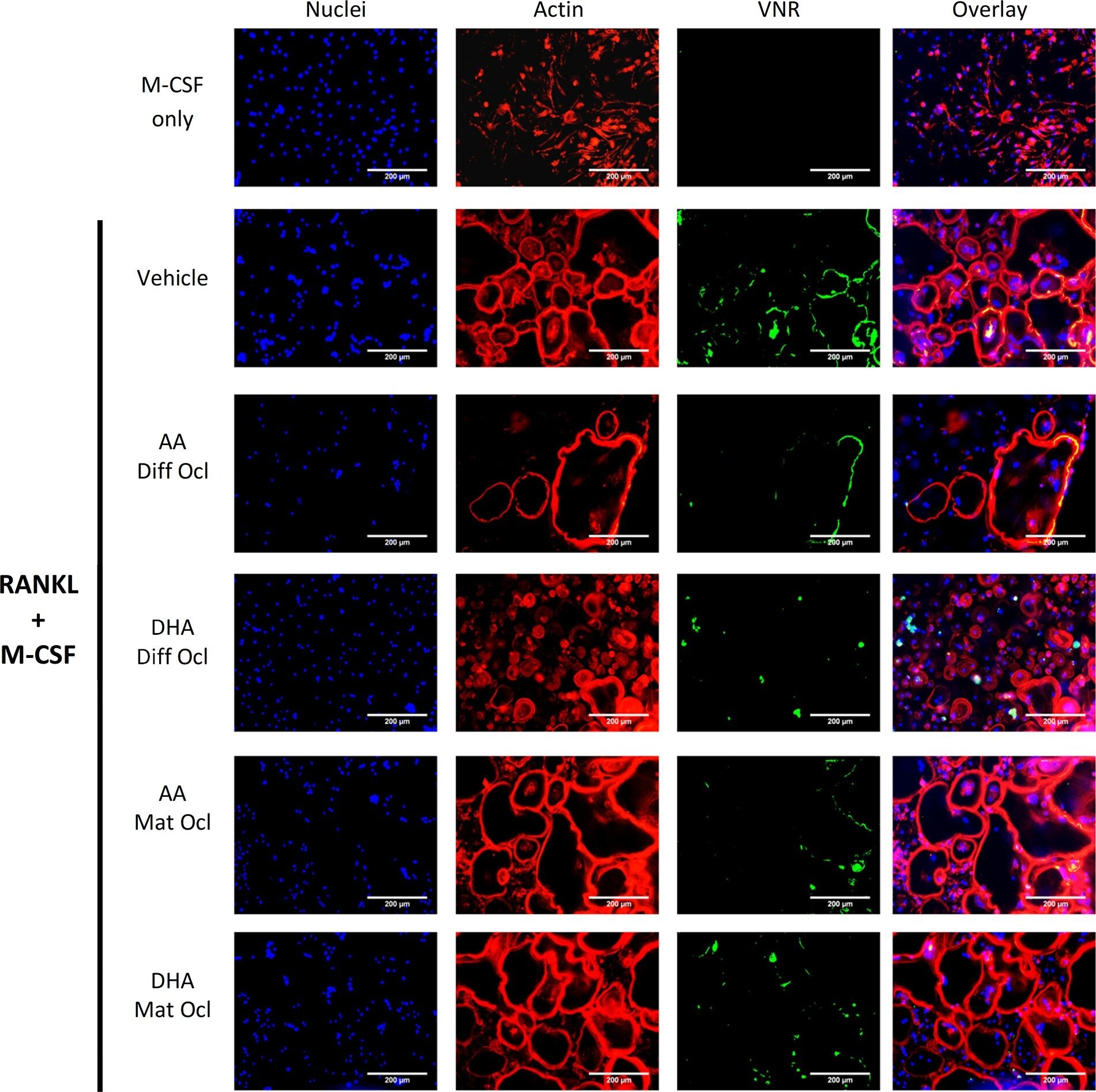
Detection of Integrin alpha V beta 3 by Immunocytochemistry/ Immunofluorescence
Effects of AA and DHA on actin ring formation and VNR expression.Osteoclast differentiation was stimulated in CD14+ monocytes by the addition of 25 ng ml-1 M-CSF and 30 ng ml-1 RANKL as described in Materials and Methods. Vehicle (0.08% ethanol) or LCPUFAs (40 μorption (day 12–14) in mature osteoclasts. After 3 weeks of culture, cells were fixed and stained with Hoechst (blue) for nuclei, phalloidin (red) for actin rings, and anti-VNR antibody (green). The results are representative of three independent experiments conducted in triplicate. Scale bar = 200 μm. Diff Ocl—differentiating osteoclasts. Mat Ocl—mature osteoclasts. Image collected and cropped by CiteAb from the following open publication (https://pubmed.ncbi.nlm.nih.gov/25867515), licensed under a CC-BY license. Not internally tested by R&D Systems.Applications for Human Integrin alpha V beta 3 Antibody (23C6)
Adhesion Blockade
CyTOF-ready
Flow Cytometry
Sample: HT1080 human fibrocarsoma cell line
Immunoprecipitation
Flow Cytometry Panel Builder
Bio-Techne Knows Flow Cytometry
Save time and reduce costly mistakes by quickly finding compatible reagents using the Panel Builder Tool.
Advanced Features
- Spectra Viewer - Custom analysis of spectra from multiple fluorochromes
- Spillover Popups - Visualize the spectra of individual fluorochromes
- Antigen Density Selector - Match fluorochrome brightness with antigen density

Formulation, Preparation, and Storage
Purification
Reconstitution
Reconstitute at 0.5 mg/mL in sterile PBS. For liquid material, refer to CoA for concentration.
Formulation
Shipping
Stability & Storage
- 12 months from date of receipt, -20 to -70 °C as supplied.
- 1 month, 2 to 8 °C under sterile conditions after reconstitution.
- 6 months, -20 to -70 °C under sterile conditions after reconstitution.
Calculators
Background: Integrin alpha V beta 3
References
- Hynes, R. O. (2002) Cell 110:673.
- Serini, G. et al. (2006) Exp. Cell Res. 312:651.
- Ross, F. P. and S. L. Teitelbaum (2005) Immunol. Rev. 208:88.
- Xiong, J. et al. (2001) Science 294:339.
- Kumar, C. S. et al. (1987) J. Biol. Chem. 272:16390.
- vanKuppevelt, H. et al. (1989) Proc. Natl. Acad. Sci. USA 86:5415.
- Djaffar, I. et al. (1994) Biochem. J. 300:67.
- McHugh, K. P. et al. (2000) J. Clin. Invest. 105:433.
- Faccio, R. et al. (2003) J. Clin. Invest. 111:749.
- Chu, J. J. and M. Ng (2004) J. Biol. Chem. 279:54533.
- Suzuki, S. et al. (1987) J. Biol. Chem. 262:14060.
- Fitzgerald, L. A. et al. (1987) J. Biol. Chem. 262:3936.
Alternate Names
Entrez Gene IDs
Gene Symbol
Additional Integrin alpha V beta 3 Products
Product Documents for Human Integrin alpha V beta 3 Antibody (23C6)
Certificate of Analysis
To download a Certificate of Analysis, please enter a lot or batch number in the search box below.
Note: Certificate of Analysis not available for kit components.
Product Specific Notices for Human Integrin alpha V beta 3 Antibody (23C6)
For research use only
Citations for Human Integrin alpha V beta 3 Antibody (23C6)
 Powered by Bioz
Customer Reviews for Human Integrin alpha V beta 3 Antibody (23C6)
There are currently no reviews for this product. Be the first to review Human Integrin alpha V beta 3 Antibody (23C6) and earn rewards!
Have you used Human Integrin alpha V beta 3 Antibody (23C6)?
Submit a review and receive an Amazon gift card!
$25/€18/£15/$25CAN/¥2500 for a review with an image
$10/€7/£6/$10CAN/¥1110 for a review without an image
Submit a review
Protocols
Find general support by application which include: protocols, troubleshooting, illustrated assays, videos and webinars.
- 7-Amino Actinomycin D (7-AAD) Cell Viability Flow Cytometry Protocol
- Extracellular Membrane Flow Cytometry Protocol
- Flow Cytometry Protocol for Cell Surface Markers
- Flow Cytometry Protocol for Staining Membrane Associated Proteins
- Flow Cytometry Staining Protocols
- Flow Cytometry Troubleshooting Guide
- Immunoprecipitation Protocol
- Intracellular Flow Cytometry Protocol Using Alcohol (Methanol)
- Intracellular Flow Cytometry Protocol Using Detergents
- Intracellular Nuclear Staining Flow Cytometry Protocol Using Detergents
- Intracellular Staining Flow Cytometry Protocol Using Alcohol Permeabilization
- Intracellular Staining Flow Cytometry Protocol Using Detergents to Permeabilize Cells
- Propidium Iodide Cell Viability Flow Cytometry Protocol
- Protocol for Liperfluo
- Protocol for the Characterization of Human Th22 Cells
- Protocol for the Characterization of Human Th9 Cells
- Protocol: Annexin V and PI Staining by Flow Cytometry
- Protocol: Annexin V and PI Staining for Apoptosis by Flow Cytometry
- Troubleshooting Guide: Fluorokine Flow Cytometry Kits
- View all Protocols, Troubleshooting, Illustrated assays and Webinars