Axl (Ufo, Ark), Dtk (Sky, Tyro3, Rse, Brt) and Mer (human and mouse homologues of chicken c-Eyk) constitute a receptor tyrosine kinase subfamily. The extracellular domains of these proteins contain two Ig-like motifs and two fibronectin type III motifs. This characteristic topology is also found in neural cell adhesion molecules and in receptor tyrosine phosphatases. These receptors bind the vitamin K-dependent protein growth-arrest-specific gene 6 (Gas6) which is structurally related to the anticoagulation factor protein S. Binding of Gas6 induces receptor autophosphorylation and downstream signaling pathways that can lead to cell proliferation, migration or the prevention of apoptosis. Recent studies suggest that this family of tyrosine kinase receptors may be involved in hematopoiesis, embryonic development, tumorigenesis and regulation of testicular functions.

Key Product Details
Validated by
Knockout/Knockdown
Species Reactivity
Validated:
Human
Cited:
Human, Mouse, Primate
Applications
Validated:
Knockout Validated, Western Blot, Flow Cytometry, CyTOF-ready
Cited:
Immunohistochemistry, Immunohistochemistry-Frozen, Western Blot, Neutralization, Flow Cytometry, Cell Culture
Label
Unconjugated
Antibody Source
Polyclonal Goat IgG
Loading...
Product Specifications
Immunogen
S. frugiperda insect ovarian cell line Sf 21-derived recombinant human Mer
Arg26-Ala499
Accession # Q12866
Arg26-Ala499
Accession # Q12866
Specificity
Detects human Mer in direct ELISAs and Western blots. In direct ELISAs, approximately 20% cross-reactivity with recombinant mouse Mer and less than 1% cross-reactivity with recombinant human (rh) Axl and rhDtk is observed.
Clonality
Polyclonal
Host
Goat
Isotype
IgG
Scientific Data Images for Human Mer Antibody
Detection of Human Mer by Western Blot.
Western blot shows lysates of U937 human histiocytic lymphoma cell line and PANC-1 human pancreatic carcinoma cell line. PVDF membrane was probed with 1 µg/mL of Goat Anti-Human Mer Antigen Affinity-purified Polyclonal Antibody (Catalog # AF891) followed by HRP-conjugated Anti-Goat IgG Secondary Antibody (Catalog # HAF017). A specific band was detected for Mer at approximately 170-230 kDa (as indicated). This experiment was conducted under reducing conditions and using Immunoblot Buffer Group 1.
Western Blot Shows Human Mer Specificity by Using Knockout Cell Line.
Western blot shows lysates of HepG2 human hepatocellular carcinoma parental cell line and Mer knockout HepG2 cell line (KO). PVDF membrane was probed with 1 µg/mL of Goat Anti-Human Mer Antigen Affinity-purified Polyclonal Antibody (Catalog # AF891) followed by HRP-conjugated Anti-Goat IgG Secondary Antibody (Catalog # HAF017). A specific band was detected for Mer at approximately 175 kDa (as indicated) in the parental HepG2 cell line, but is not detectable in knockout HepG2 cell line. GAPDH (Catalog # AF5718) is shown as a loading control. This experiment was conducted under reducing conditions and using Immunoblot Buffer Group 1.
Detection of Human Mer by Western Blot
Implication of both MERTK and TYRO3 receptors in PROS1-induced Src and VEC phosphorylation. (A,B) represent Western blot analysis (A) and subsequent quantification (B) of VEC phosphorylation on Tyr685, c-Src phosphorylation on Tyr416 or MLC phosphorylation. (B) Integrated intensity for the target proteins was divided by that for actin, and all conditions were normalized to their corresponding untreated controls. Analysis was performed with the Fiji software. (C) Proposed mechanism through which PROS1 interferes with EC permeability. PROS1 activates its tyrosine kinase receptors MERTK and TYRO3, leading to the phosphorylation of c- Src on Tyr416 and PAK-1 on Ser144 which in turn phosphorylate, respectively, VEC at Tyr685 and Ser665, two specific sites involved in the cleavage and internalization of VEC, respectively. The PROS1/MERTK-TYRO3/c-Src-Tyr416/VEC-Tyr685 pathway activates the cleavage of VEC extracellular domain, which destabilizes cell–cell junctions. The PROS1/MERTK-TYRO3/ PAK-1-Ser144/VEC-Ser665 pathway activates VEC internalization thus preventing VEC recognition between two adjacent endothelial cells. The Rho/ROCK/MLC pathway involved in the regulation of endothelial permeability is also activated by human PROS1. * p < 0.05; *** p < 0.001. Image collected and cropped by CiteAb from the following open publication (https://www.mdpi.com/1467-3045/46/4/205), licensed under a CC-BY license. Not internally tested by R&D Systems.
Human Mer ELISA Standard Curve
Recombinant Human Mer Fc Chimera (Catalog # 891-MR) was serially diluted and captured by Mouse Anti-Human Mer Monoclonal Antibody (Catalog # MAB8912) coated on a Clear Polystyrene Microplate (Catalog # DY990). Goat Anti-Human Mer Antigen Affinity-purified Polyclonal Antibody (Catalog # AF891) was biotinylated and incubated with the protein captured on the plate. Detection of the standard curve was achieved by incubating Streptavidin-HRP (Catalog # DY998)Applications for Human Mer Antibody
Application
Recommended Usage
CyTOF-ready
Ready to be labeled using established conjugation methods. No BSA or other carrier proteins that could interfere with conjugation.
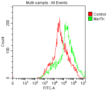
Flow Cytometry
0.25 µg/106 cells
Sample: Human peripheral blood monocytes
Sample: Human peripheral blood monocytes
Knockout Validated
Mer is
specifically detected in HepG2 human hepatocellular carcinoma parental cell line but is not detectable in Mer
knockout HepG2 cell line.
Western Blot
1 µg/mL
Sample: U937 human histiocytic lymphoma cell line and PANC‑1 human pancreatic carcinoma cell line
Sample: U937 human histiocytic lymphoma cell line and PANC‑1 human pancreatic carcinoma cell line
Reviewed Applications
Read 2 reviews rated 5 using AF891 in the following applications:
Flow Cytometry Panel Builder
Bio-Techne Knows Flow Cytometry
Save time and reduce costly mistakes by quickly finding compatible reagents using the Panel Builder Tool.
Advanced Features
- Spectra Viewer - Custom analysis of spectra from multiple fluorochromes
- Spillover Popups - Visualize the spectra of individual fluorochromes
- Antigen Density Selector - Match fluorochrome brightness with antigen density

Formulation, Preparation, and Storage
Purification
Antigen Affinity-purified
Reconstitution
Reconstitute at 0.2 mg/mL in sterile PBS. For liquid material, refer to CoA for concentration.
Loading...
Formulation
Lyophilized from a 0.2 μm filtered solution in PBS with Trehalose. *Small pack size (SP) is supplied either lyophilized or as a 0.2 µm filtered solution in PBS.
Shipping
Lyophilized product is shipped at ambient temperature. Liquid small pack size (-SP) is shipped with polar packs. Upon receipt, store immediately at the temperature recommended below.
Stability & Storage
Use a manual defrost freezer and avoid repeated freeze-thaw cycles.
- 12 months from date of receipt, -20 to -70 °C as supplied.
- 1 month, 2 to 8 °C under sterile conditions after reconstitution.
- 6 months, -20 to -70 °C under sterile conditions after reconstitution.
Calculators
Background: Mer
References
- Nagata, K. et al. (1996) J. Biol. Chem. 22:30022.
- Crosier, K.E. and P.S Crosier (1997) Pathology 29:131.
Long Name
Receptor Tyrosine Protein Kinase Mer
Alternate Names
c-Eyk, C-mer, MerTK
Gene Symbol
MERTK
UniProt
Additional Mer Products
Product Documents for Human Mer Antibody
Certificate of Analysis
To download a Certificate of Analysis, please enter a lot or batch number in the search box below.
Note: Certificate of Analysis not available for kit components.
Product Specific Notices for Human Mer Antibody
For research use only
Citations for Human Mer Antibody
 Powered by Bioz
Customer Reviews for Human Mer Antibody (2)
5 out of 5
2 Customer Ratings
Have you used Human Mer Antibody?
Submit a review and receive an Amazon gift card!
$25/€18/£15/$25CAN/¥2500 Yen for a review with an image
$10/€7/£6/$10CAN/¥1110 Yen for a review without an image
Submit a review
Customer Images
Showing
1
-
2 of
2 reviews
Showing All
Filter By:
-
Application: Flow CytometrySample Tested: Neuroblastoma cells (SHSY-5Y)Species: HumanVerified Customer | Posted 05/06/2026Image shows secondary antibody control only in red (Rabbit anti-Goat IgG Recombinant Secondary Antibody Alexa Fluor Plus 488 1ug/ml invitrogen). MER (AF891 R&D Systems 1ug/ml) is shown in green.Neuroblastoma cells were treated and fixed to observe changes that occur to extracellular Mer after treatments using flow cytometry. Concentration used: 1ug/ml In-vitro application

-
Application: Immunocytochemistry/ImmunofluorescenceSample Tested: HUMAN MACROPHAGESSpecies: HumanVerified Customer | Posted 07/03/2016
There are no reviews that match your criteria.
Protocols
Find general support by application which include: protocols, troubleshooting, illustrated assays, videos and webinars.
- 7-Amino Actinomycin D (7-AAD) Cell Viability Flow Cytometry Protocol
- Cellular Response to Hypoxia Protocols
- Extracellular Membrane Flow Cytometry Protocol
- Flow Cytometry Protocol for Cell Surface Markers
- Flow Cytometry Protocol for Staining Membrane Associated Proteins
- Flow Cytometry Staining Protocols
- Flow Cytometry Troubleshooting Guide
- Intracellular Flow Cytometry Protocol Using Alcohol (Methanol)
- Intracellular Flow Cytometry Protocol Using Detergents
- Intracellular Nuclear Staining Flow Cytometry Protocol Using Detergents
- Intracellular Staining Flow Cytometry Protocol Using Alcohol Permeabilization
- Intracellular Staining Flow Cytometry Protocol Using Detergents to Permeabilize Cells
- Propidium Iodide Cell Viability Flow Cytometry Protocol
- Protocol for Liperfluo
- Protocol for the Characterization of Human Th22 Cells
- Protocol for the Characterization of Human Th9 Cells
- Protocol: Annexin V and PI Staining by Flow Cytometry
- Protocol: Annexin V and PI Staining for Apoptosis by Flow Cytometry
- R&D Systems Quality Control Western Blot Protocol
- Troubleshooting Guide: Fluorokine Flow Cytometry Kits
- Troubleshooting Guide: Western Blot Figures
- Western Blot Conditions
- Western Blot Protocol
- Western Blot Protocol for Cell Lysates
- Western Blot Troubleshooting
- Western Blot Troubleshooting Guide
- View all Protocols, Troubleshooting, Illustrated assays and Webinars
Loading...