Müllerian inhibiting substance (MIS), also named anti-Müllerian hormone (AMH), is a tissue-specific TGF-beta superfamily growth factor. Its expression is restricted to the Sertoli cells of fetal and postnatal testis, and to the granulosa cells of postnatal ovary (1). The human MIS gene encodes a 553 amino acid residue (aa) prepropeptide containing a signal a sequence (1-24), a pro-region (25-455), and the carboxyl-terminal bioactive protein (446-553) (2-4). MIS is synthesized and secreted as a disulfide-linked homodimeric pro-protein. Proteolytic cleavage is required to generate the N-terminal pro-region and the C-terminal bioactive protein, which remain associated in a non-covalent complex. Recombinant C-terminal MIS has been shown to be bioactive. However, the complex with the N-terminal pro-region showed enhanced activity (3, 5). The C-terminal region contains the seven canonical cysteine residues found in TGF-beta superfamily members. These cysteine residues are involved in inter- and intra-molecular disulfide bonds, which forms the cysteine knot structure. Human and mouse MIS share 73% and 90% aa sequence identity in their pro-region and C-terminal region, respectively. MIS induces Mullerian duct (female reproductive tract) regression during sexual differentiation in the male embryo (6). Postnatally, MIS has been shown to regulate gonadal functions (1). MIS inhibits Leydig cell proliferation and is a regulator of the initial and cyclic recruitment of ovarian follicles. MIS has also been found to have anti-proliferative effects on breast, ovarian and prostate tumor cells (7-9). Like other TGF-beta superfamily members, MIS signals via a heteromeric receptor complex consisting of a type I and a type II receptor serine/threonine kinase. Depending on the cell context, different type I receptors (including Act RIA/ALK2, BMP RIA/ALK3, and BMP RIB/ALK6) that are shared by other TGF-beta superfamily members, have been implicated in MIS signaling (10-12). In contrast, the type II MIS receptor (MIS RII) is unique and does not bind other TGF-beta superfamily members. Upon ligand binding, MIS RII recruits the non‑ligand binding type I receptor into the complex, resulting in phosphorylation the BMP-like signaling pathway effector proteins Smad1, Smad5, and Smad 8 (10-12).

Key Product Details
Species Reactivity
Human
Applications
Immunocytochemistry
Label
Unconjugated
Antibody Source
Monoclonal Mouse IgG1 Clone # 805531
Loading...
Product Specifications
Immunogen
Mouse myeloma cell line NS0-derived recombinant human MIS/AMH
Leu19-Gln450 (predicted)
Accession # P03971
Leu19-Gln450 (predicted)
Accession # P03971
Specificity
Detects human MIS/AMH in direct ELISAs. In direct ELISAs, no cross-reactivity with recombinant
rat MIS/AMH is observed.
Clonality
Monoclonal
Host
Mouse
Isotype
IgG1
Scientific Data Images for Human MIS/AMH Antibody (805531)
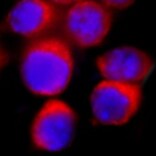
MIS/AMH in LNCaP Human Cell Line.
MIS/AMH was detected in immersion fixed LNCaP human prostate cancer cell line using Mouse Anti-Human MIS/AMH Monoclonal Antibody (Catalog # MAB17371) at 25 µg/mL for 3 hours at room temperature. Cells were stained using the NorthernLights™ 557-conjugated Anti-Mouse IgG Secondary Antibody (red; Catalog # NL007) and counterstained with DAPI (blue). Specific staining was localized to cytoplasm. View our protocol for Fluorescent ICC Staining of Cells on Coverslips.Applications for Human MIS/AMH Antibody (805531)
Application
Recommended Usage
Immunocytochemistry
8-25 µg/mL
Sample: Immersion fixed LNCaP human prostate cancer cell line
Sample: Immersion fixed LNCaP human prostate cancer cell line
Reviewed Applications
Read 1 review rated 5 using MAB17371 in the following applications:
Formulation, Preparation, and Storage
Purification
Protein A or G purified from hybridoma culture supernatant
Reconstitution
Sterile PBS to a final concentration of 0.5 mg/mL. For liquid material, refer to CoA for concentration.
Loading...
Formulation
Lyophilized from a 0.2 μm filtered solution in PBS with Trehalose. *Small pack size (SP) is supplied either lyophilized or as a 0.2 µm filtered solution in PBS.
Shipping
Lyophilized product is shipped at ambient temperature. Liquid small pack size (-SP) is shipped with polar packs. Upon receipt, store immediately at the temperature recommended below.
Stability & Storage
Use a manual defrost freezer and avoid repeated freeze-thaw cycles.
- 12 months from date of receipt, -20 to -70 °C as supplied.
- 1 month, 2 to 8 °C under sterile conditions after reconstitution.
- 6 months, -20 to -70 °C under sterile conditions after reconstitution.
Calculators
Background: MIS/AMH
References
- Teixeira, et al. (2001) Endocrine Rev. 22:657.
- Pepinsky, R.et al. (1988) J. Biol. Chem. 263:18961.
- Wilson, C.A. et al. (1993) Mol. Endocrinol. 7:247.
- Kurian, M.S. et al. (1995) Clin. Cancer Res. 1:343.
- Nachtigal, J.S. and H.A. Ingraham (1996) Proc. Natl. Acad. Sci. USA 93:7711.
- MacLaughlin, D.T. et al. (1991) Methods Enzymol. 35:358.
- Laurich, V.M. et al. (2002) Endocrinology 143:3351.
- McGee, E.A. et al. (2001) Biol. Reprod. 64:293.
- Segev, D.L. et al. (2002) Proc. Natl. Acad. Sci. USA 99:239.
- Josso, N and N. diClemente (2003) Trends Endo. Met. 14:91.
- Clarke, T.R. et al. (2001) Mol. Endocrinol. 15:946.
- Visser, J.A. (2003) Mol. Cell. Endocrinol. 211:65.
Long Name
Mullerian-inhibiting Substance/Anti-Mullerian Hormone
Alternate Names
AMH
Gene Symbol
AMH
UniProt
Additional MIS/AMH Products
Product Documents for Human MIS/AMH Antibody (805531)
Certificate of Analysis
To download a Certificate of Analysis, please enter a lot or batch number in the search box below.
Note: Certificate of Analysis not available for kit components.
Product Specific Notices for Human MIS/AMH Antibody (805531)
For research use only
Customer Reviews for Human MIS/AMH Antibody (805531) (1)
5 out of 5
1 Customer Rating
Have you used Human MIS/AMH Antibody (805531)?
Submit a review and receive an Amazon gift card!
$25/€18/£15/$25CAN/¥2500 Yen for a review with an image
$10/€7/£6/$10CAN/¥1110 Yen for a review without an image
Submit a review
Customer Images
Showing
1
-
1 of
1 review
Showing All
Filter By:
-
Application: Immunocytochemistry/ImmunofluorescenceSample Tested: LNCaP Human Cell LineSpecies: HumanVerified Customer | Posted 06/06/2022

There are no reviews that match your criteria.
Protocols
Find general support by application which include: protocols, troubleshooting, illustrated assays, videos and webinars.
- Appropriate Fixation of IHC/ICC Samples
- Cellular Response to Hypoxia Protocols
- ClariTSA™ Fluorophore Kits
- Detection & Visualization of Antibody Binding
- ICC Cell Smear Protocol for Suspension Cells
- ICC Immunocytochemistry Protocol Videos
- ICC for Adherent Cells
- Immunocytochemistry (ICC) Protocol
- Immunocytochemistry Troubleshooting
- Immunofluorescence of Organoids Embedded in Cultrex Basement Membrane Extract
- Immunohistochemistry (IHC) and Immunocytochemistry (ICC) Protocols
- Preparing Samples for IHC/ICC Experiments
- Preventing Non-Specific Staining (Non-Specific Binding)
- Primary Antibody Selection & Optimization
- Protocol for VisUCyte™ HRP Polymer Detection Reagent
- Protocol for the Fluorescent ICC Staining of Cell Smears - Graphic
- Protocol for the Fluorescent ICC Staining of Cultured Cells on Coverslips - Graphic
- Protocol for the Preparation and Fluorescent ICC Staining of Cells on Coverslips
- Protocol for the Preparation and Fluorescent ICC Staining of Non-adherent Cells
- Protocol for the Preparation and Fluorescent ICC Staining of Stem Cells on Coverslips
- Protocol for the Preparation of a Cell Smear for Non-adherent Cell ICC - Graphic
- TUNEL and Active Caspase-3 Detection by IHC/ICC Protocol
- The Importance of IHC/ICC Controls
- View all Protocols, Troubleshooting, Illustrated assays and Webinars
Loading...