Pro-MMP-9 is an inactive proenzyme secreted by multiple cell types. The N-terminal pro region is proteolytically removed, resulting in active MMP-9 which can degrade collagens and elastin as well as several non-ECM molecules.

Key Product Details
Species Reactivity
Validated:
Human
Cited:
Human, Mouse, Rabbit, Xenograft
Applications
Validated:
Immunohistochemistry, Western Blot, Immunocytochemistry, Immunoprecipitation
Cited:
Immunohistochemistry, Immunohistochemistry-Paraffin, Western Blot, Immunocytochemistry, ELISA Microarray Development, Proximity Ligation Assay, Zymography
Label
Unconjugated
Antibody Source
Polyclonal Goat IgG
Loading...
Product Specifications
Immunogen
Chinese hamster ovary cell line CHO-derived recombinant human MMP-9
Specificity
Detects human and mouse MMP-9 in Western blots, dot blots, and human MMP-9 by immunocytochemistry. In Western blots, less than 1% cross-reactivity with recombinant human MMP-1, -2, -3, -7, -8, -10, -12, and -13 is observed.
Clonality
Polyclonal
Host
Goat
Isotype
IgG
Scientific Data Images for Human MMP-9 Antibody
MMP‑9 in NS0 Mouse Cell Line.
MMP‑9 was detected in immersion fixed NS0 mouse myeloma cell line transfected with MMP-9 using 5 µg/mL Human MMP‑9 Antigen Affinity-purified Polyclonal Antibody (Catalog # AF911) for 3 hours at room temperature. Cells were stained (red) and counterstained (green). View our protocol for Fluorescent ICC Staining of Cells on Coverslips.
MMP‑9 in Human Ovarian Cancer Tissue.
MMP-9 was detected in immersion fixed paraffin-embedded sections of human ovarian cancer tissue using Human MMP-9 Antigen Affinity-purified Polyclonal Antibody (Catalog # AF911) at 10 µg/mL overnight at 4 °C. Tissue was stained using the Anti-Goat HRP-DAB Cell & Tissue Staining Kit (brown; Catalog # CTS008) and counterstained with hematoxylin (blue). View our protocol for Chromogenic IHC Staining of Paraffin-embedded Tissue Sections.
MMP‑9 in MCF‑7 Human Cell Line.
MMP-9 was detected in immersion fixed MCF-7 human breast cancer cell line using Human MMP-9 Antigen Affinity-purified Polyclonal Antibody (Catalog # AF911) at 10 µg/mL for 3 hours at room temperature. Cells were stained using the NorthernLights™ 557-conjugated Anti-Goat IgG Secondary Antibody (yellow; Catalog # NL001) and counterstained with DAPI (blue). View our protocol for Fluorescent ICC Staining of Cells on Coverslips.
MMP‑9 in Human Ovarian Cancer Tissue.
MMP-9 was detected in immersion fixed paraffin-embedded sections of human ovarian cancer tissue using Human MMP-9 Antigen Affinity-purified Polyclonal Antibody (Catalog # AF911) at 10 µg/mL overnight at 4 °C. Tissue was stained using the Anti-Goat HRP-DAB Cell & Tissue Staining Kit (brown; Catalog # CTS008) and counterstained with hematoxylin (blue). Lower panel shows a lack of labeling if primary antibodies are omitted and tissue is stained only with secondary antibody followed by incubation with detection reagents. View our protocol for Chromogenic IHC Staining of Paraffin-embedded Tissue Sections.
Detection of Human MMP-9 by Proximity Ligation Assay
Univariate survival analysis of disease free survival (DFS) in plasma for all 465 patients. A) MMP-9:TIMP-1 measured by ELISA. B) MMP-9:TIMP-1 measured by PLA. Patients are divided into four groups of equal size (Q1-Q4) according to increasing plasma MMP-9:TIMP-1 levels; Q1 being the group with the lowest level. Image collected and cropped by CiteAb from the following open publication (https://bmccancer.biomedcentral.com/articles/10.1186/1471-2407-13-598), licensed under a CC-BY license. Not internally tested by R&D Systems.
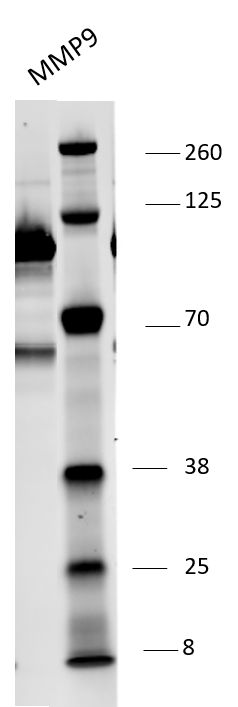
Detection of Human MMP-9 by Western Blot
Co-culture of THP1-derived macrophages with Panc89 hA8 WT and KO cells. (A) The schematic model depicts the interactions of THP1-derived macrophages (green, M0) and Panc89 cells with or without ADAM8 (red). Created with BioRender.com. (B) ADAM8 mRNA expression in both Panc89 hA8 WT and KO is not affected by M0, whereas LCN2 mRNA expression. Data are presented as mean values ± S.D. *** p < 0.001. (C) is upregulated after co-culture in an ADAM8-dependent manner. Data are presented as mean values ± S.D. *** p < 0.001. (D) The graph illustrates the upregulation of MMP-9 mRNA expression in both Panc89 hA8 WT and KO after co-culture (n = 2). Data are presented as mean values ± S.D. *** p < 0.001. (E) Representative immunoblot shows the detection of ADAM8, MMP-9, and LCN2 with or without co-culture. In addition to the qPCR results, MMP-9 and LCN2 are upregulated after co-culture at the protein level (n = 2). (F) Representative zymography of Panc89 hA8 WT and KO cells with or without co-culture demonstrates less active MMP-9 in Panc89 hA8 KO cells than in Panc89 hA8 WT cells after co-culture. (G) Quantification of active MMP-9 refers to total MMP-9 in zymography of Panc89 hA8 WT and KO cells after co-culture (n = 2). Data are presented as mean values ± S.D. * p < 0.05. Representative images of Panc89 cells before and after co-culture are shown in (H); scale bar, 100 μm. After co-culture, morphological changes are visible in both Panc89 hA8 WT and KO cells. Image collected and cropped by CiteAb from the following open publication (https://pubmed.ncbi.nlm.nih.gov/35216088), licensed under a CC-BY license. Not internally tested by R&D Systems.
Detection of Human MMP-9 by Western Blot
ADAM8 and LCN2 levels correlate in Panc89- and MB-231-derived extracellular vesicles (EV). Representative Western blots (A) of EVs derived from either Panc89 hA8 WT or KO cells, and cell lysate (CL) of Panc89 hA8 WT cells, and (B) of EVs derived from either MB-231 hA8 WT or MB-231 KO cells show the detection of ADAM8, MMP-9, LCN2, and Flotillin-1 in the upper part. Diagrams below illustrate the quantification and downregulation of LCN2 secretion (relative to Flotillin-1 secretion) in EVs isolated from Panc89 (hA8 WT or hA8 KO 1) or MB-231 (hA8 WT or hA8 KO) cells (n = 3). Data are presented as mean values ± S.D. ** p < 0.01. Image collected and cropped by CiteAb from the following open publication (https://pubmed.ncbi.nlm.nih.gov/35216088), licensed under a CC-BY license. Not internally tested by R&D Systems.
Detection of Human MMP-9 by Western Blot
Co-culture of THP1-derived and polarized macrophages with Panc89 hA8 WT and KO cells. (A) Western blot illustrates the detection of ADAM8, MMP-9, and LCN2 in Panc89 hA8 WT and KO control cells (Ø) and after co-culture with M0, M1, and M2 macrophages (two time points: 0 h and 1 h). ADAM8 is upregulated in Panc89 hA8 WT cells after co-culture with M2-polarized macrophages. Panc89 cells show the highest MMP-9 expression after co-culture with M0, but M1 macrophages also upregulate MMP-9. LCN2 is dependent on ADAM8 when upregulated in Panc89 cells after co-culture with M0 and M2 macrophages but independent of ADAM8 in Panc89 cells co-cultured with M1 macrophages. (B) ADAM8, (C) MMP-9, and (D) LCN2 ELISA of Panc89 hA8 WT and KO cell-derived supernatants of control cells and after co-culture with M0, M1, and M2 (two time points: 0 h and 1 h). In accordance with the immunoblot results of (A), ADAM8 is upregulated in supernatants derived from Panc89 hA8 WT cells after co-culture with M2 macrophages (B). At the same time, macrophages increase MMP-9 secretion from an undetectable level to almost 80,000 pg/mL in Panc89 hA8 WT and 60,000 pg/mL in Panc89 hA8 KO cells. M1 and M2 macrophages increase MMP-9 secretion of Panc89 independent of ADAM8, but not as high as in Panc89 cells co-cultured with M0. In contrast, LCN2 is upregulated in Panc89 hA8 WT cells by M0 and M1, but not by M2 macrophages. In the absence of ADAM8, Panc89 hA8 KO cells show low LCN2 secretion in control cells and after co-culture with M0 and M2 macrophages. Only after co-culture with M1 macrophages is the LCN2 secretion level increased (n = 1). Data are presented as mean values ± S.D. Image collected and cropped by CiteAb from the following open publication (https://pubmed.ncbi.nlm.nih.gov/35216088), licensed under a CC-BY license. Not internally tested by R&D Systems.Applications for Human MMP-9 Antibody
Application
Recommended Usage
Immunocytochemistry
5-15 µg/mL
Sample: Immersion fixed NS0 mouse myeloma cell line transfected with MMP-9 and immersion fixed MCF-7 human breast cancer cell line
Sample: Immersion fixed NS0 mouse myeloma cell line transfected with MMP-9 and immersion fixed MCF-7 human breast cancer cell line
Immunohistochemistry
5-15 µg/mL
Sample: Immersion fixed paraffin-embedded sections of human ovarian cancer tissue
Sample: Immersion fixed paraffin-embedded sections of human ovarian cancer tissue
Immunoprecipitation
25 µg/mL
Sample: Conditioned cell culture medium spiked with Recombinant Human MMP‑9 (Catalog # 911-MP), see our available Western blot detection antibodies
Sample: Conditioned cell culture medium spiked with Recombinant Human MMP‑9 (Catalog # 911-MP), see our available Western blot detection antibodies
Western Blot
0.1 µg/mL
Sample: Recombinant Human MMP‑9 Western Blot Standard (Catalog # WBC018)
Sample: Recombinant Human MMP‑9 Western Blot Standard (Catalog # WBC018)
Reviewed Applications
Read 5 reviews rated 4.2 using AF911 in the following applications:
Formulation, Preparation, and Storage
Purification
Antigen Affinity-purified
Reconstitution
Reconstitute at 0.2 mg/mL in sterile PBS. For liquid material, refer to CoA for concentration.
Loading...
Formulation
Lyophilized from a 0.2 μm filtered solution in PBS with Trehalose. *Small pack size (SP) is supplied either lyophilized or as a 0.2 µm filtered solution in PBS.
Shipping
Lyophilized product is shipped at ambient temperature. Liquid small pack size (-SP) is shipped with polar packs. Upon receipt, store immediately at the temperature recommended below.
Stability & Storage
Use a manual defrost freezer and avoid repeated freeze-thaw cycles.
- 12 months from date of receipt, -20 to -70 °C as supplied.
- 1 month, 2 to 8 °C under sterile conditions after reconstitution.
- 6 months, -20 to -70 °C under sterile conditions after reconstitution.
Calculators
Background: MMP-9
Long Name
Matrix Metalloproteinase 9
Alternate Names
CLG4B, Gelatinase B, GELB, MANDP2, MMP9
Gene Symbol
MMP9
Additional MMP-9 Products
Product Documents for Human MMP-9 Antibody
Certificate of Analysis
To download a Certificate of Analysis, please enter a lot or batch number in the search box below.
Note: Certificate of Analysis not available for kit components.
Product Specific Notices for Human MMP-9 Antibody
For research use only
Citations for Human MMP-9 Antibody
 Powered by Bioz
Customer Reviews for Human MMP-9 Antibody (5)
4.2 out of 5
5 Customer Ratings
Have you used Human MMP-9 Antibody?
Submit a review and receive an Amazon gift card!
$25/€18/£15/$25CAN/¥2500 Yen for a review with an image
$10/€7/£6/$10CAN/¥1110 Yen for a review without an image
Submit a review
Customer Images
Showing
1
-
5 of
5 reviews
Showing All
Filter By:
-
Application: Western BlotSample Tested: HeLa cellsSpecies: HumanVerified Customer | Posted 07/13/2018
-
Application: Western BlotSample Tested: Purified MMP9 and Purified Human MMP9Species: HumanVerified Customer | Posted 07/02/2018

-
Application: ELISASample Tested: EDTA PlasmaSpecies: HumanVerified Customer | Posted 12/06/2017
-
Application: ELISASample Tested: Serum and PlasmaSpecies: HumanVerified Customer | Posted 11/10/2017This antibody was used as the detection in an ELISA built to measure MMP-9 in human serum samples. MAB936 was used as the capture antibody in the immunoassay.
-
Application: Western BlotSample Tested: Human cell conditioned mediumSpecies: HumanVerified Customer | Posted 02/27/2016
There are no reviews that match your criteria.
Protocols
Find general support by application which include: protocols, troubleshooting, illustrated assays, videos and webinars.
- Antigen Retrieval Protocol (PIER)
- Antigen Retrieval for Frozen Sections Protocol
- Appropriate Fixation of IHC/ICC Samples
- Cellular Response to Hypoxia Protocols
- Chromogenic IHC Staining of Formalin-Fixed Paraffin-Embedded (FFPE) Tissue Protocol
- Chromogenic Immunohistochemistry Staining of Frozen Tissue
- ClariTSA™ Fluorophore Kits
- Detection & Visualization of Antibody Binding
- Fluorescent IHC Staining of Frozen Tissue Protocol
- Graphic Protocol for Heat-induced Epitope Retrieval
- Graphic Protocol for the Preparation and Fluorescent IHC Staining of Frozen Tissue Sections
- Graphic Protocol for the Preparation and Fluorescent IHC Staining of Paraffin-embedded Tissue Sections
- Graphic Protocol for the Preparation of Gelatin-coated Slides for Histological Tissue Sections
- ICC Cell Smear Protocol for Suspension Cells
- ICC Immunocytochemistry Protocol Videos
- ICC for Adherent Cells
- IHC Sample Preparation (Frozen sections vs Paraffin)
- Immunocytochemistry (ICC) Protocol
- Immunocytochemistry Troubleshooting
- Immunofluorescence of Organoids Embedded in Cultrex Basement Membrane Extract
- Immunofluorescent IHC Staining of Formalin-Fixed Paraffin-Embedded (FFPE) Tissue Protocol
- Immunohistochemistry (IHC) and Immunocytochemistry (ICC) Protocols
- Immunohistochemistry Frozen Troubleshooting
- Immunohistochemistry Paraffin Troubleshooting
- Immunoprecipitation Protocol
- Preparing Samples for IHC/ICC Experiments
- Preventing Non-Specific Staining (Non-Specific Binding)
- Primary Antibody Selection & Optimization
- Protocol for Heat-Induced Epitope Retrieval (HIER)
- Protocol for Making a 4% Formaldehyde Solution in PBS
- Protocol for VisUCyte™ HRP Polymer Detection Reagent
- Protocol for the Fluorescent ICC Staining of Cell Smears - Graphic
- Protocol for the Fluorescent ICC Staining of Cultured Cells on Coverslips - Graphic
- Protocol for the Preparation & Fixation of Cells on Coverslips
- Protocol for the Preparation and Chromogenic IHC Staining of Frozen Tissue Sections
- Protocol for the Preparation and Chromogenic IHC Staining of Frozen Tissue Sections - Graphic
- Protocol for the Preparation and Chromogenic IHC Staining of Paraffin-embedded Tissue Sections
- Protocol for the Preparation and Chromogenic IHC Staining of Paraffin-embedded Tissue Sections - Graphic
- Protocol for the Preparation and Fluorescent ICC Staining of Cells on Coverslips
- Protocol for the Preparation and Fluorescent ICC Staining of Non-adherent Cells
- Protocol for the Preparation and Fluorescent ICC Staining of Stem Cells on Coverslips
- Protocol for the Preparation and Fluorescent IHC Staining of Frozen Tissue Sections
- Protocol for the Preparation and Fluorescent IHC Staining of Paraffin-embedded Tissue Sections
- Protocol for the Preparation of Gelatin-coated Slides for Histological Tissue Sections
- Protocol for the Preparation of a Cell Smear for Non-adherent Cell ICC - Graphic
- R&D Systems Quality Control Western Blot Protocol
- TUNEL and Active Caspase-3 Detection by IHC/ICC Protocol
- The Importance of IHC/ICC Controls
- Troubleshooting Guide: Immunohistochemistry
- Troubleshooting Guide: Western Blot Figures
- Western Blot Conditions
- Western Blot Protocol
- Western Blot Protocol for Cell Lysates
- Western Blot Troubleshooting
- Western Blot Troubleshooting Guide
- View all Protocols, Troubleshooting, Illustrated assays and Webinars


