
Key Product Details
Species Reactivity
Validated:
Human, Mouse
Cited:
Human, Mouse
Applications
Validated:
Immunohistochemistry, Western Blot, Immunoprecipitation
Cited:
Immunohistochemistry, Immunohistochemistry-Paraffin, Western Blot
Label
Unconjugated
Antibody Source
Monoclonal Rat IgG2A Clone # 204101
Loading...
Product Specifications
Immunogen
Mouse myeloma cell line NS0-derived recombinant human Cathepsin L
Glu113-Val333
Accession # P07711
Glu113-Val333
Accession # P07711
Specificity
Detects human and mouse Cathepsin L in direct ELISAs and Western blots. In direct ELISAs and Western blots, no cross-reactivity with recombinant human Cathepsin B, C, L2, O, S, or X/Z/P is observed. In Western blots, both the pro and active forms of recombinant human and mouse Cathspsin L are recognized.
Clonality
Monoclonal
Host
Rat
Isotype
IgG2A
Scientific Data Images for Cathepsin L Antibody (204101)
Detection of Recombinant Human and Mouse Cathepsin L by Western Blot.
Western blot shows 100 ng of Recombinant Human Cathepsin L (Catalog # 952-CY), Recombinant Mouse Cathepsin L (Catalog # 1515-CY), Recombinant Human Cathepsin V (Catalog # 1080-CY), Recombinant Human Cathepsin K, Recombinant Human Cathepsin S (Catalog # 1183-CY), Recombinant Human Cathepsin H (Catalog # 7516-CY), and Recombinant Human Cathepsin F. PVDF Membrane was probed with 1 µg/mL of Rat Anti-Human/Mouse Cathepsin L Monoclonal Antibody (Catalog # MAB9521) followed by HRP-conjugated Anti-Rat IgG Secondary Antibody (Catalog # HAF005). A specific band was detected for Cathepsin L at approximately 35 kDa (as indicated). This experiment was conducted under reducing conditions and using Immunoblot Buffer Group 3. For natural samples, we recommend the use of Catalog # AF1515.
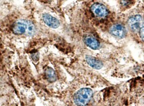
Cathepsin L in Human Kidney.
Cathepsin L was detected in immersion fixed paraffin-embedded sections of human kidney using Rat Anti-Human/Mouse Cathepsin L Monoclonal Antibody (Catalog # MAB9521) at 5 µg/mL overnight at 4 °C. Tissue was stained using the Anti-Rat HRP-DAB Cell & Tissue Staining Kit (brown; Catalog # CTS017) and counterstained with hematoxylin (blue). Specific staining was localized to cytoplasm in tubular epithelial cells. View our protocol for Chromogenic IHC Staining of Paraffin-embedded Tissue Sections.
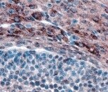
Cathepsin L in Mouse Kidney.
Cathepsin L was detected in immersion fixed frozen sections of mouse kidney using Rat Anti-Human/Mouse Cathepsin L Monoclonal Antibody (Catalog # MAB9521) at 8 µg/mL overnight at 4 °C. Tissue was stained using the Anti-Rat HRP-DAB Cell & Tissue Staining Kit (brown; Catalog # CTS017) and counterstained with hematoxylin (blue). Specific staining was localized to cytoplasm in tubular epithelial cells. View our protocol for Chromogenic IHC Staining of Frozen Tissue Sections.Applications for Cathepsin L Antibody (204101)
Application
Recommended Usage
Immunohistochemistry
5-25 µg/mL
Sample: Immersion fixed paraffin-embedded sections of human kidney and perfusion fixed frozen sections of mouse kidney
Sample: Immersion fixed paraffin-embedded sections of human kidney and perfusion fixed frozen sections of mouse kidney
Immunoprecipitation
25 µg/mL
Sample: Conditioned cell culture medium spiked with Recombinant Human Cathepsin L (Catalog # 952-CY) or Recombinant Mouse Cathepsin L (Catalog # 1515-CY), see our available Western blot detection antibodies. This antibody did not immunoprecipitate the pro form of recombinant mouse Cathepsin L.
Sample: Conditioned cell culture medium spiked with Recombinant Human Cathepsin L (Catalog # 952-CY) or Recombinant Mouse Cathepsin L (Catalog # 1515-CY), see our available Western blot detection antibodies. This antibody did not immunoprecipitate the pro form of recombinant mouse Cathepsin L.
Western Blot
1 µg/mL
Sample: Recombinant Human Cathepsin L (Catalog # 952-CY)
Recombinant Mouse Cathepsin L (Catalog # 1515-CY)
For natural samples, we recommend the use of Catalog # AF1515
Sample: Recombinant Human Cathepsin L (Catalog # 952-CY)
Recombinant Mouse Cathepsin L (Catalog # 1515-CY)
For natural samples, we recommend the use of Catalog # AF1515
Reviewed Applications
Read 4 reviews rated 4.8 using MAB9521 in the following applications:
Formulation, Preparation, and Storage
Purification
Protein A or G purified from hybridoma culture supernatant
Reconstitution
Reconstitute at 0.5 mg/mL in sterile PBS. For liquid material, refer to CoA for concentration.
Loading...
Formulation
Lyophilized from a 0.2 μm filtered solution in PBS with Trehalose. *Small pack size (SP) is supplied either lyophilized or as a 0.2 µm filtered solution in PBS.
Shipping
Lyophilized product is shipped at ambient temperature. Liquid small pack size (-SP) is shipped with polar packs. Upon receipt, store immediately at the temperature recommended below.
Stability & Storage
Use a manual defrost freezer and avoid repeated freeze-thaw cycles.
- 12 months from date of receipt, -20 to -70 °C as supplied.
- 1 month, 2 to 8 °C under sterile conditions after reconstitution.
- 6 months, -20 to -70 °C under sterile conditions after reconstitution.
Calculators
Background: Cathepsin L
References
- Goretzki, L. et al. (1992) FEBS Lett. 297:112.
- Taggart, C.C. et al. (2001) J. Biol. Chem. 276:33345.
- Gottesman, M.M. and F. Cabral (1981) Biochemistry 20:1659.
Alternate Names
CTSL, CTSL1
Gene Symbol
CTSL
UniProt
Additional Cathepsin L Products
Product Documents for Cathepsin L Antibody (204101)
Certificate of Analysis
To download a Certificate of Analysis, please enter a lot or batch number in the search box below.
Note: Certificate of Analysis not available for kit components.
Product Specific Notices for Cathepsin L Antibody (204101)
For research use only
Related Research Areas
Citations for Cathepsin L Antibody (204101)
 Powered by Bioz
Customer Reviews for Cathepsin L Antibody (204101) (4)
4.8 out of 5
4 Customer Ratings
Have you used Cathepsin L Antibody (204101)?
Submit a review and receive an Amazon gift card!
$25/€18/£15/$25CAN/¥2500 Yen for a review with an image
$10/€7/£6/$10CAN/¥1110 Yen for a review without an image
Submit a review
Customer Images
Showing
1
-
4 of
4 reviews
Showing All
Filter By:
-
Application: ImmunohistochemistrySample Tested: Liver tissueSpecies: MouseVerified Customer | Posted 07/09/2022

-
Application: ImmunohistochemistrySample Tested: Ovarian folliclesSpecies: MouseVerified Customer | Posted 10/04/2021
-
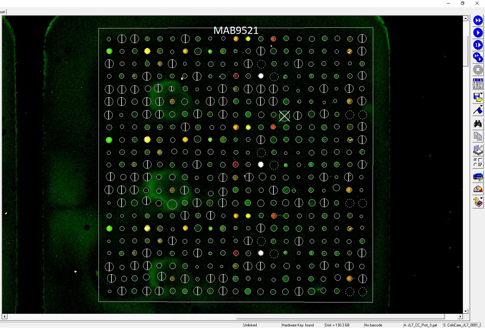
Application: MicroarraysSample Tested: EDTA PlasmaSpecies: HumanVerified Customer | Posted 03/17/2021
-
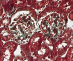
Application: ImmunohistochemistrySample Tested: Kidney tissueSpecies: MouseVerified Customer | Posted 08/05/2020
There are no reviews that match your criteria.
Protocols
Find general support by application which include: protocols, troubleshooting, illustrated assays, videos and webinars.
- Antigen Retrieval Protocol (PIER)
- Antigen Retrieval for Frozen Sections Protocol
- Appropriate Fixation of IHC/ICC Samples
- Cellular Response to Hypoxia Protocols
- Chromogenic IHC Staining of Formalin-Fixed Paraffin-Embedded (FFPE) Tissue Protocol
- Chromogenic Immunohistochemistry Staining of Frozen Tissue
- ClariTSA™ Fluorophore Kits
- Detection & Visualization of Antibody Binding
- Fluorescent IHC Staining of Frozen Tissue Protocol
- Graphic Protocol for Heat-induced Epitope Retrieval
- Graphic Protocol for the Preparation and Fluorescent IHC Staining of Frozen Tissue Sections
- Graphic Protocol for the Preparation and Fluorescent IHC Staining of Paraffin-embedded Tissue Sections
- Graphic Protocol for the Preparation of Gelatin-coated Slides for Histological Tissue Sections
- IHC Sample Preparation (Frozen sections vs Paraffin)
- Immunofluorescent IHC Staining of Formalin-Fixed Paraffin-Embedded (FFPE) Tissue Protocol
- Immunohistochemistry (IHC) and Immunocytochemistry (ICC) Protocols
- Immunohistochemistry Frozen Troubleshooting
- Immunohistochemistry Paraffin Troubleshooting
- Immunoprecipitation Protocol
- Preparing Samples for IHC/ICC Experiments
- Preventing Non-Specific Staining (Non-Specific Binding)
- Primary Antibody Selection & Optimization
- Protocol for Heat-Induced Epitope Retrieval (HIER)
- Protocol for Making a 4% Formaldehyde Solution in PBS
- Protocol for VisUCyte™ HRP Polymer Detection Reagent
- Protocol for the Preparation & Fixation of Cells on Coverslips
- Protocol for the Preparation and Chromogenic IHC Staining of Frozen Tissue Sections
- Protocol for the Preparation and Chromogenic IHC Staining of Frozen Tissue Sections - Graphic
- Protocol for the Preparation and Chromogenic IHC Staining of Paraffin-embedded Tissue Sections
- Protocol for the Preparation and Chromogenic IHC Staining of Paraffin-embedded Tissue Sections - Graphic
- Protocol for the Preparation and Fluorescent IHC Staining of Frozen Tissue Sections
- Protocol for the Preparation and Fluorescent IHC Staining of Paraffin-embedded Tissue Sections
- Protocol for the Preparation of Gelatin-coated Slides for Histological Tissue Sections
- R&D Systems Quality Control Western Blot Protocol
- TUNEL and Active Caspase-3 Detection by IHC/ICC Protocol
- The Importance of IHC/ICC Controls
- Troubleshooting Guide: Immunohistochemistry
- Troubleshooting Guide: Western Blot Figures
- Western Blot Conditions
- Western Blot Protocol
- Western Blot Protocol for Cell Lysates
- Western Blot Troubleshooting
- Western Blot Troubleshooting Guide
- View all Protocols, Troubleshooting, Illustrated assays and Webinars
Loading...