Human/Mouse PDX-1/IPF1 Antibody Summary
Ala91-Arg283
Accession # P52945
*Small pack size (-SP) is supplied either lyophilized or as a 0.2 µm filtered solution in PBS.
Applications
Please Note: Optimal dilutions should be determined by each laboratory for each application. General Protocols are available in the Technical Information section on our website.
Scientific Data
 View Larger
Detection of Mouse PDX‑1/IPF1 by Western Blot. Western blot shows lysates of beta TC-6 mouse beta cell insulinoma cell line. PVDF membrane was probed with 1 µg/mL of Mouse Anti-Human/Mouse PDX-1/IPF1 Monoclonal Antibody (Catalog # MAB2419) followed by HRP-conjugated Anti-Mouse IgG Secondary Antibody (Catalog # HAF007). A specific band was detected for PDX-1/IPF1 at approximately 46 kDa (as indicated). This experiment was conducted under reducing conditions and using Immunoblot Buffer Group 1.
 View Larger
PDX‑1/IPF1 in beta TC‑6 Mouse Cell Line. PDX-1/IPF1 was detected in immersion fixed beta TC-6 mouse beta cell insulinoma cell line using Human/Mouse PDX-1/IPF1 Monoclonal Antibody (Catalog # MAB2419) at 10 µg/mL for 3 hours at room temperature. Cells were stained using the NorthernLights™ 557-conjugated Anti-Mouse IgG Secondary Antibody (yellow, upper panel; Catalog # NL007) and counterstained with DAPI (blue, lower panel). View our protocol for Fluorescent ICC Staining of Cells on Coverslips.
 View Larger
PDX‑1/IPF1 in BG01V Human Embryonic Stem Cells. PDX-1/IPF1 was detected in immersion fixed BG01V human embryonic stem cells differentiated into pancreatic progenitor cells using Mouse Anti-Human/Mouse PDX-1/IPF1 Monoclonal Antibody (Catalog # MAB2419) at 10 µg/mL for 3 hours at room temperature. Cells were stained using the NorthernLights™ 557-conjugated Anti-Mouse IgG Secondary Antibody (red; Catalog # NL007) and counterstained with DAPI (blue). Specific staining was localized to nuclei. View our protocol for Fluorescent ICC Staining of Stem Cells on Coverslips.
 View Larger
Detection of PDX‑1/IPF1 in beta TC‑6 Mouse Cell Line by Flow Cytometry. beta TC-6 mouse beta cell insulinoma cell line was stained with Mouse Anti-Human/Mouse PDX-1/IPF1 Monoclonal Antibody (Catalog # MAB2419, filled histogram) or isotype control antibody (MAB0041, open histogram) followed by anti-Mouse IgG PE-conjugated secondary antibody (F0102B). To facilitate intracellular staining, cells were fixed and permeabilized with FlowX FoxP3 Fixation & Permeabilization Buffer Kit (FC012). View our protocol for Staining Intracellular Molecules.
 View Larger
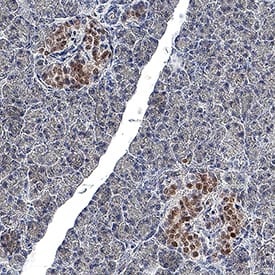
Detection of PDX‑1/IPF1 in Human Pancreas. PDX‑1/IPF1 was detected in immersion fixed paraffin-embedded sections of human pancreas using Mouse Anti-Human/Mouse PDX‑1/IPF1 Monoclonal Antibody (Catalog # MAB2419) at 5 µg/ml for 1 hour at room temperature followed by incubation with the Anti-Mouse IgG VisUCyte™ HRP Polymer Antibody (Catalog # VC001). Before incubation with the primary antibody, tissue was subjected to heat-induced epitope retrieval using VisUCyte Antigen Retrieval Reagent-Basic (Catalog # VCTS021). Tissue was stained using DAB (brown) and counterstained with hematoxylin (blue). Specific staining was localized to the nucleus in islet cells. View our protocol for IHC Staining with VisUCyte HRP Polymer Detection Reagents.
Reconstitution Calculator
Preparation and Storage
- 12 months from date of receipt, -20 to -70 °C as supplied.
- 1 month, 2 to 8 °C under sterile conditions after reconstitution.
- 6 months, -20 to -70 °C under sterile conditions after reconstitution.
Background: PDX-1/IPF1
Pancreatic-Duodenal Homeobox Factor-1 (PDX-1), also known as Insulin Promoter Factor 1 (IPF1), is a homeodomain transcription factor essential for pancreatic development and mature pancreatic beta cell function.
Product Datasheets
Citations for Human/Mouse PDX-1/IPF1 Antibody
R&D Systems personnel manually curate a database that contains references using R&D Systems products. The data collected includes not only links to publications in PubMed, but also provides information about sample types, species, and experimental conditions.
12
Citations: Showing 1 - 10
Filter your results:
Filter by:
-
YAP1 and TAZ Control Pancreatic Cancer Initiation in Mice by Direct Up-regulation of JAK–STAT3 Signaling
Authors: Ralph Gruber, Richard Panayiotou, Emma Nye, Bradley Spencer-Dene, Gordon Stamp, Axel Behrens
Gastroenterology
-
DNA methylation reveals distinct cells of origin for pancreatic neuroendocrine carcinomas and pancreatic neuroendocrine tumors
Authors: Tincy Simon, Pamela Riemer, Armin Jarosch, Katharina Detjen, Annunziata Di Domenico, Felix Bormann et al.
Genome Medicine
-
Intrinsically disordered substrates dictate SPOP subnuclear localization and ubiquitination activity
Authors: ET Usher, N Sabri, R Rohac, AK Boal, T Mittag, SA Showalter
The Journal of Biological Chemistry, 2021-04-22;0(0):100693.
Species: Human
Sample Types: Cell Lysates
Applications: Western Blot -
A programmable synthetic lineage-control network that differentiates human IPSCs into glucose-sensitive insulin-secreting beta-like cells
Authors: P Saxena, BC Heng, P Bai, M Folcher, H Zulewski, M Fussenegge
Nat Commun, 2016-04-11;7(0):11247.
Species: Human
Sample Types: Whole Cells
Applications: IHC -
Redifferentiation of adult human beta cells expanded in vitro by inhibition of the WNT pathway.
Authors: Lenz A, Toren-Haritan G, Efrat S
PLoS ONE, 2014-11-13;9(11):e112914.
Species: Human
Sample Types: Whole Cells
Applications: ICC -
Peroxisome proliferator-activated receptor gamma activation restores islet function in diabetic mice through reduction of endoplasmic reticulum stress and maintenance of euchromatin structure.
Authors: Evans-Molina C, Robbins RD, Kono T, Tersey SA, Vestermark GL, Nunemaker CS, Garmey JC, Deering TG, Keller SR, Maier B, Mirmira RG
Mol. Cell. Biol., 2009-02-23;29(8):2053-67.
Species: Mouse
Sample Types: Whole Tissue
Applications: IHC-P -
Betacellulin and nicotinamide sustain PDX1 expression and induce pancreatic beta-cell differentiation in human embryonic stem cells.
Authors: Cho YM, Lim JM, Yoo DH, Kim JH, Chung SS, Park SG, Kim TH, Oh SK, Choi YM, Moon SY, Park KS, Lee HK
Biochem. Biophys. Res. Commun., 2007-12-04;366(1):129-34.
Species: Human
Sample Types: Whole Cells
Applications: ICC -
Generation of insulin-producing cells from PDX-1 gene-modified human mesenchymal stem cells.
Authors: Li Y, Zhang R, Qiao H, Zhang H, Wang Y, Yuan H, Liu Q, Liu D, Chen L, Pei X
J. Cell. Physiol., 2007-04-01;211(1):36-44.
Species: Human
Sample Types: Whole Cells
Applications: ICC -
Epigenetic landscape of pancreatic neuroendocrine tumours reveals distinct cells of origin and means of tumour progression
Authors: Annunziata Di Domenico, Christodoulos P. Pipinikas, Renaud S. Maire, Konstantin Bräutigam, Cedric Simillion, Matthias S. Dettmer et al.
Communications Biology
-
The combined effect of PDX1, epidermal growth factor and poly-L-ornithine on human amnion epithelial cells’ differentiation
Authors: Shruti Balaji, Yu Zhou, Anasuya Ganguly, Emmanuel C. Opara, Shay Soker
BMC Developmental Biology
-
Coxsackievirus B Type 4 Infection in beta Cells Downregulates the Chaperone Prefoldin URI to Induce a MODY4-like Diabetes via Pdx1 Silencing
Authors: Bernard H, Teijeiro A, Chaves-Perez A et al.
Cell Reports Medicine
-
Pdx1 Expression in Pancreatic Precursor Lesions and Neoplasms
Authors: Jason Y. Park, Seung-Mo Hong, David S. Klimstra, Michael G. Goggins, Anirban Maitra, Ralph H. Hruban
Applied Immunohistochemistry & Molecular Morphology
FAQs
No product specific FAQs exist for this product, however you may
View all Antibody FAQsReviews for Human/Mouse PDX-1/IPF1 Antibody
Average Rating: 5 (Based on 1 Review)
Have you used Human/Mouse PDX-1/IPF1 Antibody?
Submit a review and receive an Amazon gift card.
$25/€18/£15/$25CAN/¥75 Yuan/¥2500 Yen for a review with an image
$10/€7/£6/$10 CAD/¥70 Yuan/¥1110 Yen for a review without an image
Filter by:


