
Key Product Details
Species Reactivity
Human, Mouse, Rat
Applications
Western Blot, Simple Western
Label
Unconjugated
Antibody Source
Monoclonal Mouse IgG1 Clone # 328605
Loading...
Product Specifications
Immunogen
E. coli-derived recombinant human FABP1/L-FABP
Met1-Ile127
Accession # P07148
Met1-Ile127
Accession # P07148
Specificity
Detects human FABP1/L-FABP in direct ELISAs and Western blots. In direct ELISAs and Western blots, no cross-reactivity with rhFABP2, -3, -4, -5, -6, -7, or rmFABP9 is observed.
Clonality
Monoclonal
Host
Mouse
Isotype
IgG1
Scientific Data Images for FABP1/L-FABP Antibody (328605)
Detection of Human FABP1/L‑FABP by Western Blot.
Western blot shows lysates of human liver tissue, human colon tissue, and human kidney tissue. PVDF membrane was probed with 0.25 µg/mL of Mouse Anti-Human/Mouse/Rat FABP1/L-FABP Monoclonal Antibody (Catalog # MAB29641) followed by HRP-conjugated Anti-Mouse IgG Secondary Antibody (Catalog # HAF018). A specific band was detected for FABP1/ L-FABP at approximately 14 kDa (as indicated). This experiment was conducted under reducing conditions and using Immunoblot Buffer Group 1.
Detection of Human, Mouse, and Rat FABP1/L‑FABP by Western Blot.
Western blot shows lysates of HepG2 human hepatocellular carcinoma cell line, mouse liver tissue, and rat liver tissue. PVDF membrane was probed with 0.25 µg/mL of Mouse Anti-Human/Mouse/Rat FABP1/ L-FABP Monoclonal Antibody (Catalog # MAB29641) followed by HRP-conjugated Anti-Mouse IgG Secondary Antibody (Catalog # HAF018). A specific band was detected for FABP1/L-FABP at approximately 14 kDa (as indicated). This experiment was conducted under reducing conditions and using Immunoblot Buffer Group 1.
Detection of Human FABP1/L‑FABP by Simple WesternTM.
Simple Western lane view shows lysates of human liver tissue, loaded at 0.2 mg/mL. A specific band was detected for FABP1/L‑FABP at approximately 14 kDa (as indicated) using 10 µg/mL of Mouse Anti-Human/Mouse/Rat FABP1/ L‑FABP Monoclonal Antibody (Catalog # MAB29641). This experiment was conducted under reducing conditions and using the 12-230 kDa separation system.
Detection of Human and Mouse FABP1/L‑FABP by Simple WesternTM.
Simple Western lane view shows lysates of HepG2 human hepatocellular carcinoma cell line and mouse liver tissue, loaded at 0.2 mg/mL. A specific band was detected for FABP1/L‑FABP at approximately 14-16 kDa (as indicated) using 5 µg/mL of Mouse Anti-Human/Mouse/Rat FABP1/L‑FABP Monoclonal Antibody (Catalog # MAB29641). This experiment was conducted under reducing conditions and using the 12-230 kDa separation system.Applications for FABP1/L-FABP Antibody (328605)
Application
Recommended Usage
Simple Western
5-10 µg/mL
Sample: Human liver tissue, HepG2 human hepatocellular carcinoma cell line, and mouse liver tissue
Sample: Human liver tissue, HepG2 human hepatocellular carcinoma cell line, and mouse liver tissue
Western Blot
0.25 µg/mL
Sample: Human liver tissue, human colon tissue, human kidney tissue, HepG2 human hepatocellular carcinoma cell line, mouse liver tissue, and rat liver tissue
Sample: Human liver tissue, human colon tissue, human kidney tissue, HepG2 human hepatocellular carcinoma cell line, mouse liver tissue, and rat liver tissue
Reviewed Applications
Read 3 reviews rated 4.3 using MAB29641 in the following applications:
Formulation, Preparation, and Storage
Purification
Protein A or G purified from hybridoma culture supernatant
Reconstitution
Reconstitute at 0.5 mg/mL in sterile PBS. For liquid material, refer to CoA for concentration.
Loading...
Formulation
Lyophilized from a 0.2 μm filtered solution in PBS with Trehalose. *Small pack size (SP) is supplied either lyophilized or as a 0.2 µm filtered solution in PBS.
Shipping
Lyophilized product is shipped at ambient temperature. Liquid small pack size (-SP) is shipped with polar packs. Upon receipt, store immediately at the temperature recommended below.
Stability & Storage
Use a manual defrost freezer and avoid repeated freeze-thaw cycles.
- 12 months from date of receipt, -20 to -70 °C as supplied.
- 1 month, 2 to 8 °C under sterile conditions after reconstitution.
- 6 months, -20 to -70 °C under sterile conditions after reconstitution.
Calculators
Background: FABP1/L-FABP
Long Name
Fatty Acid-Binding Protein 1
Alternate Names
L-FABP, LFABP
Gene Symbol
FABP1
UniProt
Additional FABP1/L-FABP Products
Product Documents for FABP1/L-FABP Antibody (328605)
Certificate of Analysis
To download a Certificate of Analysis, please enter a lot or batch number in the search box below.
Note: Certificate of Analysis not available for kit components.
Product Specific Notices for FABP1/L-FABP Antibody (328605)
For research use only
Related Research Areas
Citations for FABP1/L-FABP Antibody (328605)
 Powered by Bioz
Customer Reviews for FABP1/L-FABP Antibody (328605) (3)
4.3 out of 5
3 Customer Ratings
Have you used FABP1/L-FABP Antibody (328605)?
Submit a review and receive an Amazon gift card!
$25/€18/£15/$25CAN/¥2500 Yen for a review with an image
$10/€7/£6/$10CAN/¥1110 Yen for a review without an image
Submit a review
Customer Images

Showing
1
-
3 of
3 reviews
Showing All
Filter By:
-
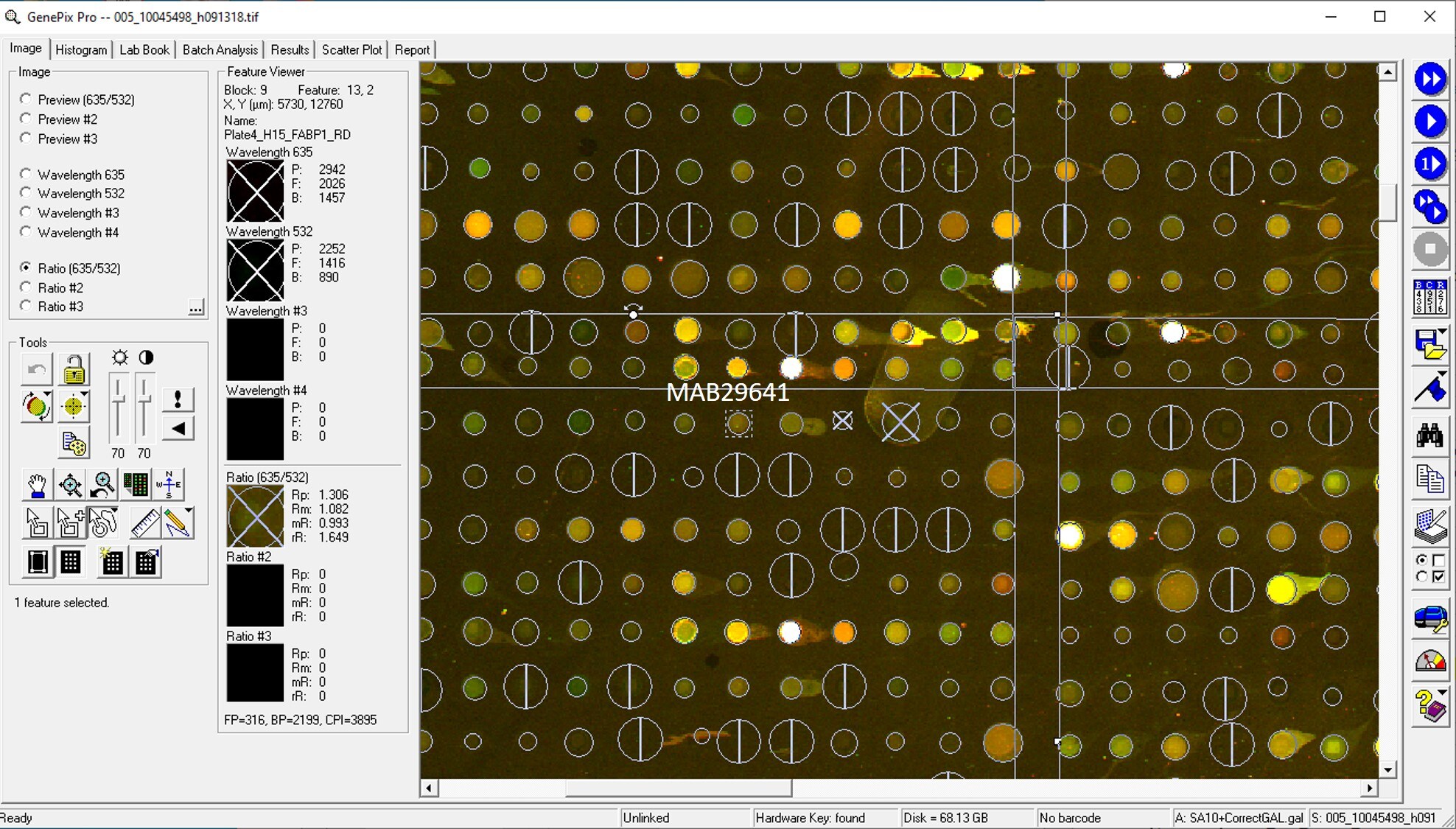
Application: MicroarraysSample Tested: EDTA PlasmaSpecies: HumanVerified Customer | Posted 06/10/2020

-
Application: MicroarraySample Tested: EDTA PlasmaSpecies: HumanVerified Customer | Posted 02/07/2020Antibody was printed on custom arrays and incubated with fluorescently labeled human EDTA plasma
-
Application: MicroarraysSample Tested: EDTA PlasmaSpecies: HumanVerified Customer | Posted 11/07/2018
There are no reviews that match your criteria.
Protocols
Find general support by application which include: protocols, troubleshooting, illustrated assays, videos and webinars.
- Cellular Response to Hypoxia Protocols
- R&D Systems Quality Control Western Blot Protocol
- Troubleshooting Guide: Western Blot Figures
- Western Blot Conditions
- Western Blot Protocol
- Western Blot Protocol for Cell Lysates
- Western Blot Troubleshooting
- Western Blot Troubleshooting Guide
- View all Protocols, Troubleshooting, Illustrated assays and Webinars
Loading...