GLG1 (Golgi complex-Localized Glycoprotein 1; also CFR1, E-Selectin ligand-1/ESL-1, MG-160 and Cys-rich FGF receptor) is a 150-160 kDa (reducing; 130 kDa nonreducing) glycoprotein. It is expressed in both Golgi and/or the cell membrane of multiple cell types, including neutrophils (from rodents; not humans), liver stellate cells, neurons, cardiac myocytes, monocytes and bronchial epithelial cells. In the blood, GLG1/ESL-1 collaborates with PSGL-1 to mediate leukocyte binding to endothelial cell surfaces. PSGL-1 initiates leukocyte tethering while GLG1 promotes slow rolling. GLG1 also serves as an intra-Golgi receptor for multiple FGFs, including FGF-1, -2, -4, -18 and possibly -3, and as a component of an unusual latent TGF-beta complex. Mature human GLG1 is an 1150 amino acid (aa) type I transmembrane protein. It contains a 1116 aa extracellular/luminal region (aa 30-1145) plus a short 13 aa cytoplasmic segment. The extracellular region possesses a 16 aa poly-Gln segment followed by 16 Cys-rich repeats (aa 116-1101). There are three potential isoform variants, one of which possess a 24 aa extension at the C‑terminus, a second that couples the aforementioned C‑terminal extension to a deletion of aa 147-157, and a third that contains a 14 aa substitution for aa 685-1179. It is suggested that the longer C‑terminus retains GLG1 in the Golgi, while shorter cytoplasmic segments allow for presentation at the cell membrane. Over aa 1048‑1145, human and mouse are identical in aa sequence.

Key Product Details
Species Reactivity
Human, Mouse, Rat
Applications
Immunohistochemistry, Western Blot, Immunocytochemistry
Label
Unconjugated
Antibody Source
Polyclonal Sheep IgG
Loading...
Product Specifications
Immunogen
E. coli-derived recombinant human Golgi Glycoprotein 1/GLG1
Lys1048-Asn1145
Accession # Q92896
Lys1048-Asn1145
Accession # Q92896
Specificity
Detects human Golgi Glycoprotein 1/GLG1 in direct ELISAs and Western blots.
Clonality
Polyclonal
Host
Sheep
Isotype
IgG
Scientific Data Images for Golgi Glycoprotein 1/GLG1 Antibody
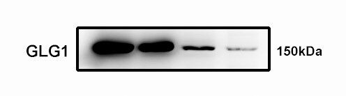
Detection of Human Golgi Glycoprotein 1/GLG1 by Western Blot.
Western blot shows lysates of HepG2 human hepatocellular carcinoma cell line and NTera-2 human testicular embryonic carcinoma cell line. PVDF membrane was probed with 2 µg/mL of Sheep Anti-Human/Mouse/Rat Golgi Glycoprotein 1/GLG1 Antigen Affinity-purified Polyclonal Antibody (Catalog # AF7879) followed by HRP-conjugated Anti-Sheep IgG Secondary Antibody (HAF016). A specific band was detected for Golgi Glycoprotein 1/GLG1 at approximately 150 kDa (as indicated). This experiment was conducted under reducing conditions and using Immunoblot Buffer Group 8.
Golgi Glycoprotein 1/GLG1 in PC‑12 Rat Cell Line.
Golgi Glycoprotein 1/GLG1 was detected in immersion fixed PC-12 rat adrenal pheochromocytoma cell line using Sheep Anti-Human/Mouse/Rat Golgi Glycoprotein 1/GLG1 Antigen Affinity-purified Polyclonal Antibody (Catalog # AF7879) at 1.7 µg/mL for 3 hours at room temperature. Cells were stained using the NorthernLights™ 557-conjugated Anti-Sheep IgG Secondary Antibody (red; NL010) and counterstained with DAPI (blue). Specific staining was localized to the Golgi complex. View our protocol for Fluorescent ICC Staining of Cells on Coverslips.
Golgi Glycoprotein 1/GLG1 in Human Astrocytoma.
Golgi Glycoprotein 1/GLG1 was detected in immersion fixed paraffin-embedded sections of human astrocytoma using Sheep Anti-Human/Mouse/Rat Golgi Glycoprotein 1/GLG1 Antigen Affinity-purified Polyclonal Antibody (Catalog # AF7879) at 3 µg/mL for 1 hour at room temperature followed by incubation with the Anti-Sheep IgG VisUCyte™ HRP Polymer Antibody (Catalog # VC006). Before incubation with the primary antibody, tissue was subjected to heat-induced epitope retrieval using Antigen Retrieval Reagent-Basic (Catalog # CTS013). Tissue was stained using DAB (brown) and counterstained with hematoxylin (blue). Specific staining was localized to Golgi apparatus. View our protocol for IHC Staining with VisUCyte HRP Polymer Detection Reagents. & NIH‑3T3 mouse embryonic fibroblast cell line (Negative).")
Detection of Golgi Glycoprotein 1/GLG1 in HepG2 human hepatocellular carcinoma cell line (Positive) & NIH‑3T3 mouse embryonic fibroblast cell line (Negative).
Golgi Glycoprotein 1/GLG1 was detected in immersion fixed HepG2 human hepatocellular carcinoma cell line (Positive) & NIH‑3T3 mouse embryonic fibroblast cell line (Negative) using Sheep Anti-Human/Mouse/Rat Golgi Glycoprotein 1/GLG1 Antigen Affinity-purified Polyclonal Antibody (Catalog # AF7879) at 5 µg/mL for 3 hours at room temperature. Cells were stained using the NorthernLights™ 557-conjugated Anti-Sheep IgG Secondary Antibody (red; Catalog # NL010) and counterstained with DAPI (blue). Specific staining was localized to cytoplasm. View our protocol for Fluorescent ICC Staining of Cells on Coverslips.Applications for Golgi Glycoprotein 1/GLG1 Antibody
Application
Recommended Usage
Immunocytochemistry
5-15 µg/mL
Sample: Immersion fixed PC‑12 rat adrenal pheochromocytoma cell line, HepG2 human hepatocellular carcinoma cell line (Positive) & NIH‑3T3 mouse embryonic fibroblast cell line (Negative)
Sample: Immersion fixed PC‑12 rat adrenal pheochromocytoma cell line, HepG2 human hepatocellular carcinoma cell line (Positive) & NIH‑3T3 mouse embryonic fibroblast cell line (Negative)
Immunohistochemistry
3-25 µg/mL
Sample: Immersion fixed paraffin-embedded sections of human astrocytoma
Sample: Immersion fixed paraffin-embedded sections of human astrocytoma
Western Blot
2 µg/mL
Sample: HepG2 human hepatocellular carcinoma cell line and NTera‑2 human testicular embryonic carcinoma cell line
Sample: HepG2 human hepatocellular carcinoma cell line and NTera‑2 human testicular embryonic carcinoma cell line
Reviewed Applications
Read 2 reviews rated 2.5 using AF7879 in the following applications:
Formulation, Preparation, and Storage
Purification
Antigen Affinity-purified
Reconstitution
Sterile PBS to a final concentration of 0.2 mg/mL. For liquid material, refer to CoA for concentration.
Loading...
Formulation
Lyophilized from a 0.2 μm filtered solution in PBS with Trehalose. *Small pack size (SP) is supplied either lyophilized or as a 0.2 µm filtered solution in PBS.
Shipping
Lyophilized product is shipped at ambient temperature. Liquid small pack size (-SP) is shipped with polar packs. Upon receipt, store immediately at the temperature recommended below.
Stability & Storage
Use a manual defrost freezer and avoid repeated freeze-thaw cycles.
- 12 months from date of receipt, -20 to -70 °C as supplied.
- 1 month, 2 to 8 °C under sterile conditions after reconstitution.
- 6 months, -20 to -70 °C under sterile conditions after reconstitution.
Calculators
Background: Golgi Glycoprotein 1/GLG1
Alternate Names
CFR-1, ESL-1, GLG1, MG160
Gene Symbol
GLG1
UniProt
Additional Golgi Glycoprotein 1/GLG1 Products
Product Documents for Golgi Glycoprotein 1/GLG1 Antibody
Certificate of Analysis
To download a Certificate of Analysis, please enter a lot or batch number in the search box below.
Note: Certificate of Analysis not available for kit components.
Product Specific Notices for Golgi Glycoprotein 1/GLG1 Antibody
For research use only
Related Research Areas
Customer Reviews for Golgi Glycoprotein 1/GLG1 Antibody (2)
2.5 out of 5
2 Customer Ratings
Have you used Golgi Glycoprotein 1/GLG1 Antibody?
Submit a review and receive an Amazon gift card!
$25/€18/£15/$25CAN/¥2500 Yen for a review with an image
$10/€7/£6/$10CAN/¥1110 Yen for a review without an image
Submit a review
Customer Images
Showing
1
-
2 of
2 reviews
Showing All
Filter By:
-
Application: Western BlotSample Tested: 4T1 mouse breast cancer cell lineSpecies: MouseVerified Customer | Posted 02/16/2020

-
Application: Western BlotSample Tested: MDA-MB-231 human breast cancer cell lineSpecies: HumanVerified Customer | Posted 12/10/2018Cells that had verified Glg1 overexpression or knockdown/knockout were lysed and probed with this antibody at 1:1000 in combination with Beta-actin. Glg1 was not detectable even at a very high exposure and image.
Bio-Techne ResponseThank you for reviewing our product. We are sorry to hear that this antibody did not perform as expected. We have been in touch with the customer to resolve this issue according to our Product Guarantee and to the customer’s satisfaction.
There are no reviews that match your criteria.
Protocols
Find general support by application which include: protocols, troubleshooting, illustrated assays, videos and webinars.
- Antigen Retrieval Protocol (PIER)
- Antigen Retrieval for Frozen Sections Protocol
- Appropriate Fixation of IHC/ICC Samples
- Cellular Response to Hypoxia Protocols
- Chromogenic IHC Staining of Formalin-Fixed Paraffin-Embedded (FFPE) Tissue Protocol
- Chromogenic Immunohistochemistry Staining of Frozen Tissue
- ClariTSA™ Fluorophore Kits
- Detection & Visualization of Antibody Binding
- Fluorescent IHC Staining of Frozen Tissue Protocol
- Graphic Protocol for Heat-induced Epitope Retrieval
- Graphic Protocol for the Preparation and Fluorescent IHC Staining of Frozen Tissue Sections
- Graphic Protocol for the Preparation and Fluorescent IHC Staining of Paraffin-embedded Tissue Sections
- Graphic Protocol for the Preparation of Gelatin-coated Slides for Histological Tissue Sections
- ICC Cell Smear Protocol for Suspension Cells
- ICC Immunocytochemistry Protocol Videos
- ICC for Adherent Cells
- IHC Sample Preparation (Frozen sections vs Paraffin)
- Immunocytochemistry (ICC) Protocol
- Immunocytochemistry Troubleshooting
- Immunofluorescence of Organoids Embedded in Cultrex Basement Membrane Extract
- Immunofluorescent IHC Staining of Formalin-Fixed Paraffin-Embedded (FFPE) Tissue Protocol
- Immunohistochemistry (IHC) and Immunocytochemistry (ICC) Protocols
- Immunohistochemistry Frozen Troubleshooting
- Immunohistochemistry Paraffin Troubleshooting
- Preparing Samples for IHC/ICC Experiments
- Preventing Non-Specific Staining (Non-Specific Binding)
- Primary Antibody Selection & Optimization
- Protocol for Heat-Induced Epitope Retrieval (HIER)
- Protocol for Making a 4% Formaldehyde Solution in PBS
- Protocol for VisUCyte™ HRP Polymer Detection Reagent
- Protocol for the Fluorescent ICC Staining of Cell Smears - Graphic
- Protocol for the Fluorescent ICC Staining of Cultured Cells on Coverslips - Graphic
- Protocol for the Preparation & Fixation of Cells on Coverslips
- Protocol for the Preparation and Chromogenic IHC Staining of Frozen Tissue Sections
- Protocol for the Preparation and Chromogenic IHC Staining of Frozen Tissue Sections - Graphic
- Protocol for the Preparation and Chromogenic IHC Staining of Paraffin-embedded Tissue Sections
- Protocol for the Preparation and Chromogenic IHC Staining of Paraffin-embedded Tissue Sections - Graphic
- Protocol for the Preparation and Fluorescent ICC Staining of Cells on Coverslips
- Protocol for the Preparation and Fluorescent ICC Staining of Non-adherent Cells
- Protocol for the Preparation and Fluorescent ICC Staining of Stem Cells on Coverslips
- Protocol for the Preparation and Fluorescent IHC Staining of Frozen Tissue Sections
- Protocol for the Preparation and Fluorescent IHC Staining of Paraffin-embedded Tissue Sections
- Protocol for the Preparation of Gelatin-coated Slides for Histological Tissue Sections
- Protocol for the Preparation of a Cell Smear for Non-adherent Cell ICC - Graphic
- R&D Systems Quality Control Western Blot Protocol
- TUNEL and Active Caspase-3 Detection by IHC/ICC Protocol
- The Importance of IHC/ICC Controls
- Troubleshooting Guide: Immunohistochemistry
- Troubleshooting Guide: Western Blot Figures
- Western Blot Conditions
- Western Blot Protocol
- Western Blot Protocol for Cell Lysates
- Western Blot Troubleshooting
- Western Blot Troubleshooting Guide
- View all Protocols, Troubleshooting, Illustrated assays and Webinars
Loading...